2.22 Tendinosis and Tendon Ruptures
1/56
There's no tags or description
Looks like no tags are added yet.
Name | Mastery | Learn | Test | Matching | Spaced | Call with Kai |
|---|
No analytics yet
Send a link to your students to track their progress
57 Terms
What are the flexor tendons?
Superficial Digital Flexor Tendon (SDFT)
Deep Digital Flexor Tendon (DDFT)
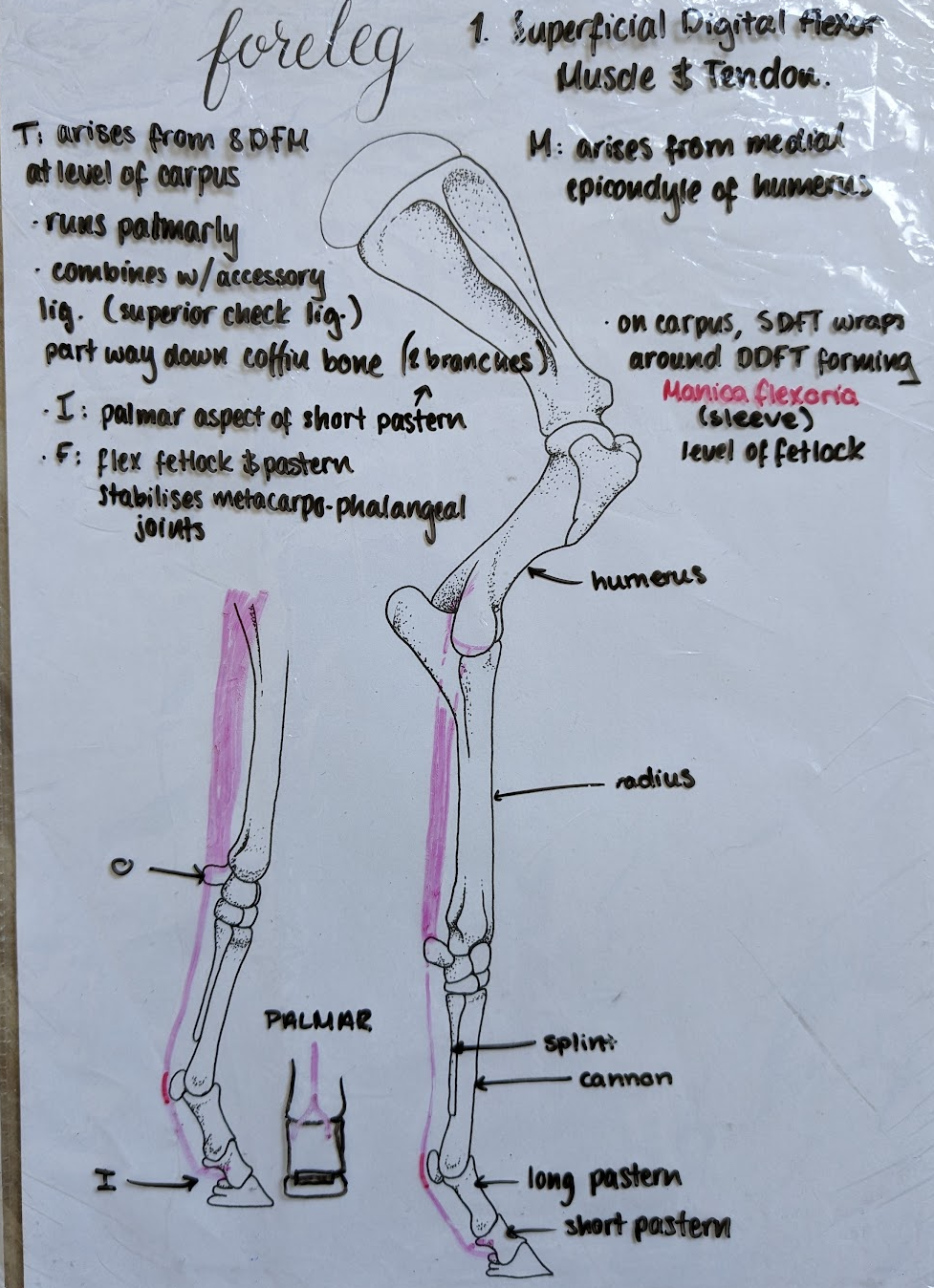
Short pastern bone.
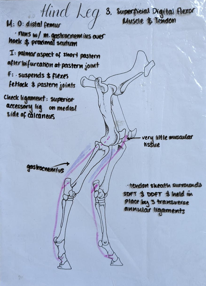
Where is the origin and insertion of the SDFT in the hind leg?
Origin: Proximal tibia
Insertion: Short pastern bone
What is the function of the superficial digital flexor tendon?
Flexes and stabilises the fetlock.
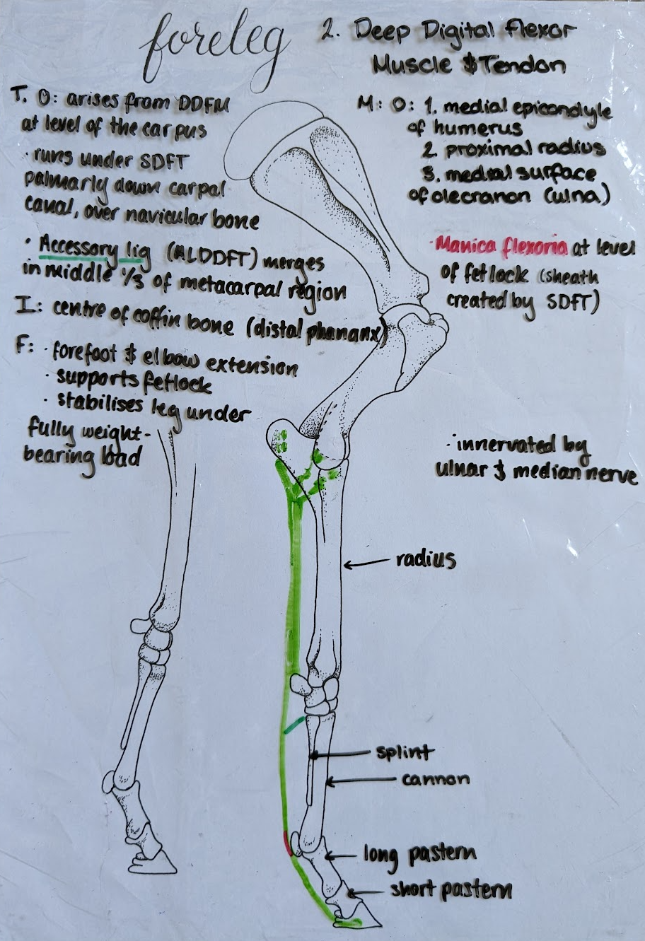
Coffin bone.
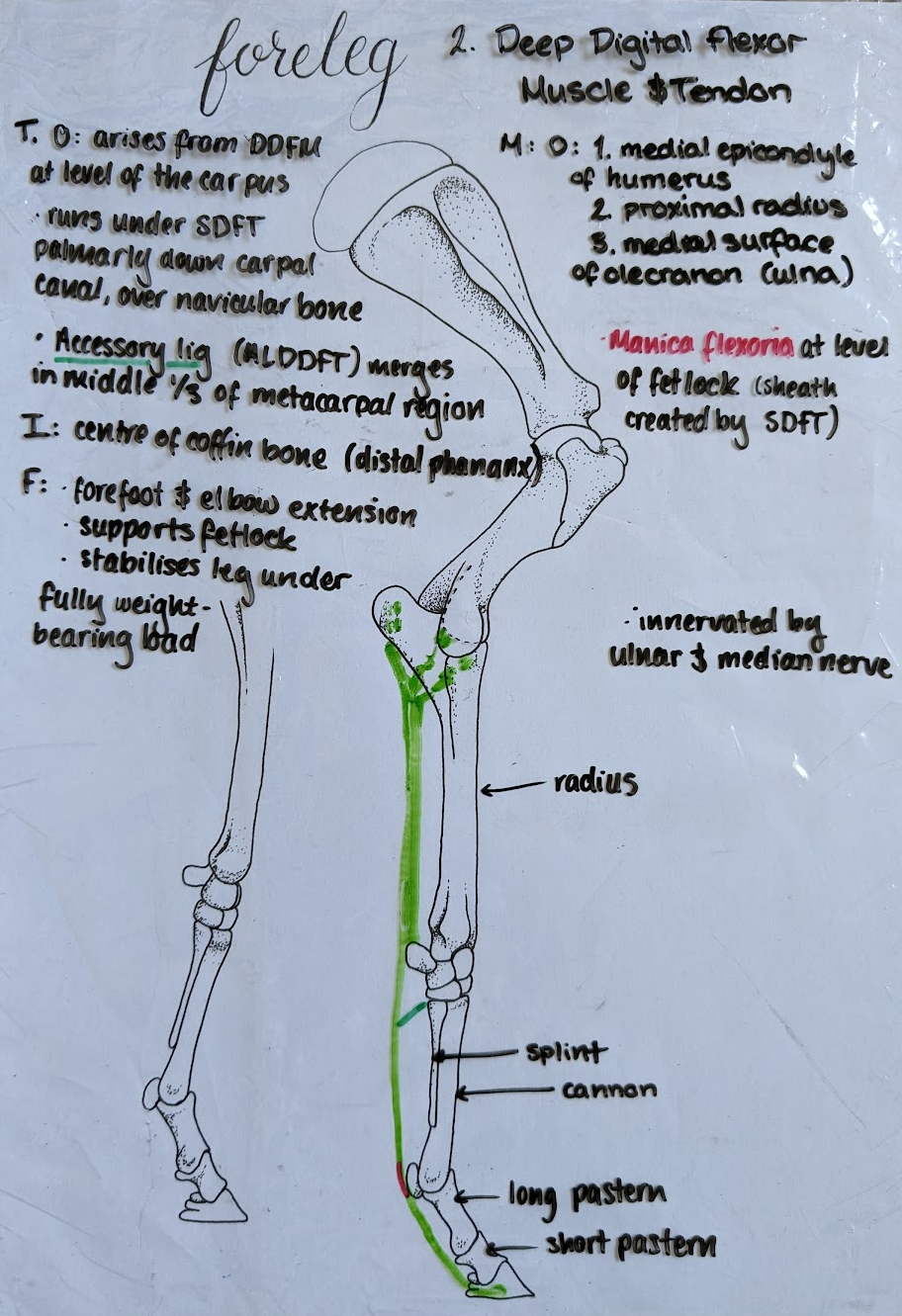
Where is the origin and insertion of the DDFT in the hind limb?
Origin: from deep digital flexor muscle at the level of the tarsus/hock
Insertion: Coffin bone
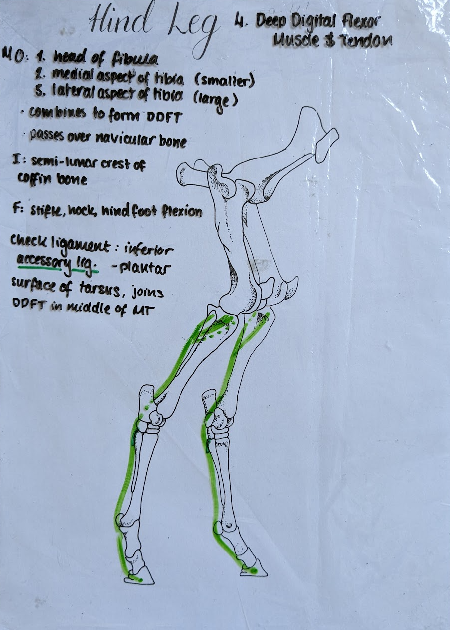
What is the function of the deep digital flexor tendon (DDFT)?
Flexes the coffin bone.
Accessory ligaments connecting the flexor tendons to bone.
What are the check ligaments, which tendon do they attach, and to where?
Proximal check ligament – belongs to SDFT, attached to radius or calcaneus.
Distal check ligament – belongs to DDFT, attached to the cannon bone.
What are the extensor tendons on the limbs?
Common digital extensor tendon
Lateral digital extensor tendon
Long digital extensor tendon
Where does the common digital extensor tendon originate and insert in the front leg?
Origin: CDE muscle originates from lateral humeral epicondyle.
Insertion: Coffin bone
What are the origins and insertions of the lateral digital extensor tendon in the front and hind limbs?
Front Origin: Proximal to carpus
Hind Origin: Proximal to hock/tarsus
Insertion: Long pastern
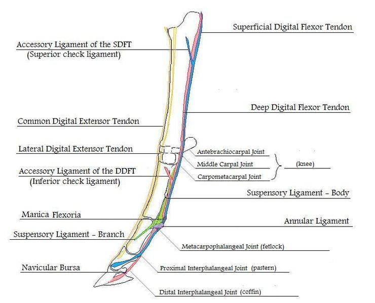
Where is the origin and insertion of the long digital extensor tendon?
Origin: LDE muscle originates from lateral femur
Insertion: Coffin bone
What does the suspensory apparatus consist of?
Suspensory ligament
DDFT
SDFT
Sesamoid bones
Extensor branches
Sesamoid ligaments
Where does the suspensory ligament originate, travel and insert in the horse?
Origin: Proximal region of 3rd metacarpal (cannon bone).
Runs down the palmar/plantar surface of meta-carpus/tarsus 3 (Cannon bone). Bifurcate medially and laterally and runs over the proximal sesamoids
Insertion: Into common/long extensor tendon near long pastern bone
What is the treatment for damage to the suspensory ligament?
None. Euthanasia.
What are the sesamoid ligaments?
Straight
Oblique
Cruciate
Short
Collateral

What is tendinosis?
A strain injury acquired from excessive training.
What is tendonitis?
The inflammatory reaction to the strain injury. Ranges from mild inflammation to disruption/rupture of tendon.
What horses is tendonitis more common in?
Race horses.
Superficial digital flexor tendon (SDFT) (also DDFT, ALDDFT, suspensory ligament).
What is the aetiology of tendinitis?
Overextension, poor conditioning, fatigue, poor racetrack conditions, persistent training with present inflammatory problems.
What is the primary pathology of tendinitis?
Central rupture of tendon fibres with associated haemorrhage and oedema.
What are the chronic clinical signs of tendinosis/tendonitis?
Lameness under hard work. Fibrosis with thickening and adhesions in the peritendinous area.
What is the diagnosis for tendinitis?
USG: see fluid effusion around the tendon, central core lesion.
MRI
Signs of inflammation (pain, heat, swelling, lameness)
How long does it take for tendons to recover from tendonitits?
Up to 18 months.
Icepacks, NSAIDs, growth factors (PRP), laser therapy, and controlled movement.
What are methods for tendon repair?
Scaffold: urinary bladder matrix (Basically a graft sewn on the ruptured tendon - like a skeleton for the cells to be healed. The scar tissue usually formed has less elasticity – this scaffold improves the scar tissue quality).
Growth factors: platelet-rich plasma (PRP) injected intra-tendon with USG control.
Stem cell therapy: from bone marrow. Impractical.
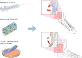
How is the recovered tendon in comparison to a healthy tendon?
The new/healed tendon is stronger and stiffer – but functionally inferior to the original. It is predisposed to injury.
What are the tendon surgeries for tendonitis?
Proximal check ligament desmotomy: Cutting of the proximal accessory ligament compensates for the scar formation, which reduces the elasticity. The cutting increases the elasticity of the scarred tendon.
Annular ligament desmotomy: Transection of proximal or distal annular ligament – the scar formation will not allow the tendon to be distended.
Fasciotomy: To improve healing. Front limb - Carpal joint, Hind limb - Plantar tarsal region.
What occurs when the superficial digital flexor tendon (SDFT) ruptures?
Hyperextension of the fetlock and heel drop.
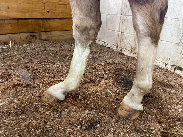
What happens when the deep digital flexor tendon (DDFT) ruptures?
Overextension and elevation of the toe.
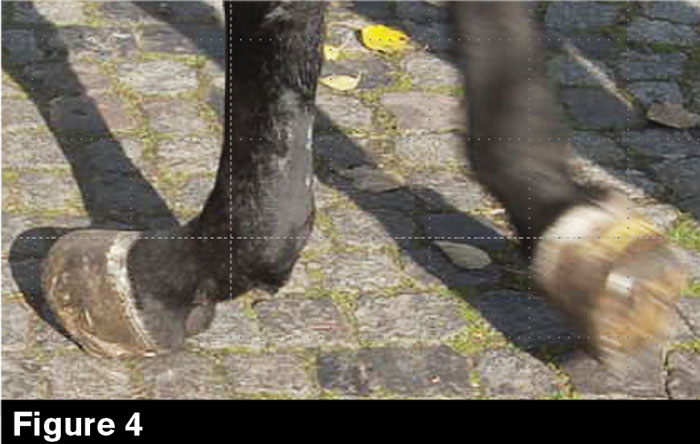
What is seen when the entire suspensory apparatus is ruptured?
The leg is broken, and the horse steps on the palmar surface of the fetlock.
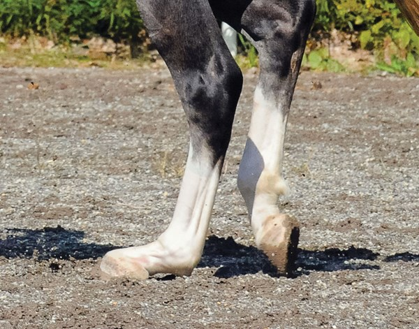
What happens when the musculus peroneus tertius is involved in tendon rupture?
The stifle joint is flexed.
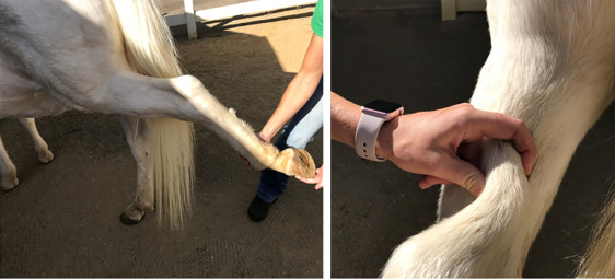
How is the diagnosis of tendon rupture made?
Palpation, comparison of contralateral limbs, and X-ray with four views to check for sesamoid bone fractures.
How can ruptured tendons be treated?
Conservative
Debridement
Surgery
Euthanasia
How should a ruptured tendon be debrided?
Debride damaged tissue, saving the remaining tendon, but do not suture.
What surgery is used for stabilisation of ruptured tendons?
Arthrodesis.