Visual Pathways and Reflexes
1/79
Earn XP
Description and Tags
These flashcards cover key concepts and details about the visual pathways, visual reflexes, and their clinical implications.
Name | Mastery | Learn | Test | Matching | Spaced |
|---|
No study sessions yet.
80 Terms
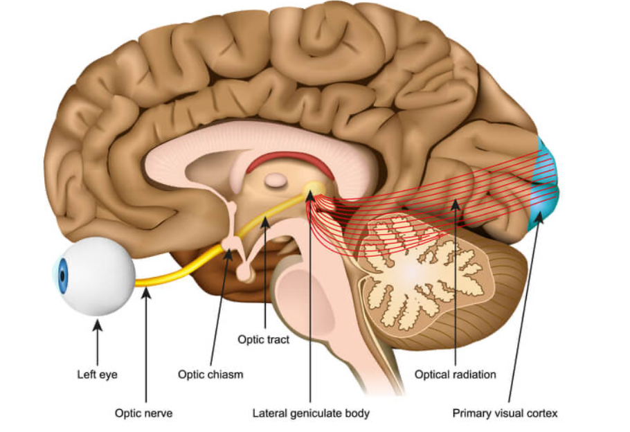
What structures make up the visual pathway from the eye to the visual cortex?
Eye, Optic Nerve, Optic Chiasm, Optic Tract, Lateral Geniculate Nucleus, Optic Radiation, Primary Visual Cortex.
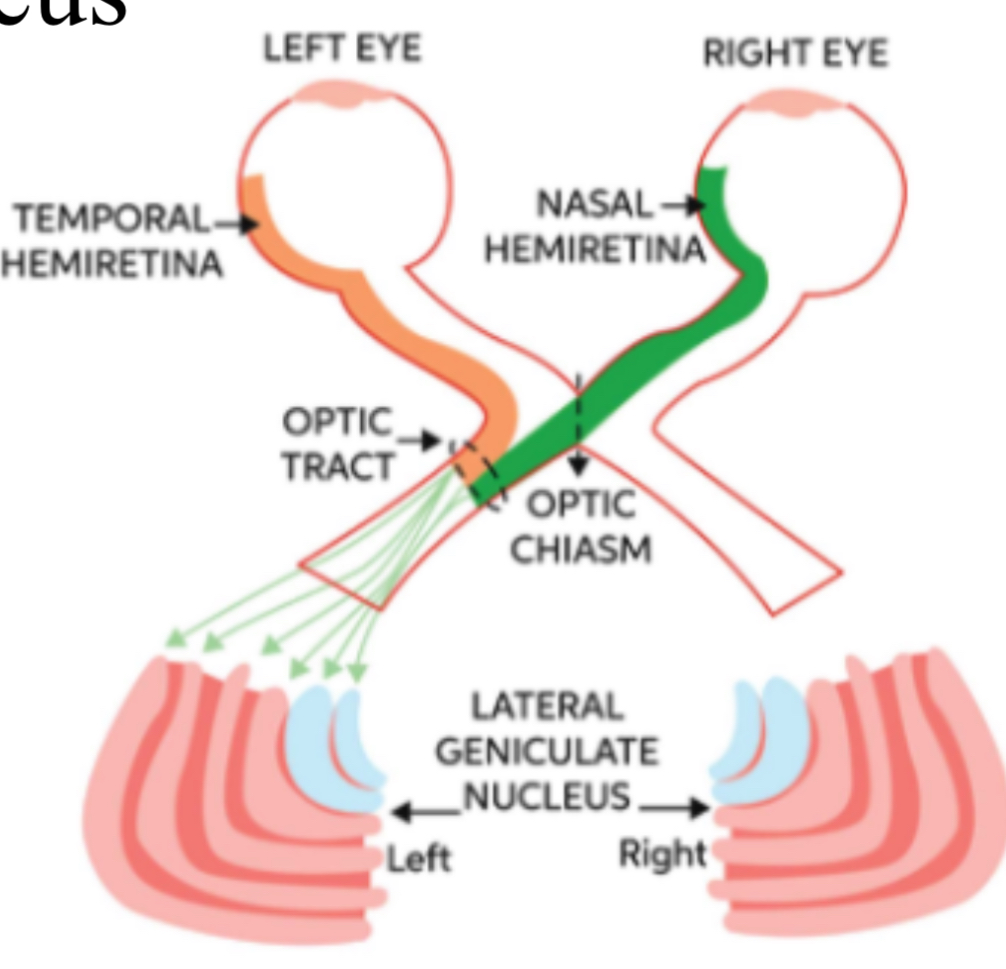
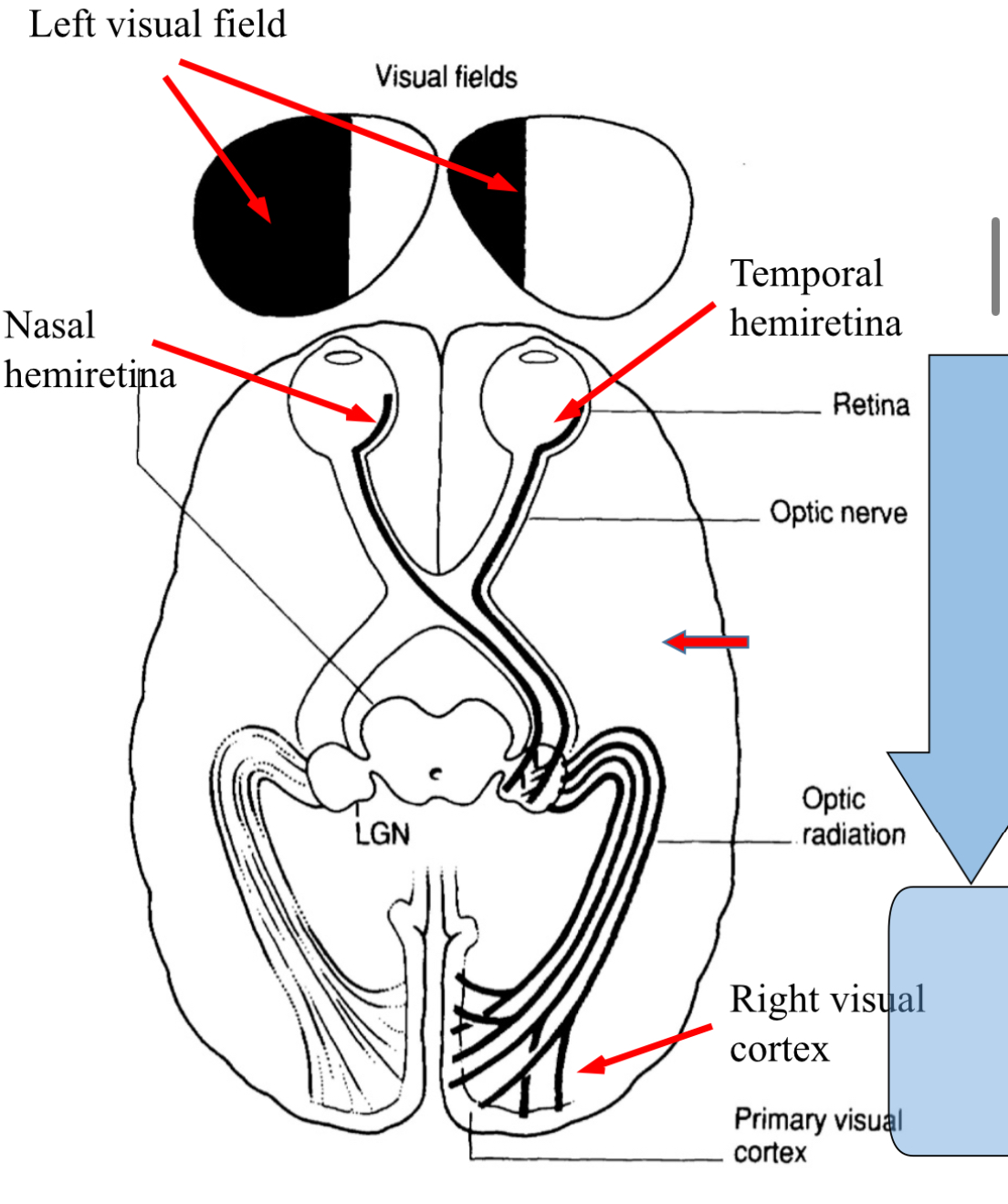
What happens at the optic chiasm?
The optic nerves from both eyes meet and fibers from the nasal hemiretina cross over.
Where do fibers from the left visual field project?
To the right visual cortex.
What effect does a pituitary tumor have on vision?
It can cause bilateral loss of peripheral receptive fields due to pressure on the optic chiasm.
What is the primary function of the Lateral Geniculate Nucleus?
It is crucial for conscious visual perception and integrates input from both eyes.
Like the gate of transmission to visual cortex , controls how much signal is allowed to pass.
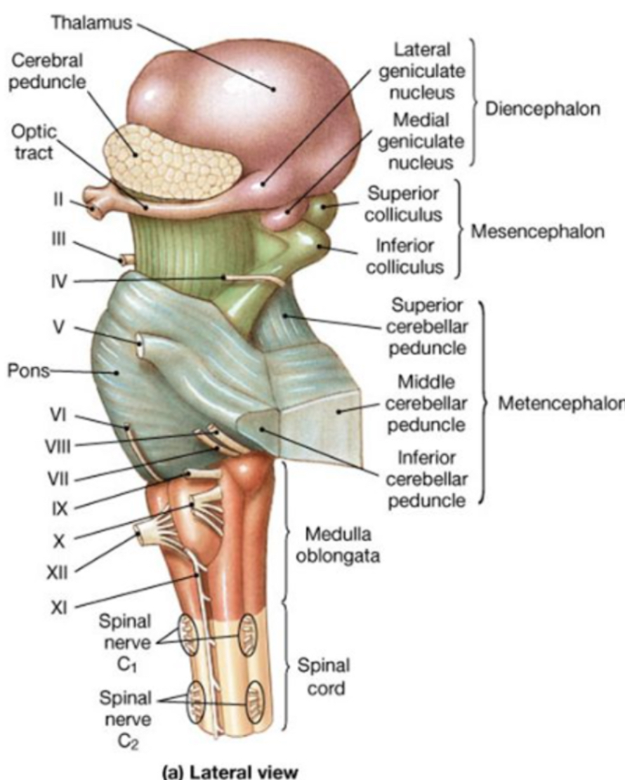
Located next to superior and inferior colliculus.
What role do optic radiation fibers play?
They transmit visual information from the lateral geniculate nucleus to the primary visual cortex.
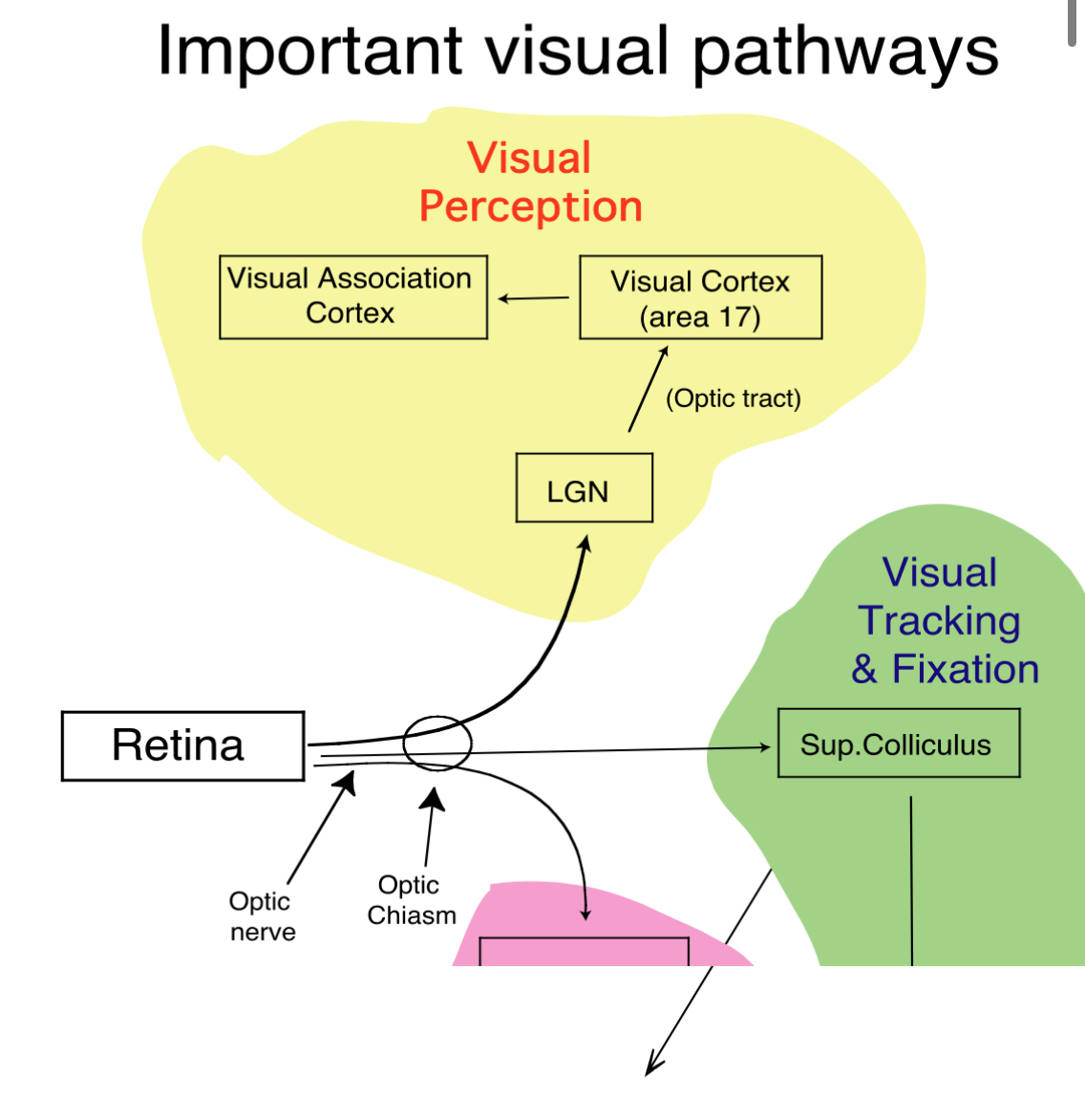
Where is data from retina converted into conscious perception
The surrounding visual association cortex in the occipital lobe. Cells in primary visual cortex send cortico-cortical axons
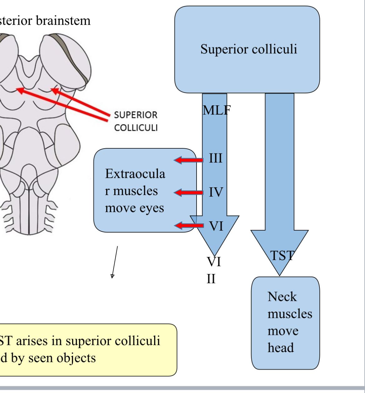
What area is visual tracking and fixation mediated by ?
the superior collisions acting on the oculomotor nuclei ( connects via the medial longitudinal fascicle) and the neck muscles by the tectospinal tract.
You can test this using the H test
What is the effect of damage to the optic tract?
Homonymous hemianopia - loss of vision in the same field from both eyes.
Why does the left visual field project to the right visual cortex and vice versa ?
Nasal hemiretina from left eye projects to right visual cortex.
The temporal hemiretina from right eye projects to the same right visual cortex.
Both view objects in the left visual field.
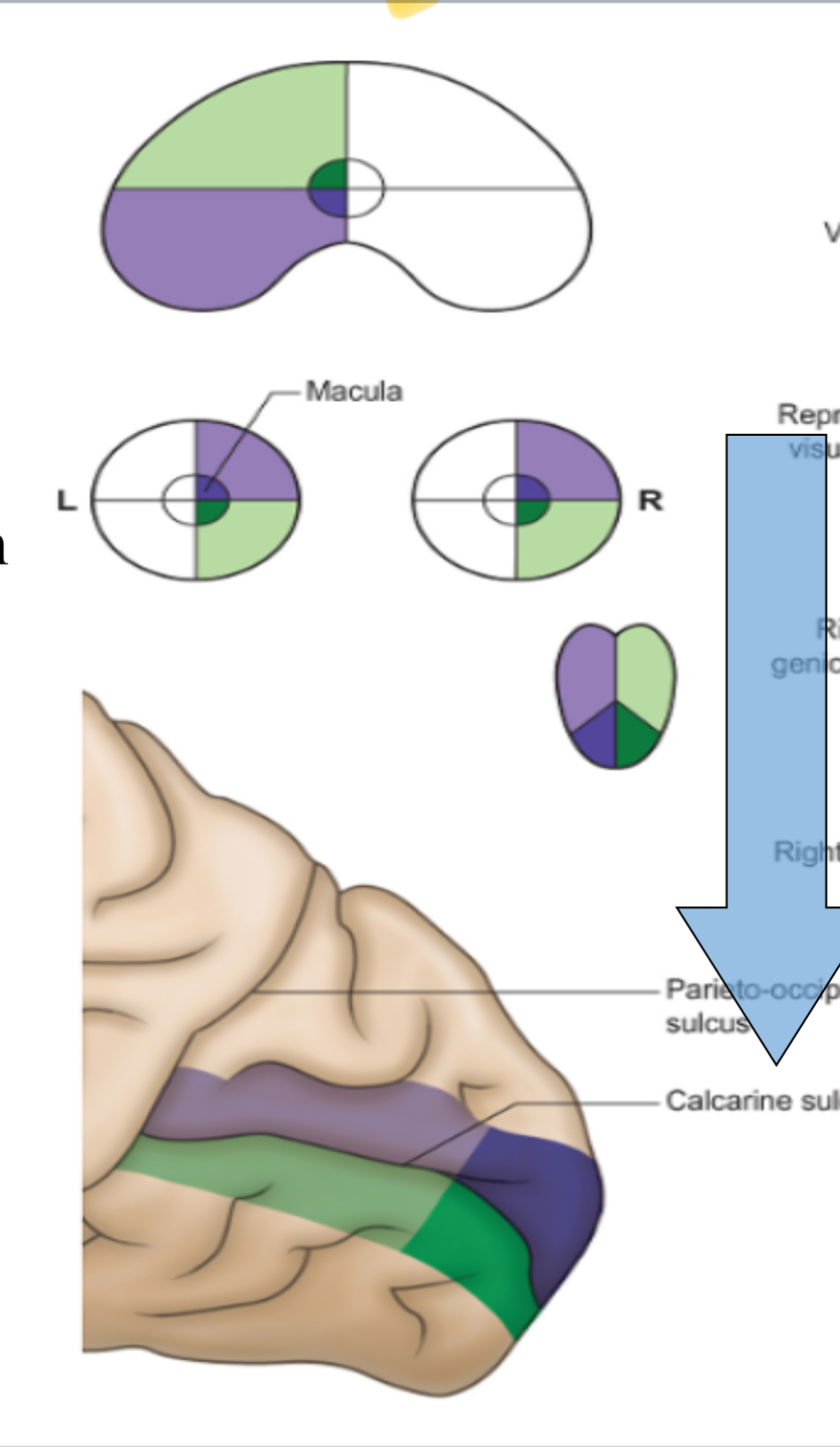
In optic radiation, where to fibres from lower and upper visual fields terminate
from lower visual fields course superiorly to terminate in the visual cortex above the calcarine sulcus
Upper visual fields terminate below calcarine sulcus
Called vertical inversion
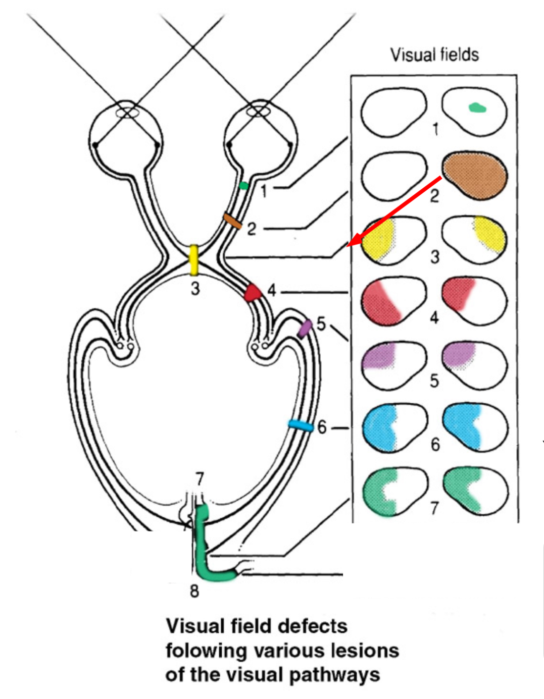
What does partial optic nerve lesion cause
An ipsilaterak scotoma, which is a patch of localised blindness in one eye, since only some fibres are damaged
What does a complete optic nerve lesion cause ?
Total blindness in that eye, called anopia. All the visual info from that eye is lost before it reaches the chasm or the brain.
What does an optic chains lesion cause ?
Bitemporal hemianopia, the loss of outer visual fields in both eyes. Nasal retinal fibres and the only fibres that cross there, which are temporal vision. So there is no side vision - this tunnel vision is caused.
What does an optic react lesion cause ?
Homonymous hemianopia, which is contralateral- loss of the same half of the visual field from both eyes.
After the chains, each optic tract carries visual info from the opposite visual field. So right tract carries left visual fields - so both left sides are gone.
What is Macular Sparing?
It is when a unilateral lesion does not affect central vision due to bilateral projections. Central vision can see
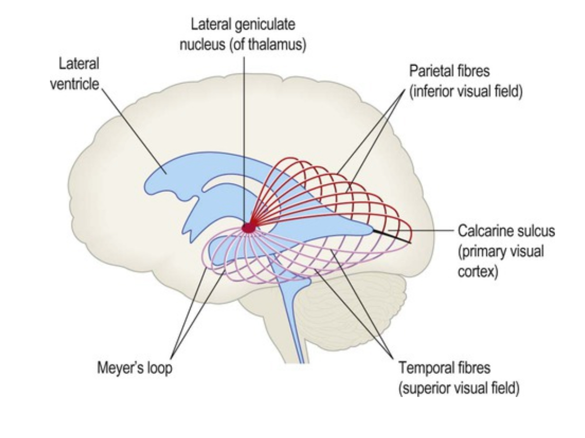
What is meyers loop
Part of the optic radiation - white matter fibres that carry visual information from lateral geniciculate nucleus in the thalamus to the primary visual cortex in the occipital lobe.
Inferior part of optic radiation, loops into temporal lobe.
Difference between fibres from upper quadrants compared to lower quadrants
for vision In upper quadrants the fibres from the optic radiation loop more anteriorly around the side of the lateral ventricle.
from lower quadrats, they travel more directly to the visual cortex.
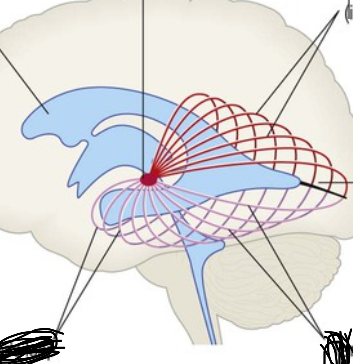
Label these structures : lateral ventricle, meyers loop , lateral geniculate nucleus from thalamus , parietal fibres, calcarine sulcus , temporal fibres
What does damage to meyers loop cause ?
homonymous superior quadrantanaopia - so the axons to upper quadrants are affected , so you can’t see the upper quadrants. Number 5 on diagram
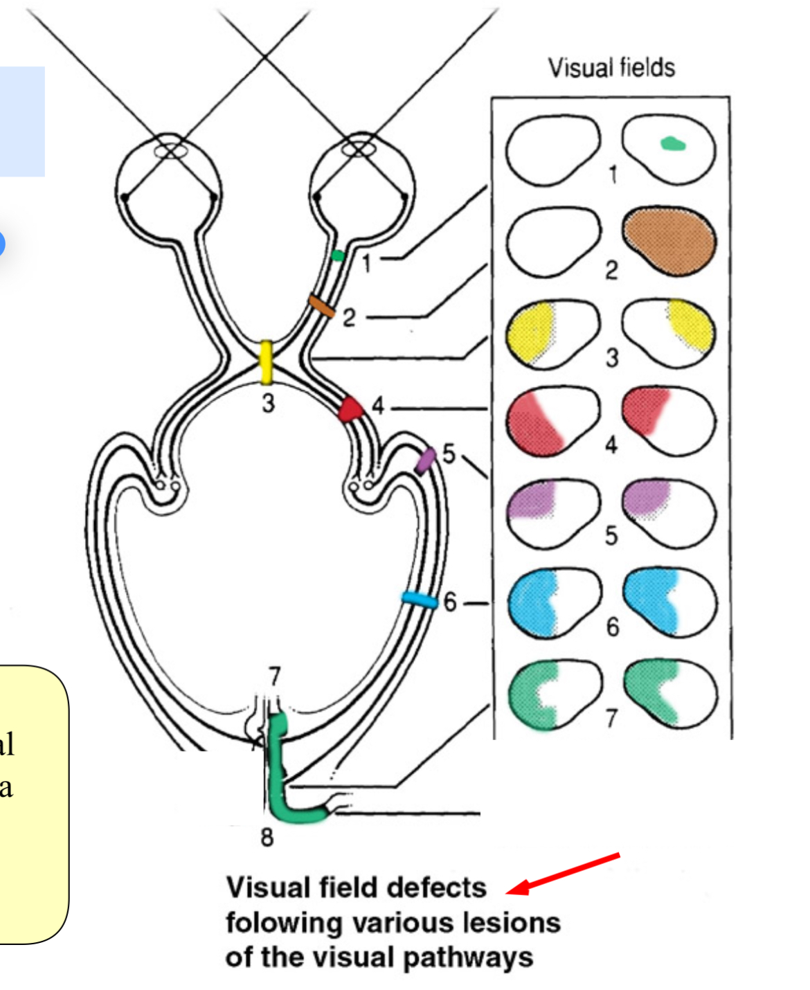
What does an optic radiation lesion cause ?
Homonymous hemianopia , number 6. ?
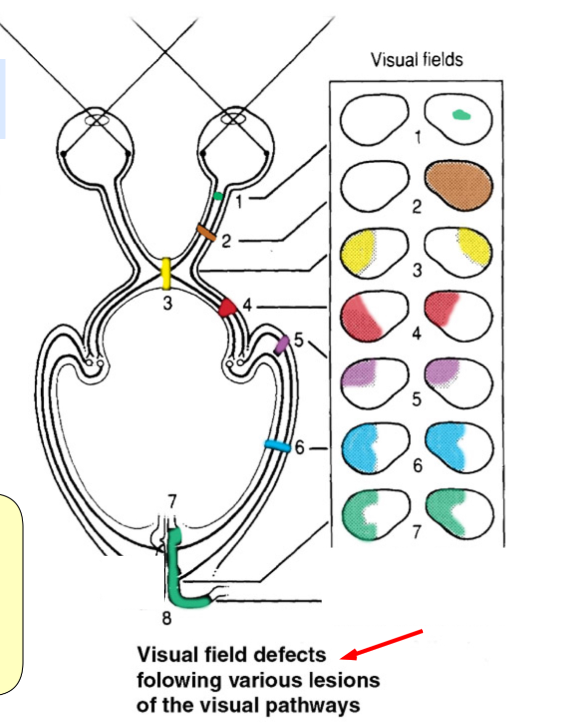
What does a visual cortex lesion cause ?
Homonymous hemianopia , number 7 and macular sparring

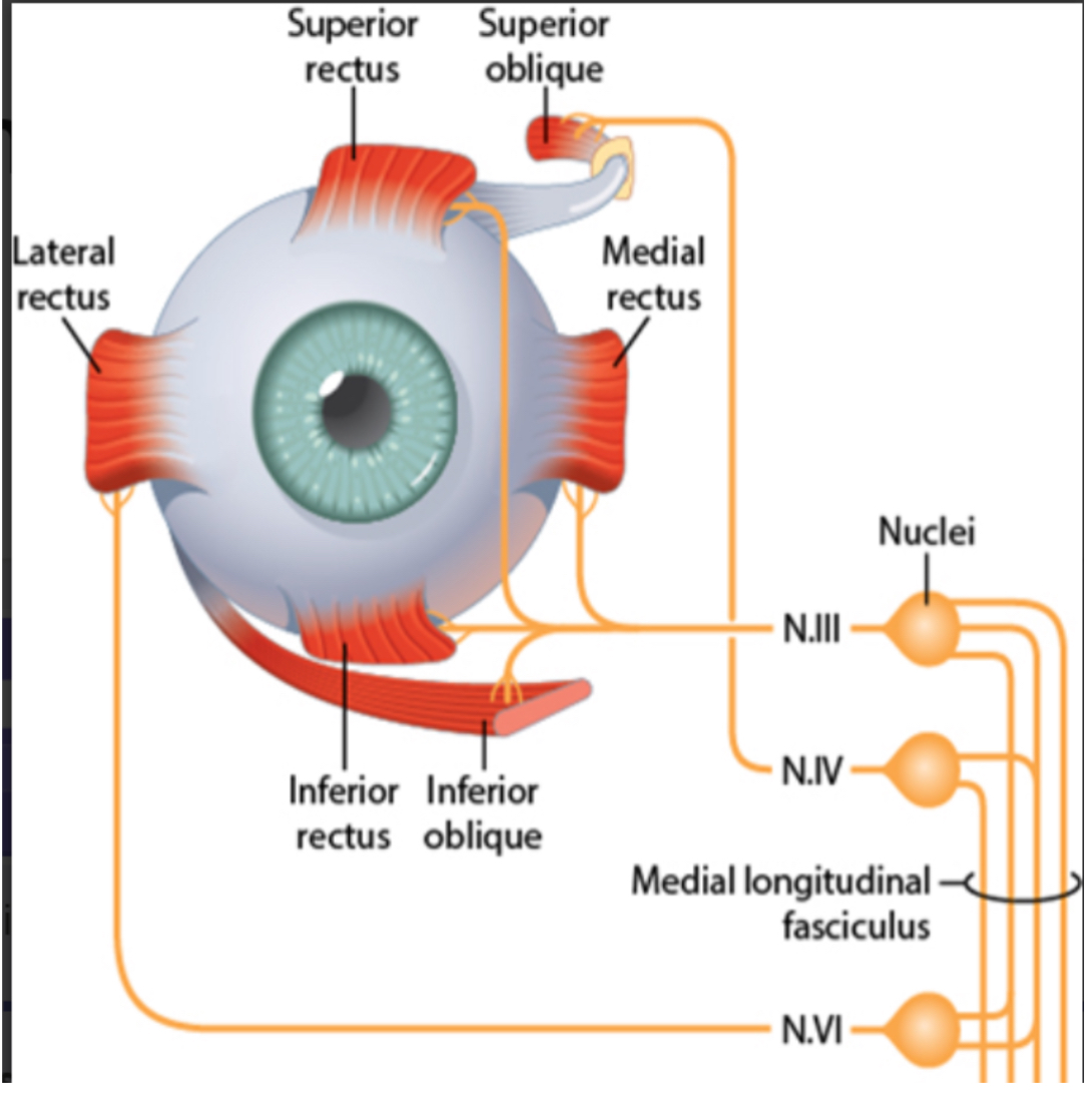
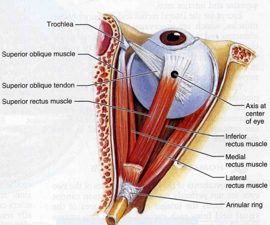
What three pairs of muscles are eye movements controlled by
Medial and lateral rectus - adduction and abduction
Superior and inferior rectus - elevation and depression
Superior oblique - internal rotation , depression and abduction
Inferior oblique - external rotation, elevation and abduction.
All innervated by oculomotor nerve
except lateral rectus which is abducens
And superior oblique trochlear
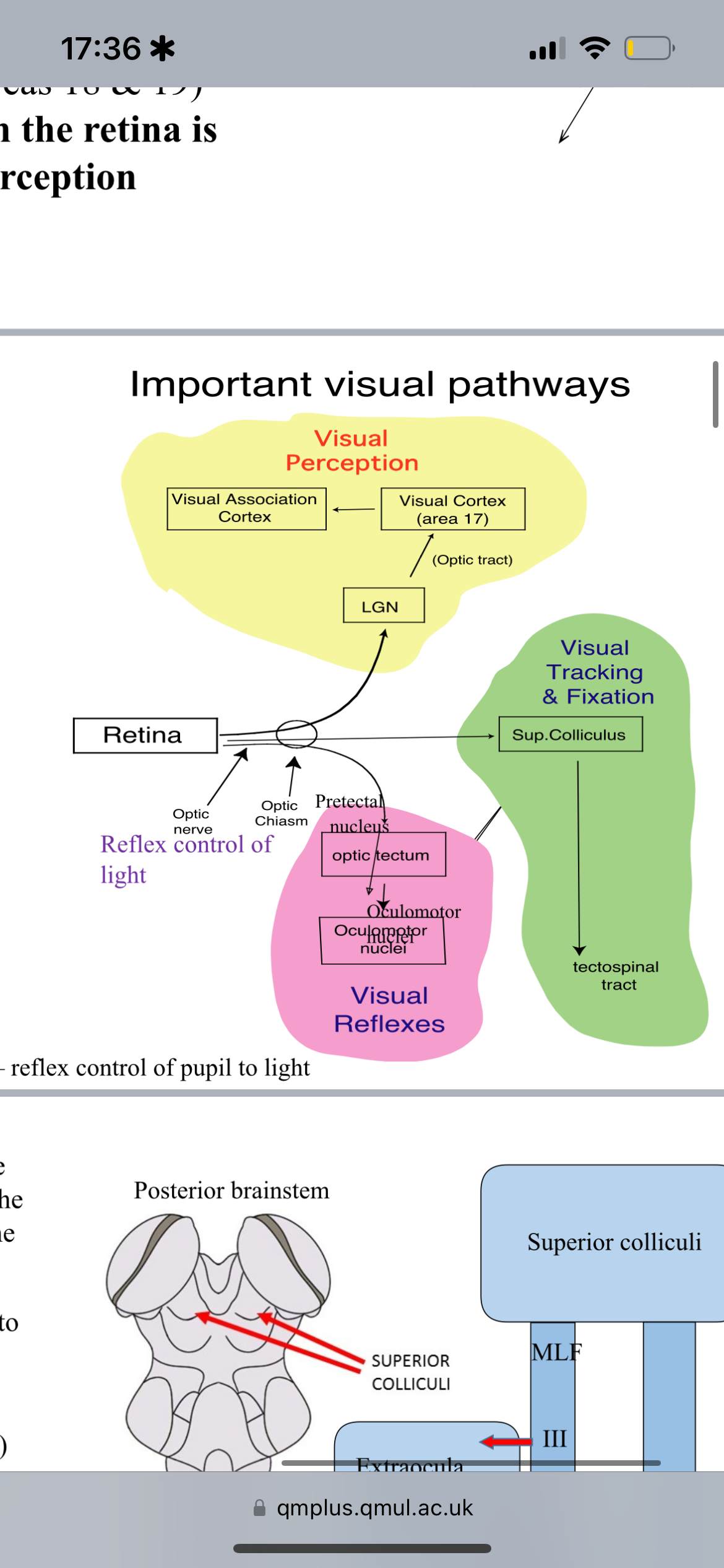
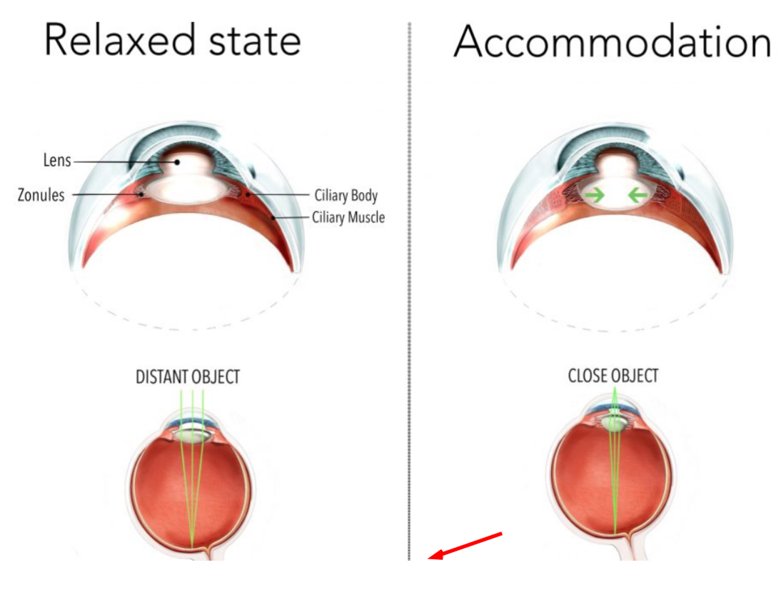
What is the accommodation reflex?
The response of the eye to focus on near objects involving lens thickening, convergence, and pupil constriction.
What cranial nerve is responsible for pupil constriction?
Cranial Nerve III (Oculomotor nerve).
What does cilliary muscles do ?
And their affect on the lens ?
What is the innervation ?
They contract reducing tension on the suspensory ligaments - allowing the lens to become rounder ( which is accommodation ) and vice versa.
Innervated by parasympathetic cranial nerve : oculomotor
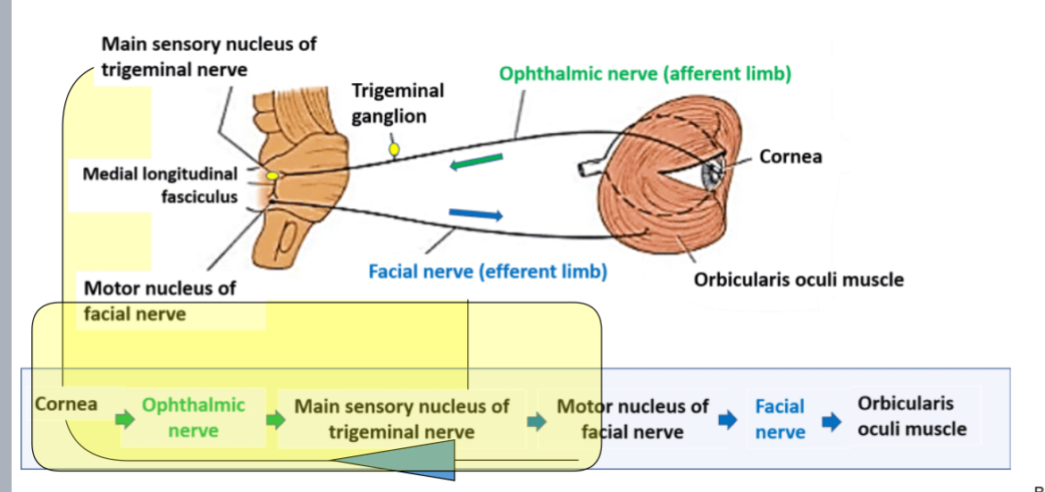
What is the blink reflex?
A protective reflex involving both CN V (afferent) and CN VII (efferent) when the cornea is stimulated.
What controls the pupillary light reflex?
Stimulation of the retina via CN II leads to bilateral pupil constriction via CN III.
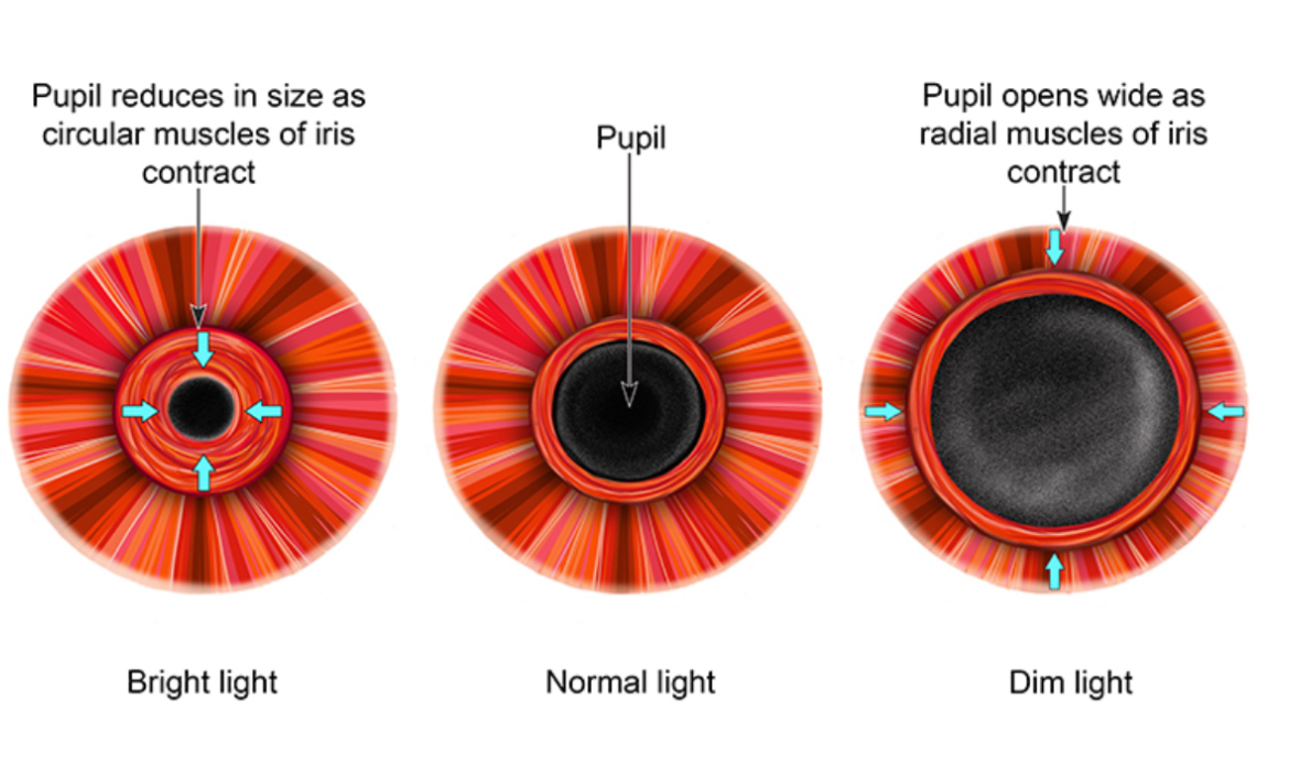
Describe the structure of the pupil/ iris
and outline innervation to constrict/ dilate.
The pupil is a hole in the iris. It has circularly arranged smooth muscle that can constrict the pupil to get smaller ( makes hole smaller )
innervation by CN3
To dilate pupil the radial fibres contract
innervation is by sympathetic T1
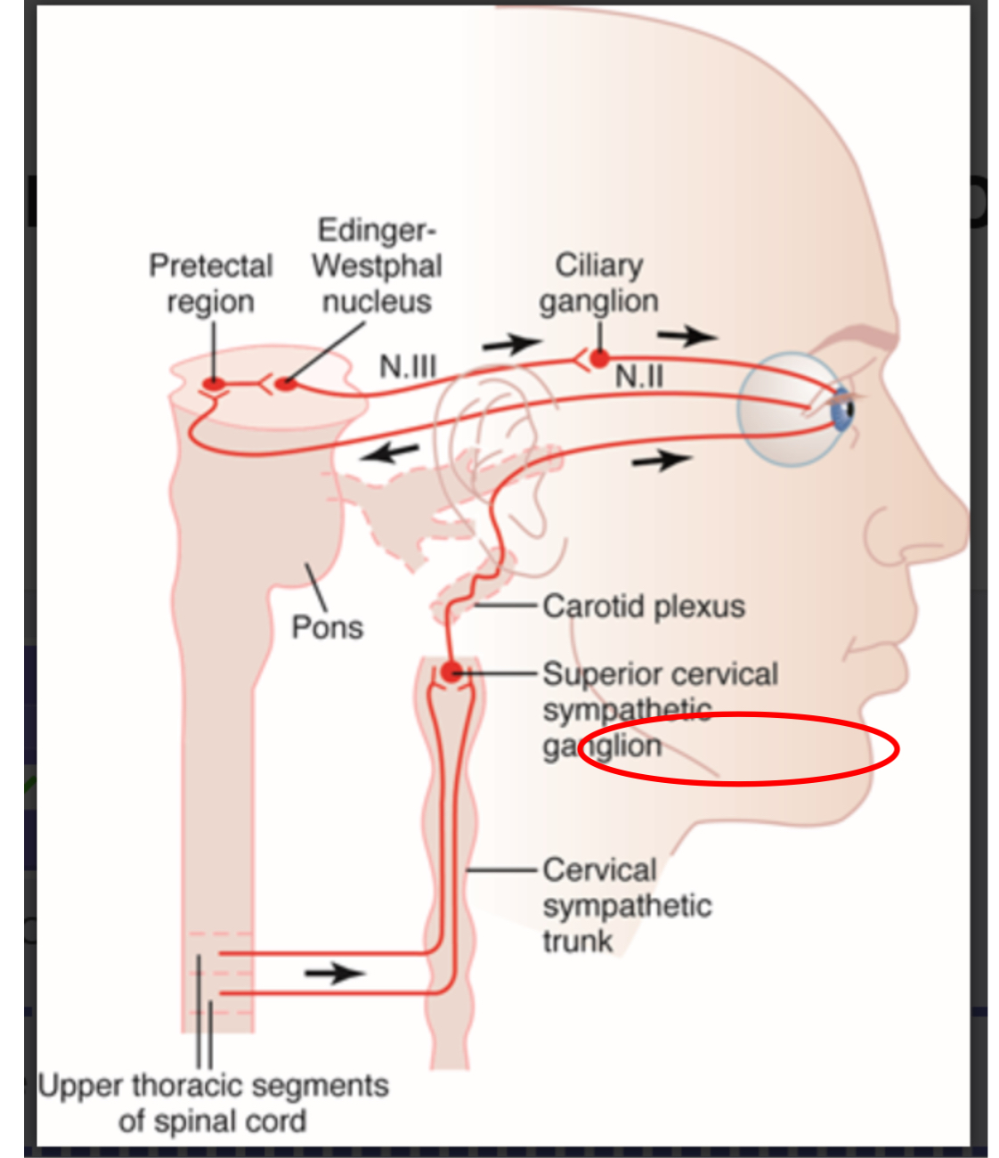
Outline ANS parasympathetic innervation of eye
parasympathetic pre-ganglion fibres in the Edinger-west fall nucleus
Then CN 3 oculomotor to the ciliary ganglion behind the eye
Changes into post ganglion parasympathetics through the ciliary nerve to eye ball
Then ciliary muscles and sphincter muscles contract in iris - called miosis
Outline ANS sympathetic innervation of eye
Lateral horn of T1 to sympathetic chain
Then superior cervical ganglion synapses with post ganglion
What is the function of the superior colliculus?
It is involved in visual reflexes such as tracking moving objects.
What happens when the Frontal Eye Fields are damaged?
Inability to direct gaze from one object to another, leading to difficulty in tracking.
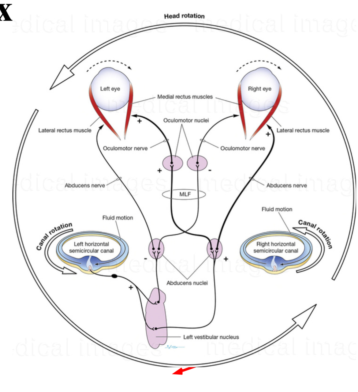
How do eye movements remain coordinated during head movement?
Through the vestibulo-ocular reflex (VOR), which stabilizes visual focus.
Which areas of the visual cortex are primarily involved in visual perception?
Primary visual cortex (Area 17) and surrounding visual association areas (Areas 18 & 19).
What are the key visual reflexes mentioned in the notes?
Pupillary light reflex, accommodation reflex, vestibulo-ocular reflex, and blink reflex.
What is the pathway of visual information from the retina to the visual cortex?
Retina → Optic Nerve → Optic Chiasm → Optic Tract → Lateral Geniculate Nucleus → Optic Radiation → Primary Visual Cortex.
What visual field defect is caused by a lesion in the optic chiasm?
Bitemporal hemianopia.
What is the role of the ciliary muscle in focusing light?
It adjusts the shape of the lens for accommodation by contracting or relaxing.
How does a complete optic nerve lesion present in clinical terms?
Blindness in the affected eye (anopia).
What defines the term 'scotoma'?
A localized area of partial or complete loss of vision in an otherwise normal visual field.
What does the visual field comprise?
Four quadrants: right/left and upper/lower, each projecting to corresponding areas in the visual cortex.
What is the Caloric Stimulation Test used for?
To test the vestibulo-ocular reflex by irrigating warm or cold water in the ear to assess eye movement.
What is the role of the medial longitudinal fasciculus (MLF)?
It connects the oculomotor nuclei for coordinating eye movements.
What is the relationship between the superior and inferior colliculus?
Both are involved in processing visual and auditory stimuli; superior for vision and movement response, inferior for sound localization.
How do both eyes contribute to vision?
Each eye has a temporal and nasal hemiretina that help visualize the left and right fields in a coordinated manner.
What is Meyer's Loop associated with?
Fibers in the optic radiation mediating vision from the upper visual quadrants.
What indicates damage to Meyer's Loop?
Homonymous superior quadrantanopia, characterized by a 'pie in the sky' defect.
What happens to visual perception when the Lateral Geniculate Nucleus is damaged?
It may lead to impaired conscious visual perception.
How is the pupillary light reflex pathway organized?
Optic nerve → Pretectal nucleus → Edinger-Westphal nuclei → Ciliary ganglion → Sphincter pupillae.
What occurs during the accommodation reflex when focusing on a near object?
Thickening of the lens, convergence of the eyes, and constriction of the pupils.
What is one effect of alcohol on the convergence part of the accommodation reflex?
It can disrupt convergence, potentially leading to diplopia (double vision).
What is the term for reflexes that do not require conscious input?
Unconscious reflexes.
Which cranial nerve mediates the vestibulo-ocular reflex?
Cranial Nerve VIII (vestibulocochlear nerve) for input and CN III and CN VI for output.
What results from damage to the optic radiation?
Homonymous hemianopia or visual field loss corresponding to the damaged side.
What indicates a partial optic nerve lesion?
Ipsilateral scotoma, a localized area of vision loss in the affected eye.
What mnemonic can help remember the pupillary light reflex pathway?
"In on II, out on III" (Cranial Nerve II input, Cranial Nerve III output).
What is the effect of Atropine on the pupillary light reflex?
It blocks the pupillary light reflex, preventing pupil constriction.
What anatomical structure is crucial for the pupillary light reflex?
The Edinger-Westphal nucleus.
What’s the afferent and efferent paths of the puppilary reflex
Afferent is CN2 to pretcetal areas to both EWN
Efferent is CN3 for direct and consensual pupil closure
What therapy can help improve visual tracking in patients with superior colliculus damage?
Rehabilitation exercises that focus on eye movement coordination.
What is the difference between the medial and lateral recti muscles?
Medial recti muscles are responsible for adduction, while lateral recti muscles facilitate abduction.
What do the terms 'upper visual field' and 'lower visual field' refer to?
Upper visual field images project to the lower retina and vice versa.
What is the significance of the 'blink reflex' in neurological examinations?
It checks the integrity of the sensory (CN V) and motor (CN VII) pathways.
How do the iris dilator and sphincter muscles respond to light?
The sphincter contracts to constrict the pupil in bright light, and the dilator contracts to enlarge it in dim light.
What occurs during the vestibulo-ocular reflex when the head turns?
Eyes move in the opposite direction to maintain focus on a fixed object.
What aspect of the visual field does the fovea cater to?
High visual acuity due to a dense concentration of photoreceptors.
Test what reflex by having someone follow your finger?
The ability to track a moving object through the function of the superior colliculus.
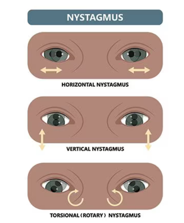
What is nystagmus and how is it caused ?
It’s a form of VOR caused by continuing rotation of fluid in the semi circulation.
It is showed as an initial slow rotation followed by a fast flick back.
Direction of nystagmus is where the fast flick back is
What is meant by 'consensual reflex' in the context of pupil response?
When one eye's stimulation leads to constriction of both pupils.
What visual processing occurs in the surrounding visual association cortex?
Conversion of raw visual data into conscious perception.
How can vestibulo-ocular reflex be tested clinically?
By using the caloric stimulation test to observe eye movement responses.
what part of brain is accommodation reflex controlled by ?
By frontal eye fields which are specialised areas of the pre motor area , dedicated to motor control of extra ocular eye muscles .
Can only happen when conscious .
What does damage to the frontal eye fields cause
Inability to make smooth rapid eye movements called saccades between objects.
Fast phase of nystagmus is affected.
3 components of accommodation
Lens thickening
Convergence of eyes
Pupil construction
How does lens thickening occur
For distant objects
cilliary muscle sphincter is relaxed and lens is under tension by a ring of suspensions ligaments - pull and flatten it - decreases refractive power
In the reflex the cilliary muscle contacts and opposes tension is suspensory ligaments. Lens thickens and gets more curved for near objects.
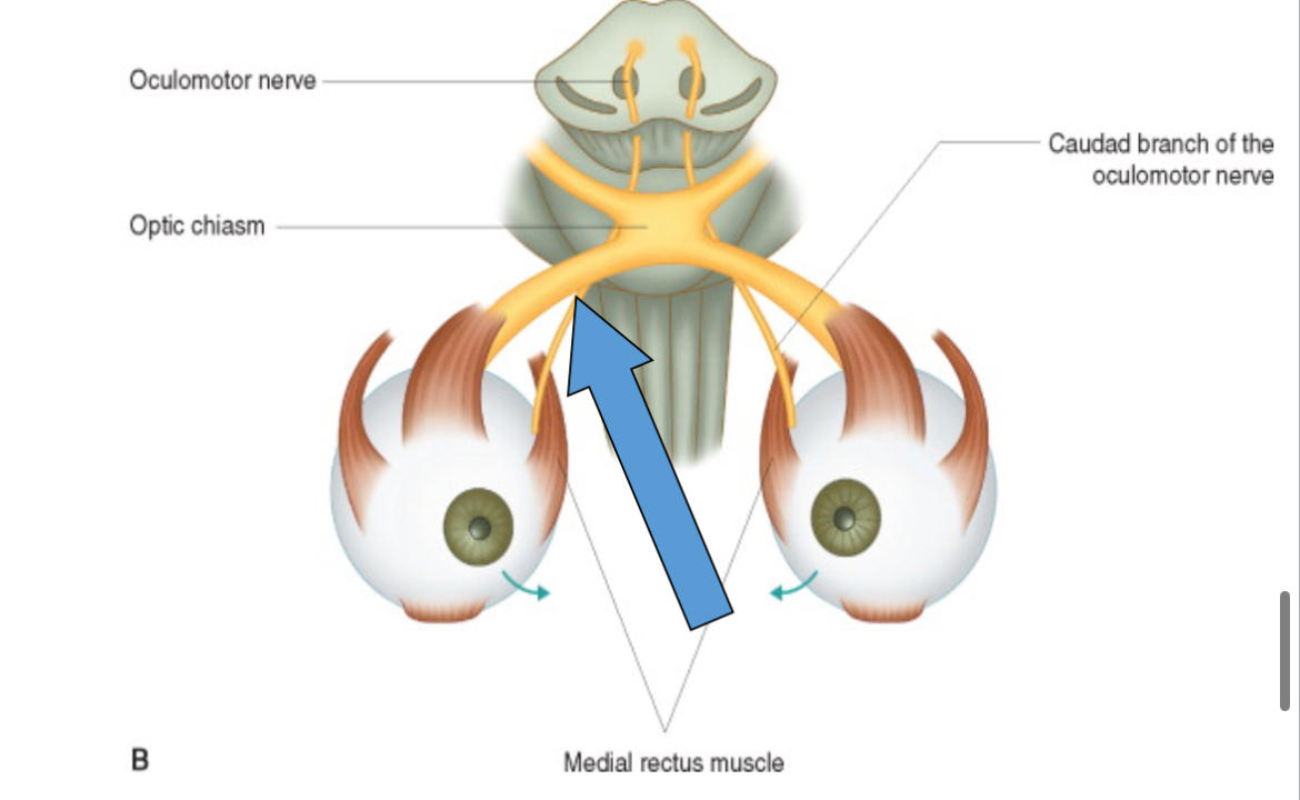
What is convergence of eyes - controlled by what nerves and muscles
It’s controlled from frontal eye fields, it’s simultaneous activity in the medial rectus.
Controlled by oculomotor
Only in conscious behaviour.
If it’s damaged then it causes double vision.
What connects the visual cortex to the subcortical structures and aids in reflexes?
Cortical pathways that project to various brain regions, especially for visual reflexes.
How to test the vestibulo cochlear reflex
Caloric stimulation.
Basically warm water put into external auditory canal and temp difference causes convection currents in endolymph of horizontal semi circular canals .
Normal results :
Warm water - head turns to water ear , same side. Both eyes turn slowly to other side. Then fast flick (nystagmus) to water ear
Cold water - the opposite
COWS
Cold other warm same
Describe path of the blink reflex