Age-related macular degeneration
1/15
Earn XP
Description and Tags
As part of posterior segment disorders
Name | Mastery | Learn | Test | Matching | Spaced |
|---|
No study sessions yet.
16 Terms
What are the risk factors for AMD?
Age
Genetics
Smoking
UV exposure
Hypertension
Female
Obesity
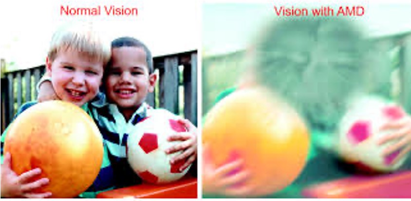
Does AMD affect central or peripheral vision?
AMD affects central vision because it involves the macula. Peripheral vision is usually unaffected.
What are the two types of AMD?
Dry - accumulation of Drusen between Bruch’s membrane and the RPE, leading to progressive photoreceptor loss called geographic atrophy. This is the most common type.
Wet - hypoxic photoreceptors release VEGF, stimulating choroidal neovascularisation. These vessels are leaky, causing fluid build-up and haemorrhage at the macula and detachment of the RPE from Bruch’s membrane. This is less common.
Can a patient have only dry or wet AMD?
No - dry AMD can develop into wet AMD and does so in 10-15% of cases.
True or false? The symptoms of dry AMD include a sudden monocular loss of central vision with severe distortion.
False, the symptoms of dry AMD typically include gradual vision loss and distortion, rather than sudden monocular loss. Patients usually experience distortion of faces and metamorphopsia. Dry AMD is usually bilateral, although it can be asymmetrical.
What are the symptoms of wet AMD?
A sudden, painless loss of central vision, usually in one eye. The VA will be significantly reduced and vision will be severely distorted.
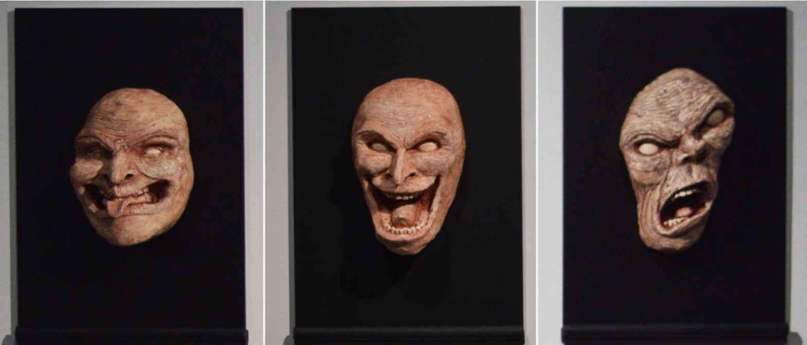
True or false? Charles-Bonnet syndrome is only associated with wet AMD.
False - this condition can occur in either dry or wet AMD.
Charles Bonnet syndrome is a symptom of the severe sight loss caused by AMD and involves vivid and often frightening visual hallucinations, particularly around human faces. Patients, especially elderly patients, will often not speak up about this condition as they are afraid of being diagnosed with psychiatric problems.
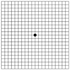
What is an Amsler chart used for in AMD?
The Amsler chart is used by the patient to monitor the progression of their visual loss.
What would be seen on fundus photography in dry AMD?
Fundus photography would typically show drusen, retinal pigment epithelial changes, and/or geographic atrophy.
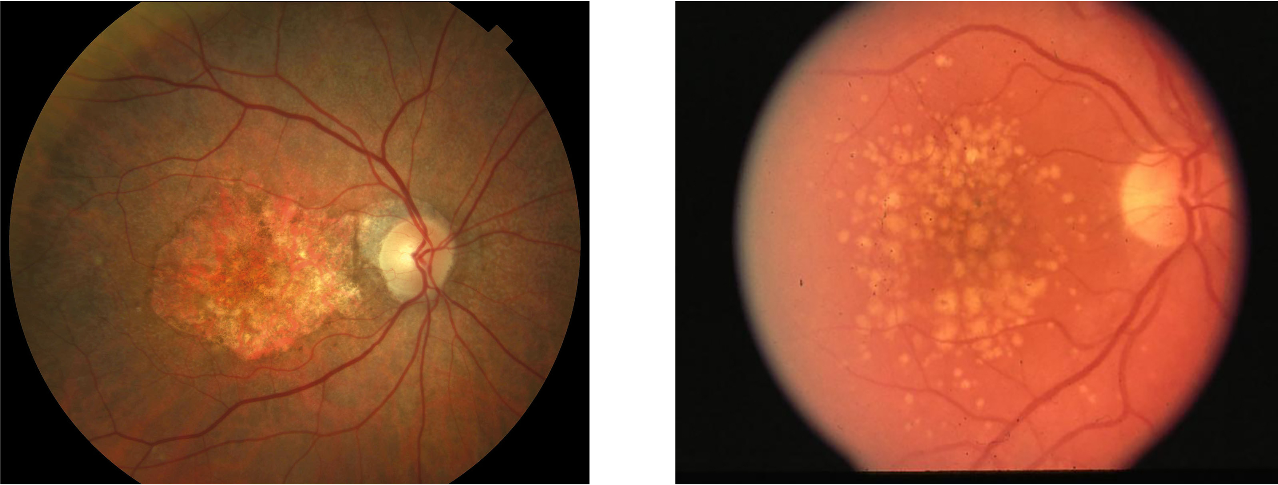
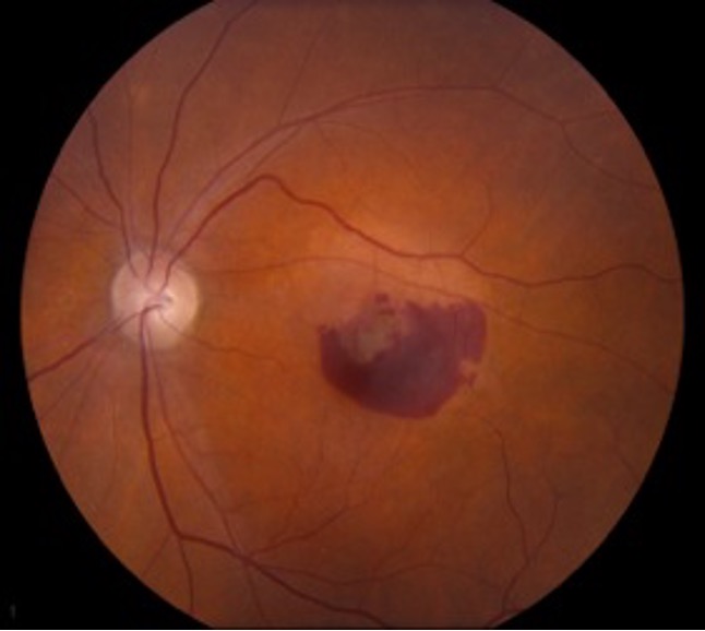
What would be seen on fundus photography in wet AMD?
Fundus photography would typically show choroidal neovascularisation and possible haemorrhages at the macula.
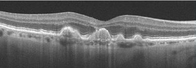
What would an OCT scan show in dry AMD?
The OCT would show accumulation of Drusen between the RPE and Bruch’s membrane at the fovea, visualised as bulging of the membrane with the bright white RPE rippled as a result.
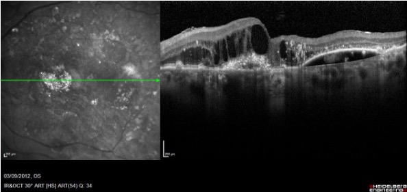
What would an OCT scan show in wet AMD?
The OCT would show the presence of fluid accumulation between the RPE and Bruch’s membrane, as well as possible separation of the RPE from the membrane.
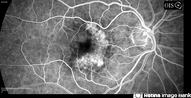
What is fluorescein angiography used for in the AMD clinic?
Fluorescein is used to visualise the choroidal vessels and pinpoint the location of leaks and haemorrhages.
What four methods are used to diagnose and monitor AMD in clinic?
Fundus photography, OCT, fluorescein angiography, and Amsler charts.
True or false? Dry AMD is the only type which can be treated.
False - there are currently no treatments for dry AMD. The patient will instead be advised on lifestyle modifications such as smoking cessation, nutrition improvements, weight loss, and the importance of sun protection. They may also be referred to the low vision clinic for low vision support and advice to maximise the vision they do have.
True or false? Anti-VEGF injections can be used to treat wet AMD.
True - anti-VEGF can be injected into the vitreous to slow the growth of new leaky choroidal vessels. This is a lifelong treatment and can only delay the progression of AMD and limit vision loss, not cure the patient.