Bio Unit 4
1/85
There's no tags or description
Looks like no tags are added yet.
Name | Mastery | Learn | Test | Matching | Spaced |
|---|
No study sessions yet.
86 Terms
Closed Circulatory System (what does it require, what are examples, and how does it flow? What is its function in humans?)
Requires encapsulated vessels to contain blood (arteries → capillaries → veins), and blood flow is high pressure and efficient.
In humans, it transports nutrients and removes waste products from the digestive system as well as exchange of gases (O2 & CO2)
How are blood vessels organized? 5 ways
1. Arteries — carry blood away from the heart
2. Arterioles — small branches of arteries
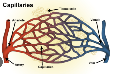
3. Capillaries — tiny, thin-walled exchange vessels
4. Venules — collect blood from capillaries
5. Veins — carry blood back to the heart
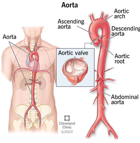
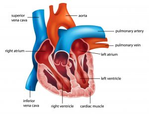
Aorta (what is its function and where does it flow to)
Largest artery in the heart, the main artery that carries blood away from your heart to the rest of your body
Aortas → arteries → arterioles (ARTERIAL SYSTEM)
Arterioles (what are they, what do they do, and what do they control)
Arterioles are smaller branches that extend out from arteries
Distribute blood to tissues by controlling blood flow and pressure by narrowing or widening
Venules (what do they do)
Small veins that collect blood from capillaries after gas/nutrient exchange. They merge to form increasingly larger veins

Vein (what is it, what is the main one?)
a blood vessel in the circulatory system of humans and most other animals that carries blood towards the heart.
The main vein in the body is the vena cava
Systolic Pressure (120/80)
the pressure in your arteries when your heart contracts (pumps blood)
Diastolic Pressure (120/80)
the pressure in your arteries when your heart relaxes (fills with blood)
Blood Volume and Pressure
As blood volume increases, blood pressure increases, pressure depends on volume
Why do arteries have thick walls and veins have thin walls?
Arteries carry blood under high pressure directly from the heart, so their walls must withstand strong force and help maintain blood pressure
Veins carry blood under low pressure back to the heart and rely on valves and skeletal muscle contractions to move blood. Their thin walls also allow veins to expand easily and act as blood reservoirs.
Veins and skeletal muscle contractions
The calf muscle acts as a pump for deep leg veins; as the muscle contracts, the increased pressure on a vein causes a valve to open pushing the blood upwards
Pulmonary Circulation (what is it and what occurs?)
Carries deoxygenated blood from the right side of the heart to the lungs.
In the lungs, the blood picks up oxygen and releases carbon dioxide.
The oxygenated blood then returns to the left side of the heart.
Pulmonary Circulation Process
Right ventricle → pulmonary artery → lungs → pulmonary veins→ left atrium
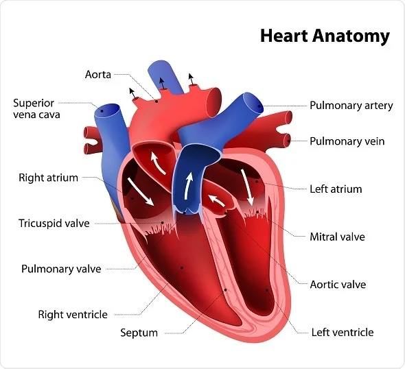
Ventricles (what is it and which ventricle starts which circuit?)
Lower chambers of the heart that act as high-pressure pumps that propel blood through the pulmonary and systemic circulation.
RIGHT VENTRICLE: Starts pulmonary circuit, pumps deoxygenated blood from the right atrium to the lungs
LEFT VENTRICLE: Starts systemic circuit, pumps oxygenated blood from the left atrium to the entire body
ATRIA ACCEPT, VENTRICLES VENT
Systemic Circulation (what is it and what occurs?)
Carries oxygenated blood from the left side of the heart to the entire body.
The body uses the oxygen, and the blood becomes deoxygenated.
The deoxygenated blood returns to the right side of the heart, completing the cycle.
Systemic Circulation Process
Left ventricle → aorta → body tissues → veins → vena cavae → right atrium
Vertebrae Circulatory System 7 Functions
Transports O2 from lungs to tissues, and CO2 from tissues to lungs
Distributes nutrients from the digestive system to the body's cells.
Transport waste and toxic substances to the liver
Distribute hormones from organs to tissues on which they act.
Regulates body temperature by adjustments in blood flow
Prevent blood loss with clotting mechanism
Protects the body from bacteria and viruses by circulating antibodies and white blood cells
Capillary Beds (what are they, function, where are they located)
Joining of arterioles and venules where deoxygenated and oxygenated blood mixes
Exchanges materials between the blood and tissue cells
Located near organs with associated high metabolic activity, such as the kidneys, brain, and lungs.
Inferior Vena Cava (what is it, what does it do, what does it connect to?)
Vein system near the artery that is the largest in your body. Carries oxygen-depleted blood back to your heart from the lower part of your body. Connects to venules
[Inferior vena cava → superior vena cava → veins → venules, and then to arterial system]
![<p>Vein system near the artery that is the <strong>largest </strong>in your body. Carries oxygen-depleted blood back to your heart from the <strong>lower part</strong> of your body. Connects to <strong>venules </strong></p><p><span style="background-color: transparent;"><span>[Inferior vena cava → superior vena cava → veins → venules, </span><strong><span>and then to arterial system]</span></strong></span></p>](https://knowt-user-attachments.s3.amazonaws.com/43d83b19-85dc-4358-8019-6c551f4782ab.png)
Superior Vena Cava (what is it, what does it do, what does it connect to?)
The second-largest vein in your body that lies above the inferior vena cava. It brings oxygen-depleted blood back to your heart from the upper part of your body.
[Inferior vena cava → superior vena cava → veins → venules, and then to arterial system]
![<p>The <strong>second-largest</strong> vein in your body that lies above the inferior vena cava. It brings oxygen-depleted blood back to your heart from the <strong>upper</strong> part of your body. </p><p>[Inferior vena cava → superior vena cava → veins → venules,<strong> and then to arterial system]</strong></p>](https://knowt-user-attachments.s3.amazonaws.com/3eee21b7-cfc5-4b93-a435-e2876433a362.png)
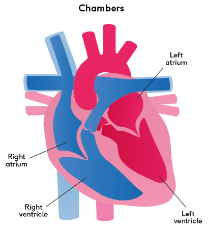
Atria (What does each side do?)
RIGHT ATRIUM: Receives deoxygenated blood from the vena cavae, then sends blood to the right ventricle
LEFT ATRIUM: Receives oxygenated blood returning from the lungs from the pulmonary veins, then sends it to the left ventricle
ATRIA ACCEPT, VENTRICLES VENT

Pulmonary Veins and Arteries
VEINS: Carries newly oxygenated blood from the lungs back to the heart's left atrium
ARTERIES: Carry deoxygenated blood from the heart's right ventricle to the lungs for oxygenation
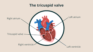
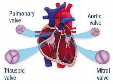
Tricuspid Valve (AV Valve)
Located between right atrium and right ventricle, prevents blood from flowing back into the right atrium during right ventricular contraction
Bicuspid/Mitral Valve (AV Valve)
Located between Left atrium and Left ventricle, it regulates blood flow from the upper left chamber (left atrium) into the lower left chamber (left ventricle)

Aortic Valve (Semilunar Valve)
Separates the left ventricle from the aorta. Opens to allow blood to leave the heart from the left ventricle through the aorta and the body.
Prevents the backflow of blood from the aorta to the left ventricle.

Pulmonary Valve (Semilunar Valve)
Separates the right ventricle from the pulmonary artery. Opens to pass blood from the right ventricle to the lungs through the pulmonary artery where it will receive oxygen. Prevents the back flow of blood from the pulmonary artery to the right ventricle
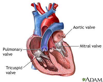
Atrioventricular vs Semilunar Valves
AV: Maintain unidirectional flow between atria and ventricles
Semilunar: Ensure one-way flow out of ventricles to arterial systems
Cardiac Cycle (0.8 seconds)
Blood enters atria
Atria contract
Valves open
Blood enters ventricles
Blood is pumped out of the heart
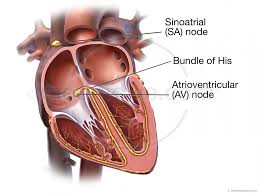
Sinoatrial (SA) Node
Located in the right atrium, is called the “pacemaker” of the heart, as it continuously generates electrical impulses, setting a healthy heart's normal rhythm and rate

Atrioventricular (AV) Node
responsible for generating impulses for the contractions of the heart and ensuring coordinated blood flow through the chambers of the heart.
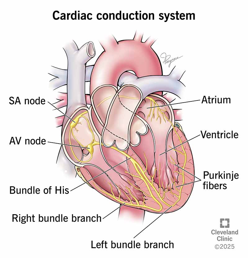
Bundle of His
carries the electrical signal from the AV node to the ventricles and helps them contract
What is happening in the circulatory system when you hear the “lub” and “dub” sound in a stethoscope?
LUB IS MADE BY THE AV VALVES WHICH CLOSE AT THE START OF SYSTOLE
DUB IS MADE BY THE SEMILUNAR VALVES DURING DIASTOLE
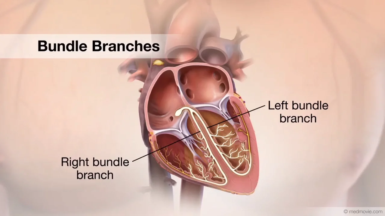
Bundle Branches
split the electrical signal and send it to both ventricles so they contract at the same time.
Purkinje Fibers
Purkinje fibers rapidly spread the electrical signal through the ventricles, causing a strong, coordinated contraction for efficient blood pumping.
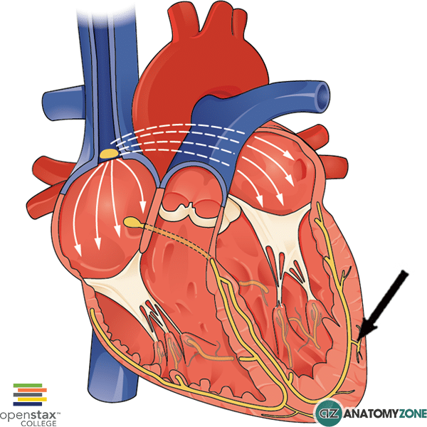
Process of heart activation
The heartbeat starts at the SA node, spreads to the AV node where it briefly pauses, then travels through the Bundle of His and the bundle branches, and finally reaches the Purkinje fibers, causing the ventricles to contract.
P wave
the first peak, produced by atrial depolarization in the right atrium

Q Wave
Represents depolarisation in the septum
Whilst the electrical stimulus passes through the bundle of His, and before it separates down the two bundle branches, it starts to depolarise the septum from left to right.

R Wave
R reflects depolarization of the main mass of the ventricles, largest wave
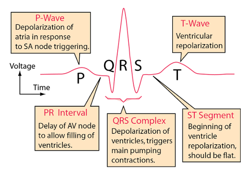
S Wave
the S wave signifies the final depolarization of the ventricles, at the base of the heart

T Wave
Indicates ventricular repolarization

Peristalsis (what is it, where does it occur)
Type of mechanical digestion that involves a series of wave-like muscle contractions that move food through the digestive tract, occurs in the esophagus
Maceration
the breakdown of food into chyme during digestion in the stomach
Segmentation (what is it and where does it occur?)
Muscles contracting in a mixing motion that churns food in the intestines with acids to help with digestion and absorption. Occurs in small intestine
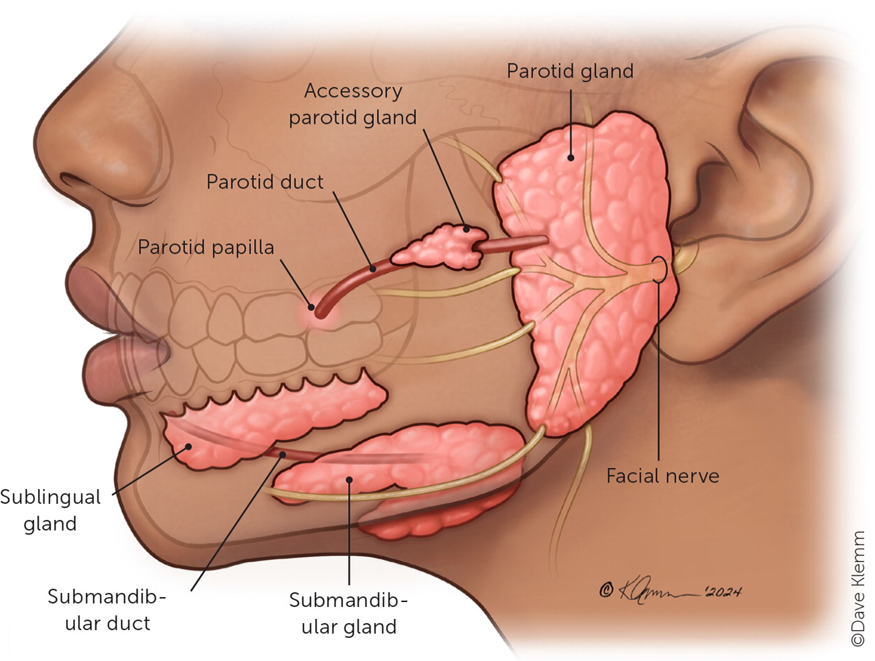
Accessory Salivary Glands
produce mucus and small amounts of saliva to keep the mouth moist, protect oral tissues, and begin digestion.
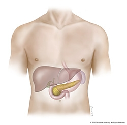
Pancreas
The pancreas helps digestion by releasing enzymes that break down carbs, fats, and proteins, and by releasing bicarbonate to neutralize stomach acid.
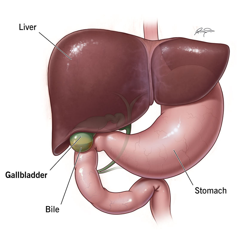
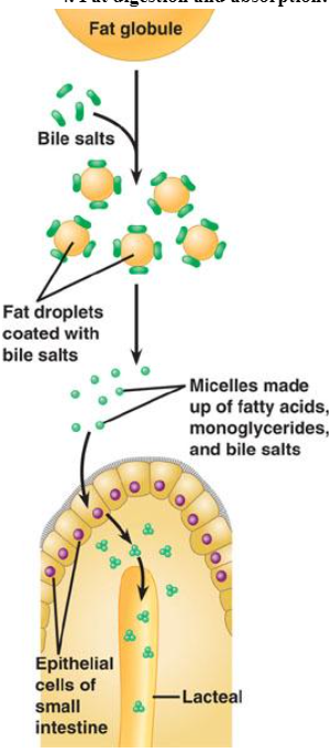
Bile Salts
They attach to fat and pull it apart into smaller droplets, allowing the pancreas to release the enzyme lipase on more surface area, which digests fatty acids and monoglycerides
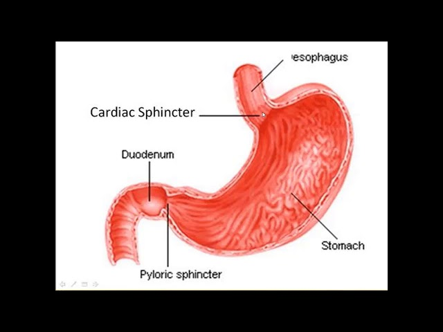
Cardiac Sphincters
The cardiac sphincter prevents acid reflux by keeping stomach acid from flowing back into the esophagus after food enters the stomach.

Pyloric Sphincter
The pyloric sphincter controls how much chyme leaves the stomach and prevents it from flowing backward from the small intestine.
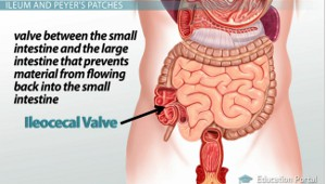
Ileocecal sphincter
Controls the movement of material into the large intestine and prevents backflow of bacteria-filled contents into the small intestine.
Bolus
a ball-like mixture of food and saliva that forms in the mouth during the process of chewing
Epiglottis
Protects your ability to breathe by protecting your larynx. It keeps food and liquid from getting into your respiratory system by folding backwards to cover the entrance of the larynx

Stomach, Chyme, and Churning
Churning is the mixing and kneading action of the stomach muscles that physically breaks food into smaller pieces.
Chyme is when these pieces are mixed with gastric juices such as hydrochloric acid (HCl), pepsin, and mucus
Pepsinogen & pepsin
Pepsinogen is an inactive form of pepsin made by cells in the stomach. Hydrochloric acid in the stomach changes pepsinogen to pepsin
Pepsin is an active enzyme which breaks down proteins during digestion into polypeptides (amino acids)
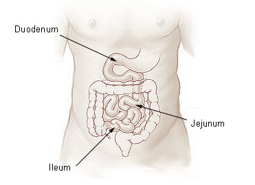
Duodenum (10% absorption)
The first part of the small intestine, receives acidic chyme from the stomach and mixes it with bile and pancreatic enzymes to continue digestion.
Neutralizes stomach acid and begins some nutrient absorption

Emulsification (what does it produce)
Breaking up large fat globules into tiny droplets so they can mix with water and be more easily digested by enzymes.
Produces micelles, which are tiny spherical structures formed during fat digestion that help transport fatty acids and monoglycerides to the intestinal lining for absorption.
Jejunum (90% absorption) Where is it located/what it do
Located in the small intestine, it absorbs nutrients (vitamins, minerals, carbohydrates, fats, proteins) and water from food so they can be used by the body.

Ileum (10% absorption)
Absorbs the remaining nutrients, especially bile salts, vitamin B12, and water. Also helps transport undigested food to the large intestine
Villi and microvilli
Villi are big finger-like projections, and microvilli are microscopic projections on those fingers, both working together to maximize nutrient absorption.
how does the liver help with digestion
Your liver continually produces bile. This is a chemical that helps turn fats into energy that your body uses.
Stomach Absorption
A small amount (10%) of substances get absorbed into the stomach, such as caffeine, aspirin, small amount of water (if dehydrated)
~10-20% of alcohol go into the stomach
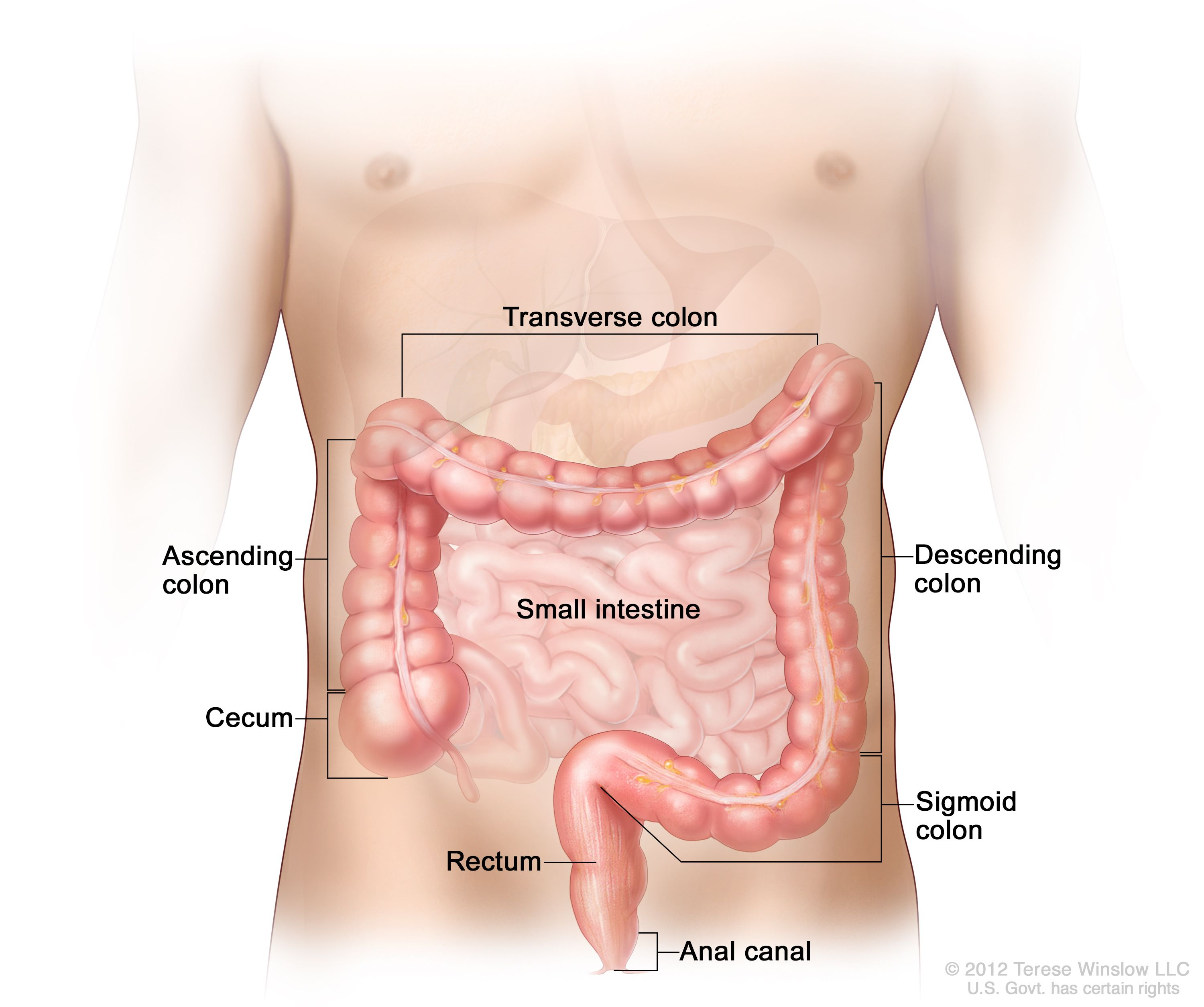
Large intestine facts
90% water absorption occurs here, it is around 5 feet long, and requires ~1.8 gallons of water for digestion
Cecum (First part of the large intestine and ascending colon)
First part of the large intestine that is a “Dead end pouch,” which means it holds digested food waste from your small intestine such as water and salts
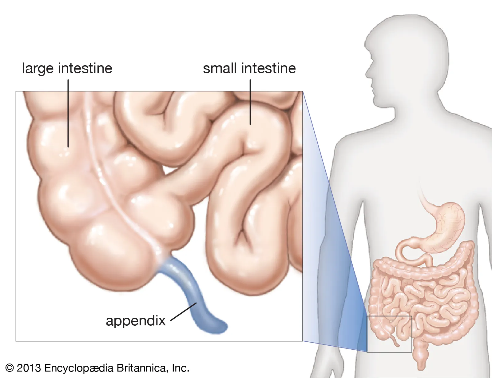
Appendix
Vestigial organ that connects to the cecum acting as a reservoir of beneficial microbes
Transverse Colon
absorbs water and nutrients that were not absorbed by the small intestine in order to prevent malnutrition and dehydration

Descending Colon
Stores the remains of digested food that will be emptied into the rectum.
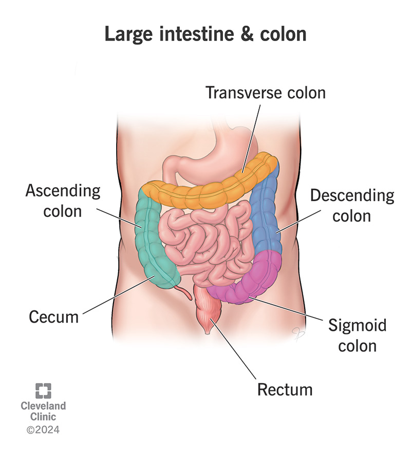
Rectum (4.7 inches long)
Defecation occurs here, and fecal matter is undigested material, as well as 60% bacteria, mucus, and cellulose.
What is the digestion Process (food moves through the stomach and small intestine within 6 to 8 hours)
Carbs are digested in the mouth by salivary amylase
Proteins are then digested in the stomach when pepsinogen, activated by HCl, turns into pepsin
Lipids are broken down when fat in the small intestine produces hormones that activate pancreatic lipase and bile salts break it down into fatty acids that can be absorbed in the duodenum
Fiber helps with pushing bulk flow
Cirrhosis of the Liver
A condition caused by alcohol damage, hepatitis B & C, and non-alcoholic fatty liver disease. This damage is characterized by the replacement of normal liver tissue by scar tissue.
Diabetes Types
Type 1: Results from the pancreas's failure to produce enough insulin
Type 2: Begins with insulin resistance, a condition in which cells fail to respond to insulin properly. Results from excessive body weight and not enough exercise.
Gestational: Occurs when pregnant women without a previous history of diabetes develop high blood sugar levels
Saliva Functions (4)
Helps prevent mouth from drying-out
Helps prevent cavities
First line of defense against disease-causing microorganisms entering mouth
Coats food and helps form bolus
Xylem
Thick walled membrane that transports water and nutrients from roots to leaves and stems, and provides structural support
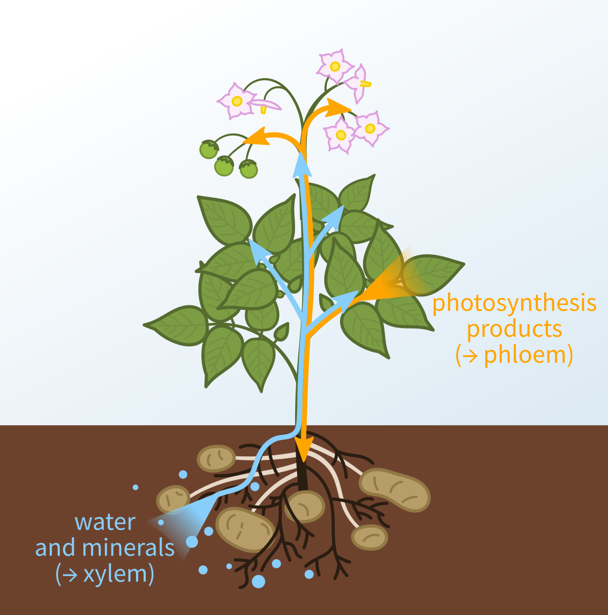
Phloem
Moves organic compounds from photosynthesis sites to other parts of the plant “food flows” (thin wall)

Suspension/Filter Feeders:
Capture and ingestion of food particles suspended in water (baleen whales, clams)
Substrate Feeders
Substrate feeders live in or on their food source and eat their way through it (caterpillars, maggots)
Deposit Feeders
Eat their way through dirt, picking up decayed organic matter (earthworms)
Bulk Feeders
Eat large pieces of food using adaptations like claws or fangs (scorpion, anaconda)
Ectomycorrhiza
A symbiotic association between fungi and plant roots, where fungi form a mantle around the root, and both partners benefit.

Endomycorrhiza
Fungal hyphae penetrate the root cortical cells of plants, forming intracellular structures known as arbuscules.
Endophytic Fungi
Exist widely inside the healthy tissue of living plants, and are important components of plant micro-ecosystems
Stomata
Allows a plant to take in carbon dioxide, which is needed for photosynthesis. They also help to reduce water loss by closing when conditions are hot or dry
CONTROLS TRANSPIRATION
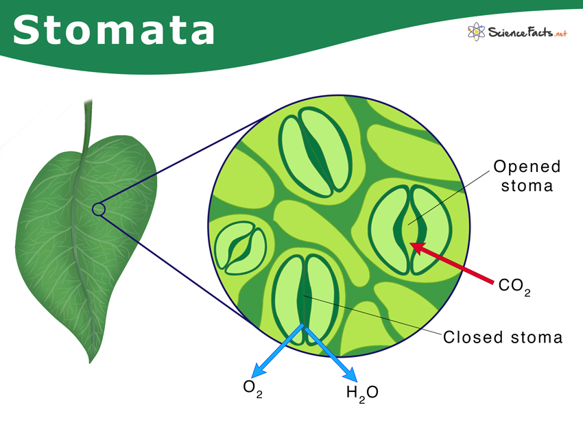
Transpiration (what is it, what controls it, and what does it lead to?)
Done by the stomata, is the process of water leaving the xylem, and this leads to the occurrence of evaporative cooling
Evaporative Cooling
the process where plants lose water as vapor through the stomata, and this evaporation removes heat from the leaf, cooling the plant down.
Translocation
the transport of materials from the leaves to other parts of the plant
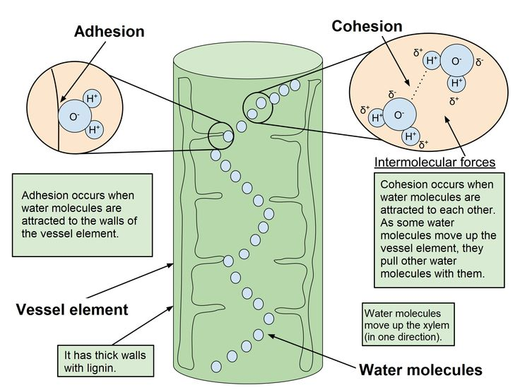
Cohesion Tension Theory
Transpiration pulls water out of the leaf’s xylem, creating tension/negative pressure. Because water molecules stick together (cohesion from hydrogen bonding), this pull is passed down through the whole column. This continuous pulling force draws water upward from the roots to the leaves, with the plant using no energy.
What percent of the water is lost by transpiration and guttation?
97-99.5%
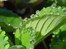
Guttation
The loss of drops of xylem sap on the tips of leaves of some vascular plants (grasses, some fungi)