3 MCQs Back, Neck, Thorax
1/238
There's no tags or description
Looks like no tags are added yet.
Name | Mastery | Learn | Test | Matching | Spaced | Call with Kai |
|---|
No analytics yet
Send a link to your students to track their progress
239 Terms
Regio scapularis is a back region.
A. Yes
B. No
Regio scapularis is a back region.
A. Yes
The anterior rami of C1 through C4 take part in the formation of cervical plexus.
A. Yes
B. No
The anterior rami of C1 through C4 take part in the formation of cervical plexus.
A. Yes
The boundaries between the thoracic region and the back are the anterior axillary lines.
A. Yes
B. No
The boundaries between the thoracic region and the back are the anterior axillary lines.
B. No
Sternocleidomastoid is a superficial muscle of the back.
A. Yes
B. No
Sternocleidomastoid is a superficial muscle of the back.
B. No
M. latissimus dorsi is a powerful extensor of the arm.
A. Yes
B. No
M. latissimus dorsi is a powerful extensor of the arm.
A. Yes
Superficial muscles of the back are supplied by dorsal branches of spinal nerves.
A. Yes
B. No
Superficial muscles of the back are supplied by dorsal branches of spinal nerves.
B. No
Auscultation triangle on the back is located medial to the scapula.
A. Yes
B. No
Auscultation triangle on the back is located medial to the scapula.
A. Yes
[triangle of auscultation is bounded by the latissimus dorsi, trapezius, and rhomboid major]
![<p>Auscultation triangle on the back is located medial to the scapula.</p><p><strong>A. Yes</strong></p><p><strong>[</strong><span>triangle of auscultation is </span><strong>bounded by the latissimus dorsi, trapezius, and rhomboid major]</strong></p><p></p>](https://knowt-user-attachments.s3.amazonaws.com/894f66a6-7e69-4db3-98d6-d569a3b0d778.png)
Serratus posterior superior muscle is a muscle of inspiration.
A. Yes
B. No
Serratus posterior superior muscle is a muscle of inspiration.
A. Yes
Muscles of the back are arranged in three groups with distinct functions.
A. Yes
B. No
Muscles of the back are arranged in three groups with distinct functions.
A. Yes
Erector spinae muscle is made of three columns.
A. Yes
B. No
Erector spinae muscle is made of three columns.
A. Yes
Intermediate muscles of the back are respiratory muscles.
A. Yes
B. No
Intermediate muscles of the back are respiratory muscles.
A. Yes
[serratus posterior superior and serratus posterior inferior are muscles of inspiration and expiration]
Platysma is innervated by a branch of the facial nerve.
A. Yes
B. No
Platysma is innervated by a branch of the facial nerve.
A. Yes
The platysma is a superficial muscle that overlaps the sternocleidomastoid.
A. Yes
B. No
The platysma is a superficial muscle that overlaps the sternocleidomastoid.
A. Yes
Accessory nerve is a branch of cervical plexus.
A. Yes
B. No
Accessory nerve is a branch of cervical plexus.
B. No
Phrenic nerve (C3-C5 (primarily C4))-innervates thoracic diaphragm.
A. Yes
B. No
Phrenic nerve (C3-C5 (primarily C4))-innervates thoracic diaphragm.
A. Yes
Internal carotid artery has two cervical branches.
A. Yes
B. No
Internal carotid artery has two cervical branches.
B. No
The upper end of internal jugular vein dilates into internal jugular fossa.
A. Yes
B. No
The upper end of internal jugular vein dilates into internal jugular fossa.
A. Yes
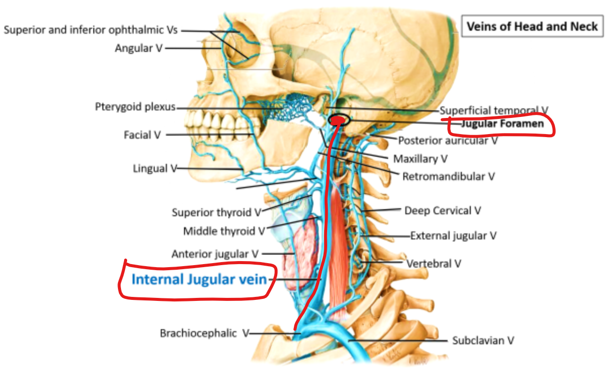
Near the termination of the internal jugular vein is a smaller dilatation, the inferior bulb.
A. Yes
B. No
Near the termination of the internal jugular vein is a smaller dilatation, the inferior bulb.
A. Yes
The inferior thyroid artery is a branch of the external carotid artery.
A. Yes
B. No
The inferior thyroid artery is a branch of the external carotid artery.
B. No
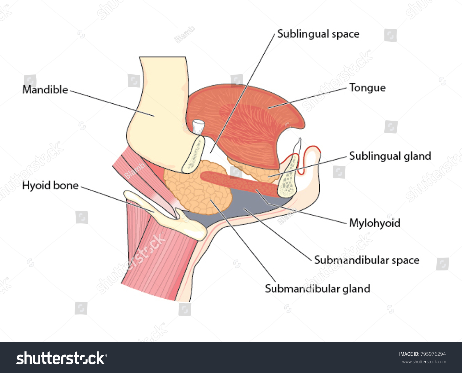
The submandibular gland is in infrahyoid region.
A. Yes
B. No
The submandibular gland is in infrahyoid region.
B. No
Which one of the listed is not a muscle of the back:
A. M. serratus posterior superior
B. M. serratus anterior
C. M. iliocostalis
D. M. longissimus
E. M. spinalis
Which one of the listed is not a muscle of the back:
A. M. serratus posterior superior
B. M. serratus anterior
C. M. iliocostalis
D. M. longissimus
E. M. spinalis
Which of the muscles listed below is a deep muscle of the back
A. Levator costae
B. Latissimus dorsi
C. Levator scapulae
D. Rhomboidei
E. Splenius
Which of the muscles listed below is a deep muscle of the back
A. Levator costae
B. Latissimus dorsi
C. Levator scapulae
D. Rhomboidei
E. Splenius
Interruption of cranial nerve XI would paralyze which muscle?
A. deltoid
B. latissimus dorsi
C. levator scapulae
D. rhomboideus major
E. trapezius
Interruption of cranial nerve XI would paralyze which muscle?
A. deltoid
B. latissimus dorsi
C. levator scapulae
D. rhomboideus major
E. trapezius - innervated by accessory nerve
If the right dorsal scapular nerve was cut near its origin, what would result:
A. Skin of the upper back on the right side would be numb
B. The point of the right shoulder would droop
C. Scapular retraction on the right would be weakened
D. Extension of the right arm would be weakened
E. Inability to adduct the right arm
The cutaneous branch of the posterior primary ramus of C2 is called the:
A. Accessory nerve
B. Great auricular nerve
C. Greater occipital nerve
D. Lesser occipital nerve
E. Superior ramus of the ansa cervicalis
The cutaneous branch of the posterior primary ramus of C2 is called the:
A. Accessory nerve
B. Great auricular nerve
C. Greater occipital nerve
D. Lesser occipital nerve
E. Superior ramus of the ansa cervicalis
Which muscle is innervated by posterior primary rami?
A. Latissimus dorsi
B. Levator scapulae
C. Rhomboideus major
D. Erector spinae
E. Trapezius
Which muscle is innervated by posterior primary rami?
A. Latissimus dorsi
B. Levator scapulae
C. Rhomboideus major
D. Erector spinae
E. Trapezius
Which of the elements listed below is not in the subcutaneous layer of the neck?
A. M. platysma
B. V. jugularis anterior
C. V. jugularis externa
D. Plexus cervicalis
E. Transverse cervical nerve
Which of the elements listed below is not in the subcutaneous layer of the neck?
A. M. platysma
B. V. jugularis anterior
C. V. jugularis externa
D. Plexus cervicalis
E. Transverse cervical nerve
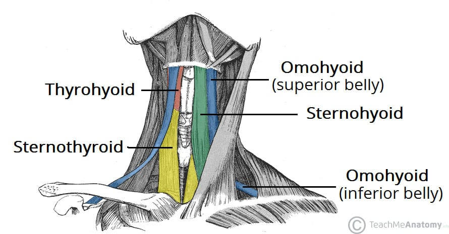
Which of the following does not belong to the infrahyoid muscles?
A. M. sternothyroideus
B. M. omohyoideus
C. M. sternocleidomastoideus
D. M. sternohyoideus
E. M. thyrohyoideus
Which of the following does not belong to the infrahyoid muscles?
A. M. sternothyroideus
B. M. omohyoideus
C. M. sternocleidomastoideus
D. M. sternohyoideus
E. M. thyrohyoideus
Which one of the following structures is NOT related to infrahyoid region?
A. gl. thyroidea
B. m. thyrohyoideus
C. n. vagus
D. m. cricothyroideus
E. v. jugularis anterior
Which one of the following structures is NOT related to infrahyoid region?
A. gl. thyroidea
B. m. thyrohyoideus
C. n. vagus
D. m. cricothyroideus
E. v. jugularis anterior
Which one of the following structures is not related to the carotid triangle?
A. hypoglossal nerve
B. superior laryngeal nerve
C. facial artery
D. thyrohyoid muscle
E. sternohyoid muscle
Which one of the following structures is not related to the carotid triangle?
A. hypoglossal nerve
B. superior laryngeal nerve
C. facial artery
D. thyrohyoid muscle
E. sternohyoid muscle
To study the compensatory response of selective suprahyoid muscles in elevating the hyoid bone, an experiment was designed in which the posterior belly of the digastric and stylohyoid muscles were paralyzed by drugs. The muscular branches of which of the following nerves must be chemically interrupted to produce paralysis in both muscles?
A. inferior alveolar
B. facial
C. hypoglossal
D. glossopharyngeal
E. lingual
The muscle which separates the submandibular triangle from the paralingual space is the:
A. Digastric, posterior belly
B. Hyoglossus
C. Mylohyoid
D. Stylohyoid
E. Styloglossus
The muscle which separates the submandibular triangle from the paralingual space is the:
A. Digastric, posterior belly
B. Hyoglossus
C. Mylohyoid
D. Stylohyoid
E. Styloglossus
Which of the following suprahyoid muscles would be paralyzed if the inferior alveolar nerve was severed at its origin?
A. Geniohyoid m.
B. Hyoglossus m.
C. Mylohyoid m.
D. Stylohyoid m.
Which of the following suprahyoid muscles would be paralyzed if the inferior alveolar nerve was severed at its origin?
A. Geniohyoid m.
B. Hyoglossus m.
C. Mylohyoid m.
D. Stylohyoid m.
In accessing the submandibular gland in the submandibular triangle, what vessel coursing through the gland and triangle would need to be protected?
A. External jugular vein
B. Facial artery
C. Maxillary artery
D. Retromandibular vein
E. Superior thyroid artery
In accessing the submandibular gland in the submandibular triangle, what vessel coursing through the gland and triangle would need to be protected?
A. External jugular vein
B. Facial artery
C. Maxillary artery
D. Retromandibular vein
E. Superior thyroid artery
T/F Lamina superficialis of the deep cervical fascia
A. Covers entire neck
B. Forms fascia masseterica
C. Extends from the skull base to the bodies of T3-T4
D. Forms fascia of submandibular gland
E. Extends posteriorly to proc. transversi
T/F Lamina superficialis of the deep cervical fascia
T - A. Covers entire neck
F - B. Forms fascia masseterica
F - C. Extends from the skull base to the bodies of T3-T4
T - D. Forms fascia of submandibular gland
T - E. extends posteriorly to proc. transversi
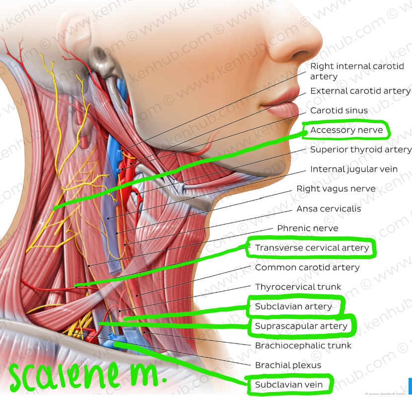
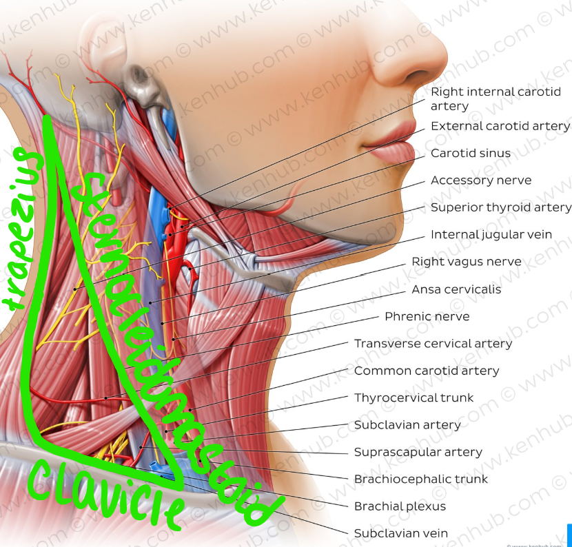
T/F Which of the following structures are boundaries of lateral cervical region?
A. Posterior border of m. sternocleidomastoideus.
B. Venter anterior of m. digastricus
C. Anterior border of m. trapezius
D. Venter superior of m. omohyoideus
E. Middle third of clavicle.
T/F Which of the following structures are boundaries of lateral cervical region?
T - A. Posterior border of m. sternocleidomastoideus.
F - B. Venter anterior of m. digastricus
T - C. Anterior border of m. trapezius
F - D. Venter superior of m. omohyoideus
T - E. Middle third of clavicle.
T/F Which of the following structures are elements of lateral cervical region?
A. Mm. scaleni
B. A. carotis communis
C. V. jugularis interna
D. A. subclavia
E. V. subclavia
T/F Which of the following structures are elements of lateral cervical region?
T - A. Mm. scaleni
F - B. A. carotis communis
F - C. V. jugularis interna
T - D. A. subclavia
T - E. V. subclavia
T/F Which of the following are from the superficial muscles of the back?
A. M. trapezius
B. M. pectoralis major
C. M. latissimus dorsi
D. M. rectus abdominis
E. M. levator scapulae
T/F Which of the following are from the superficial muscles of the back?
T - A. M. trapezius
F - B. M. pectoralis major
T - C. M. latissimus dorsi
F - D. M. rectus abdominis
T - E. M. levator scapulae
T/F The deep muscles of the back
A. divide into three subgroups
B. erect the body and the neck in bilateral contraction
C. are located dorsally to the vertebral column
D. are supplied by the ventral branches of the spinal nerves
E. are autochtonous (own) muscles of the back.
T/F The deep muscles of the back
T - A. divide into three subgroups
T - B. erect the body and the neck in bilateral contraction
T - C. are located dorsally to the vertebral column
F - D. are supplied by the ventral branches of the spinal nerves - [posterior rami of spinal nerves]
T - E. are autochtonous (own) muscles of the back.
T/F The cervical plexus of nerves
A. supplies motor branches to the infrahyoid muscles
B. supplies motor branches to the muscles of the suboccipital triangle
C. supplies branches to the trapezius muscle
D. supplies sensory branches to the diaphragm
E. supplies sensory branches to the front of the scalp.
T/F The cervical plexus of nerves
T - A. supplies motor branches to the infrahyoid muscles
F - B. supplies motor branches to the muscles of the suboccipital triangle
T - C. supplies branches to the trapezius muscle
T - D. supplies sensory branches to the diaphragm
F - E. supplies sensory branches to the front of the scalp.
T/F The sternocleidomastoid muscle
A. is attached to the temporal bone deep to the splenius capitis muscle
B. is active if the head is flexed against resistance
C. has a nerve supply from the cervical plexus
D. is an anterior relation of the scalenus anterior muscle
E. is crossed superficially by the external jugular vein.
T/F The sternocleidomastoid muscle
F - A. is attached to the temporal bone deep to the splenius capitis muscle
T - B. is active if the head is flexed against resistance
T - C. has a nerve supply from the cervical plexus
T - D. is an anterior relation of the scalenus anterior muscle
T - E. is crossed superficially by the external jugular vein.
T/F The thyroid gland
A. clasps the upper part of trachea.
B. is highly vascular.
C. doesn't move with the larynx.
D. is ductless gland.
E. consists of only one lobe.
T/F The thyroid gland
T - A. clasps the upper part of trachea.
T - B. is highly vascular.
F - C. doesn't move with the larynx.
T - D. is ductless gland.
F - E. consists of only one lobe.
T/F The brachiocephalic vein
A. collects blood only from the head and neck.
B. ends by joining the opposite one to form the superior vena cava.
C. has no valves.
D. the right one crosses the median plain.
E. the right one is laterally to the brachiocephalic artery
T/F The brachiocephalic vein
F - A. collects blood only from the head and neck.
T - B. ends by joining the opposite one to form the superior vena cava.
T - C. has no valves.
F - D. the right one crosses the median plain.
T - E. the right one is laterally to the brachiocephalic artery
T/F The vertical neurovascular bundle of the neck
A. lies on each side of the median airway and foodway.
B. extends from the base of the skull to the root of the neck.
C. contains glossopharyngeal nerve in its lower part.
D. is enclosed by the layers of the deep cervical fascia.
E. lies on the sympathetic trunk.
T/F The vertical neurovascular bundle of the neck
T - A. lies on each side of the median airway and foodway.
T - B. extends from the base of the skull to the root of the neck.
F - C. contains glossopharyngeal nerve in its lower part.
T - D. is enclosed by the layers of the deep cervical fascia.
T - E. lies on the sympathetic trunk.
T/F The internal jugular vein
A. in the upper part of the neck is posterolateral to the internal carotid a.
B. is accompanied superiorly by the last four cranial nerves.
C. is posterior to vagus nerve
D. has inferiorly the sympathetic trunk lying between the vein and common carotid artery.
E. lies on the cervical plexus.
T/F The internal jugular vein
T - A. in the upper part of the neck is posterolateral to the internal carotid a.
T - B. is accompanied superiorly by the last four cranial nerves.
F - C. is posterior to vagus nerve
F - D. has inferiorly the sympathetic trunk lying between the vein and common carotid artery.
T - E. lies on the cervical plexus.
T/F A. carotis externa:
A. is in the carotid triangle
B. gives off a. thyroidea inferior
C. supplies head and neck structures
D. has baroreceptors at its origin - the bifurcation of the common carotid artery
E. occurs at the upper border of the thyroid cartilage.
T/F A. carotis externa:
T - A. is in the carotid triangle
F - B. gives off a. thyroidea inferior
T - C. supplies head and neck structures
F - D. has baroreceptors at its origin - the bifurcation of the common carotid artery
T - E. occurs at the upper border of the thyroid cartilage.
T/F Which of the following are NOT anterior branches of external carotid artery?
A. A. pharyngea ascendens
B. A. thyroidea superior
C. A. sternocleidomastoidea
D. A. lingualis
E. A. occipitalis
T/F Which of the following are NOT anterior branches of external carotid artery?
T - A. A. pharyngea ascendens
F - B. A. thyroidea superior
T - C. A. sternocleidomastoidea
F - D. A. lingualis
T - E. A. occipitalis
T/F Anterior branches of external carotid artery are:
A. A. thyroidea superior
B. A. occipitalis
C. A. lingualis
D. A. subscapularis
E. A. facialis
T/F Anterior branches of external carotid artery are:
T - A. A. thyroidea superior
F - B. A. occipitalis
T - C. A. lingualis
F - D. A. subscapularis
T - E. A. facialis
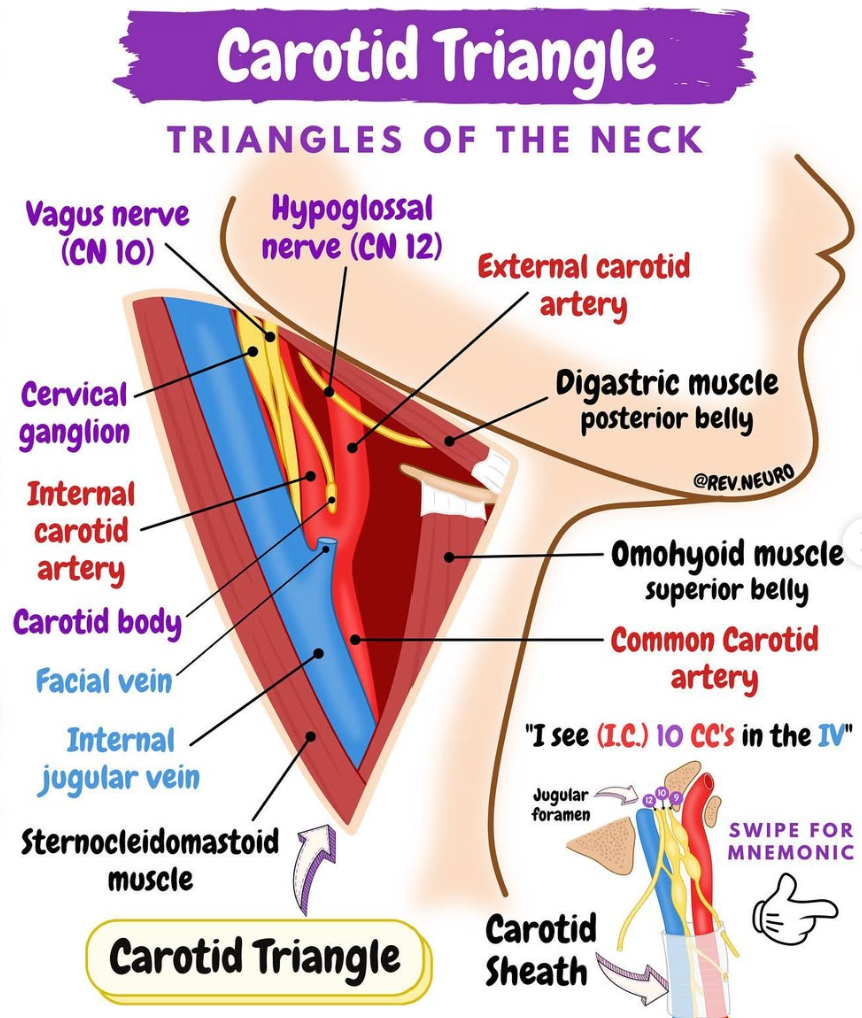
T/F The following elements are located in the carotid triangle:
A. N. laringeus superior
B. N. hypoglossus
C. Glandula thyroidea
D. Ansa cervicalis
E. A. thyroidea inferior
T/F The following elements are located in the carotid triangle:
T - A. N. laringeus superior
T - B. N. hypoglossus
F - C. Glandula thyroidea
T - D. Ansa cervicalis
F - E. A. thyroidea inferior
T/F The scalenus anterior muscle
A. is anterior to the nerves forming the brachial plexus
B. is attached to the posterior tubercles of the transverse processes of some of the cervical vertebrae
C. is medial to the vertebral artery
D. is anterior to the subclavian artery
E. is lateral to the inferior cervical ganglion.
T/F The scalenus anterior muscle
T - A. is anterior to the nerves forming the brachial plexus
F - B. is attached to the posterior tubercles of the transverse processes of some of the cervical vertebrae
F - C. is medial to the vertebral artery
T - D. is anterior to the subclavian artery
T - E. is lateral to the inferior cervical ganglion.
T/F The external carotid artery
A. is crossed anteriorly by the hypoglossal nerve
B. usually divides into its terminal branches at the level of the angle of the jaw
C. at its origin is lateral to the internal carotid artery
D. is the only source of blood to the thyroid gland
E. is superficial to the glossopharyngeal nerve.
T/F The external carotid artery
T - A. is crossed anteriorly by the hypoglossal nerve
F - B. usually divides into its terminal branches at the level of the angle of the jaw
F - C. at its origin is lateral to the internal carotid artery
F - D. is the only source of blood to the thyroid gland
T - E. is superficial to the glossopharyngeal nerve.
T/F The cricoid cartilage
A. has an anterior arch which moves upwards and backwards due to the contraction of the cricothyroid muscle
B. lengthens the vocal fold (true vocal cord) when its anterior part moves upwards and backwards
C. has the vocal folds attached to it
D. gives attachment to the inferior constrictor muscle of the pharynx
E. is at the level of the fourth cervical vertebra.
T/F The cricoid cartilage
T - A. has an anterior arch which moves upwards and backwards due to the contraction of the cricothyroid muscle
T - B. lengthens the vocal fold (true vocal cord) when its anterior part moves upwards and backwards
F - C. has the vocal folds attached to it
T - D. gives attachment to the inferior constrictor muscle of the pharynx
F - E. is at the level of the fourth cervical vertebra.
T/F The scalenus medius muscle
A. is posterior to the nerves forming the brachial plexus
B. is attached to the scalene tubercle
C. is used in deep breathing
D. is posterior to the subclavian artery
E. is crossed anteriorly by the omohyoid muscle.
T/F The scalenus medius muscle
T - A. is posterior to the nerves forming the brachial plexus
F - B. is attached to the scalene tubercle
T - C. is used in deep breathing
T - D. is posterior to the subclavian artery
T - E. is crossed anteriorly by the omohyoid muscle.
T/F The internal jugular vein
A. is, along its whole course, directly lateral to the internal carotid artery
B. has no valves
C. is anterior to the phrenic nerve
D. receives all the venous blood from the thyroid gland
E. is anterior to the thoracic duct on the left side.
T/F The internal jugular vein
F - A. is, along its whole course, directly lateral to the internal carotid artery
F - B. has no valves
T - C. is anterior to the phrenic nerve
F - D. receives all the venous blood from the thyroid gland
T - E. is anterior to the thoracic duct on the left side.
T/F The digastric muscle
A. has a motor innervation from nerves of the branchial arches
B. is inferior to the submandibular gland
C. is attached to the ramus of the mandible
D. is superficial to the hypoglossal nerve
E. is deep to the carotid sheath.
T/F The digastric muscle
T - A. has a motor innervation from nerves of the branchial arches
T - B. is inferior to the submandibular gland
F - C. is attached to the ramus of the mandible
T - D. is superficial to the hypoglossal nerve
F - E. is deep to the carotid sheath.
T/F The scalenus anterior muscle
A. is anterior to the subclavian vein
B. is anterior to the phrenic nerve
C. is anterior to the suprascapular artery
D. is used in deep respiration
E. is attached to the first and second ribs.
T/F The scalenus anterior muscle
F - A. is anterior to the subclavian vein
F - B. is anterior to the phrenic nerve
F - C. is anterior to the suprascapular artery
T - D. is used in deep respiration
F - E. is attached to the first and second ribs.
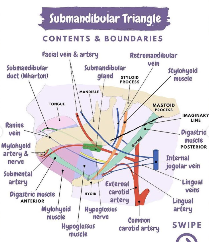
T/F Trigonum submandibulare contains:
A. glandula submandibularis
B. accessory nerve
C. phrenic nerve
D. facial artery
E. lingual nerve
T/F Trigonum submandibulare contains:
T - A. glandula submandibularis
F - B. accessory nerve
F - C. phrenic nerve
T - D. facial artery
T - E. lingual nerve
T/F The following elements are part of trigonum submandibulare:
A. n. mylohyoideus
B. n. hypoglossus
C. glandula thyroidea
D. trigonum Pirogovi
E. a. thyroidea inferior
T/F The following elements are part of trigonum submandibulare:
T - A. n. mylohyoideus
T - B. n. hypoglossus
F - C. glandula thyroidea
T - D. trigonum Pirogovi
F - E. a. thyroidea inferior
MATCH EACH NUMBERED TERM WITH THE MOST PROPER LETTERED ONE
A. M. latissimus dorsi
B. M. levator scapulae
C. M. platysma
D. M. trapezius
E. M. erector spinae
1. N. facialis
2. Rr. dorsales of nn. spinales
3. N. dorsalis scapulae
4. N. thoracodorsalis
5. N. accessories
A. M. latissimus dorsi - 4. N. thoracodorsalis
B. M. levator scapulae - 3. N. dorsalis scapulae
C. M. platysma - 1. N. facialis
D. M. trapezius - 5. N. accessories
E. M. erector spinae - 2. Rr. dorsales of nn. spinales
MATCH EACH NUMBERED TERM WITH THE MOST PROPER LETTERED ONE
Which of the following A to F supplies the muscles 1 to 6?
A. Cervical plexus
B. Spinal accessory nerve
C. Cranial accessory nerve
D. Facial nerve
E. None of these
1 Platysma
2 Infrahyoid
3 Sternocleidomastoid
4 Levator veli palatini
5 Orbicularis oculi
Which of the following A to F supplies the muscles 1 to 6?
A. Cervical plexus - 2 Infrahyoid
B. Spinal accessory nerve - 3 Sternocleidomastoid
C. Cranial accessory nerve
D. Facial nerve - 1 Platysma, 5 Orbicularis oculi
E. None of these - 4 Levator veli palatini
MATCH EACH NUMBERED TERM WITH THE MOST PROPER LETTERED ONE
A. Anterior cervical triangle
B. Posterior(lateral) cervical triangle
1. Omotrapezoid (Subclavian triangle)
2. Carotid
3. Omoclavicular (Occipital triangle)
4. Digastric triangle
A. Anterior cervical triangle - 2. Carotid, 4. Digastric triangle
B. Posterior(lateral) cervical triangle - 1. Omotrapezoid (Subclavian triangle), 3. Omoclavicular (Occipital triangle)
Erector spinae muscle comprises three muscle columns
A.
B.
C.
Erector spinae muscle comprises three muscle columns
A. spinalis
B. longissimus
C. iliocostalis
List the boundaries of lateral cervical triangle:
A.
B.
C.
List the boundaries of lateral cervical triangle:
A. trapezius m.
B. sternocleidomastoid m.
C. clavicle
Vagina carotica in the neck contains:
A.
B.
C.
Vagina carotica in the neck contains:
A. A. carotis communis (interna)
B. v. jugularis interna
C. n. vagus
List the boundaries of carotid triangle:
A.
B.
C.
List the boundaries of carotid triangle:
A. venter posterior of m. digastricus
B. m. sternocleidomastoideus
C. m. omohyoideus - venter superior
External carotid artery gives off the following branches from its anterior surface:
A.
B.
C.
External carotid artery gives off the following branches from its anterior surface:
A. superior thyroid a.
B. lingual aa.
C. facial aa.
The thyroid gland is drained by the following three pairs of veins:
A.
B.
C.
The thyroid gland is drained by the following three pairs of veins:
A. superior thyroid vv.
B. middle thyroid vv.
C. inferior thyroid vv.
Define the boundaries of trigonum submandibulare:
A.
B.
C.
Define the boundaries of trigonum submandibulare:
A. posterior bellies of digastric m.
B. anterior bellies of digastric m.
C. mandible
Pectoral nerves are branches of the medial and lateral cords of the brachial plexus.
A. Yes
B. No
Pectoral nerves are branches of the medial and lateral cords of the brachial plexus.
A. Yes
B. No
Long thoracic nerve is a direct branch of the roots of the brachial plexus.
A. Yes
B. No
Long thoracic nerve is a direct branch of the roots of the brachial plexus.
A. Yes
B. No
Dorsal scapular nerve is a direct branch of the roots of the brachial plexus.
A. Yes
B. No
Dorsal scapular nerve is a direct branch of the roots of the brachial plexus.
A. Yes
B. No
Superior trunk of the brachial plexus gives off n. suprascapularis and nerve to the subclavius.
A. Yes
B. No
Superior trunk of the brachial plexus gives off n. suprascapularis and nerve to the subclavius.
A. Yes
B. No
Mediastinum is the space in thoracic cavity where the lungs are located.
A. Yes
B. No
Mediastinum is the space in thoracic cavity where the lungs are located.
A. Yes
B. No
The lower border of parietal pleura is projected at the level of 12th rib on the back of the thorax.
A. Yes
B. No
The lower border of parietal pleura is projected at the level of 12th rib on the back of the thorax.
A. Yes
B. No
The suprasternal space is between the superficial (investing) and middle lamina of deep cervical fascia.
A. Yes
B. No
The suprasternal space is between the superficial (investing) and middle lamina of deep cervical fascia.
A. Yes
B. No
The phrenic nerve is a part of the neurovascular bundle of the neck.
A. Yes
B. No
The phrenic nerve is a part of the neurovascular bundle of the neck.
A. Yes
B. No
The cervical parietal pleura forms the dome of each pleural cavity.
A. Yes
B. No
The cervical parietal pleura forms the dome of each pleural cavity.
A. Yes
B. No
The parietal and visceral pericardium are separated by a thin film of fluid.
A. Yes
B. No
The parietal and visceral pericardium are separated by a thin film of fluid.
A. Yes
B. No
The subclavian vessels arch over the anterior surface of the dome of the pleura.
A. Yes
B. No
The subclavian vessels arch over the anterior surface of the dome of the pleura.
A. Yes
B. No
The costomediastinal recess lies along the inferior margin of the pleura.
A. Yes
B. No
The costomediastinal recess lies along the inferior margin of the pleura.
A. Yes
B. No
The costodiaphragmatic recess extends between the thoracic wall and the vertical part of the diaphragm.
A. Yes
B. No
The costodiaphragmatic recess extends between the thoracic wall and the vertical part of the diaphragm.
A. Yes
B. No
The phrenic nerve and its accompanying vessels pass anterior to the lung root.
A. Yes
B. No
The phrenic nerve and its accompanying vessels pass anterior to the lung root.
A. Yes
B. No
The vagus nerve descends posterior to the lung root.
A. Yes
B. No
The vagus nerve descends posterior to the lung root.
A. Yes
B. No
The phrenic and vagus nerves descend between the pericardium and sternum.
A. Yes
B. No
The phrenic and vagus nerves descend between the pericardium and sternum.
A. Yes
B. No
The thoracic duct enters the thoracic cavity through hiatus aorticus.
A. Yes
B. No
The thoracic duct enters the thoracic cavity through hiatus aorticus.
A. Yes
B. No
The lower boundary of mediastinum superius is the plane between angulus sterni and Th4/Th5.
A. Yes
B. No
The lower boundary of mediastinum superius is the plane between angulus sterni and Th4/Th5.
A. Yes
B. No
The upper boundary of mediastinum superius is the plane between the sternal notch and Th1.
A. Yes
B. No
The upper boundary of mediastinum superius is the plane between the sternal notch and Th1.
A. Yes
B. No
Phrenic nerve passes posteriorly to the root of the lungs.
A. Yes
B. No
Phrenic nerve passes posteriorly to the root of the lungs.
A. Yes
B. No
Vagus nerve passes anteriorly to the root of the lungs.
A. Yes
B. No
Vagus nerve passes anteriorly to the root of the lungs.
A. Yes
B. No
Phrenic and vagus nerves pass together posteriorly to the root of the lungs.
A. Yes
B. No
Phrenic and vagus nerves pass together posteriorly to the root of the lungs.
A. Yes
B. No
In the thorax sympathetic trunk is intimately related to the esophagus.
A. Yes
B. No
In the thorax sympathetic trunk is intimately related to the esophagus.
A. Yes
B. No
Aortic arch may have from 1 to 6 branches.
A. Yes
B. No
Aortic arch may have from 1 to 6 branches.
A. Yes
B. No
The pleural cavity contains:
A. Blood
B. Mucosal fluid
C. Serous fluid
D. Air
The pleural cavity contains:
A. Blood
B. Mucosal fluid
C. Serous fluid
D. Air
In lymphatic drainage of the breast, the major portion (about 75%) enters eventually into which group of nodes?
A. Central axillary
B. Deltopectoral
C. Lateral axillary
D. Parasternal
E. Subscapular
In lymphatic drainage of the breast, the major portion (about 75%) enters eventually into which group of nodes?
A. Central axillary
B. Deltopectoral
C. Lateral axillary
D. Parasternal
E. Subscapular
A woman with breast cancer subsequently develops metastases in her vertebral column. The most direct route for spread of the tumor to the vertebral column was via:
A. branches of the cephalic vein
B. branches of the lateral thoracic vein
C. branches of the thoracoacromial veins
D. lymphatic vessels draining into the axilla
E. branches of the intercostal veins
The clavipectoral fascia is penetrated by which artery?
A. Anterior circumflex humeral
B. Axillary
C. Subscapular
D. Thoracoacromial
E. Thoracodorsal
The clavipectoral fascia is penetrated by which artery?
A. Anterior circumflex humeral
B. Axillary
C. Subscapular
D. Thoracoacromial
E. Thoracodorsal
During a motorcycle accident, an 18-year-old male landed on the right lateral side of his rib cage with his right upper limb abducted. In the hospital he was found to have "winging" of the right scapula. Which nerve was likely damaged in the accident?
A. Accessory
B. Lateral pectoral
C. Long thoracic
D. Phrenic
E. Vagus
Breast cancer cells can spread directly to the cranial cavity and brain via the vertebral venous plexus. Through which route can they reach this plexus?
A. Axillary lymph nodes
B. Internal thoracic vein
C. Intercostal veins
D. Parasternal lymph nodes
E. Thoracoacromial artery
Breast cancer cells can spread directly to the cranial cavity and brain via the vertebral venous plexus. Through which route can they reach this plexus?
A. Axillary lymph nodes
B. Internal thoracic vein
C. Intercostal veins
D. Parasternal lymph nodes
E. Thoracoacromial artery
While observing a mastectomy on a 60-year-old female patient, a medical student was asked by the surgeon to help tie off the arteries that supply the medial side of the breast. The artery that gives origin to these small branches is the:
A. Internal thoracic
B. Musculophrenic
C. Posterior intercostal
D. Superior epigastric
E. Thoracoacromial
Upon finding a malignant tumor in the medial portion of the breast of a 40-year-old female, the surgeon began to search for the lymph nodes that would be the first ones reached by metastatic spread of cancer cells from this site. Which group(s) would have to be examined to determine whether metastasis had occurred?
A. Central only
B. Parasternal only
C. Parasternal and apical
D. Parasternal and lateral
E. Parasternal and pectoral