TAP - guided quizzes
1/31
There's no tags or description
Looks like no tags are added yet.
Name | Mastery | Learn | Test | Matching | Spaced | Call with Kai |
|---|
No analytics yet
Send a link to your students to track their progress
32 Terms
The thoracic wall functions to protect the heart and lungs, support the thoracic limb, and counteract the negative pressure created during respiration.
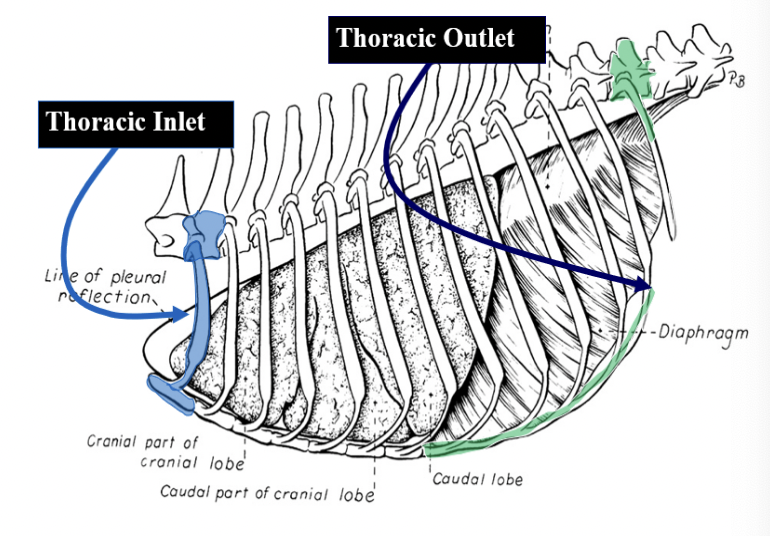
What are the dorsal, lateral, ventral, cranial, and caudal boundaries of the thoracic wall?
Dorsal - Thoracic vertebrae
Lateral - Ribs & costal cartilages
Ventral - Sternebrae
Cranial - Thoracic inlet
Caudal - Thoracic outlet
The muscular support for the thoracic wall comes from the epaxial muscles and the __________ muscles that are found between the ribs. There are two intercostal muscle groups, the __________ intercostal mm. that run perpendicular to each other. The fiber direction of the external intercostal muscles run in a __________ direction while the internal intercostal muscles run in a __________ direction.
The intercostal arteries serve as the primary blood supply to the intercostal mm. The intercostal arteries are branches of the __________. The intercostal arteries run in a neurovascular bundle, along with the intercostal veins and nerves, along the __________ aspect of each rib. The internal thoracic arteries branch off of the __________ before running parallel with one another close to midline in the ventral thoracic cavity. These arteries give off branches which travel a short distance dorsad to anastomose with the intercostal arteries. Don't worry, you will not be asked to number the intercostal arteries, veins or nerves associated with each intercostal space.
The muscular support for the thoracic wall comes from the epaxial muscles and the intercostal muscles that are found between the ribs. There are two intercostal muscle groups, the external & internal intercostal mm. that run perpendicular to each other. The fiber direction of the external intercostal muscles run in a craniodorsal to caudoventral direction while the internal intercostal muscles run in a cranioventral to caudodorsal direction.
The intercostal arteries serve as the primary blood supply to the intercostal mm. The intercostal arteries are branches of the descending aorta. The intercostal arteries run in a neurovascular bundle, along with the intercostal veins and nerves, along the caudal aspect of each rib. The internal thoracic arteries branch off of the subclavian arteries before running parallel with one another close to midline in the ventral thoracic cavity. These arteries give off branches which travel a short distance dorsad to anastomose with the intercostal arteries. Don't worry, you will not be asked to number the intercostal arteries, veins or nerves associated with each intercostal space.
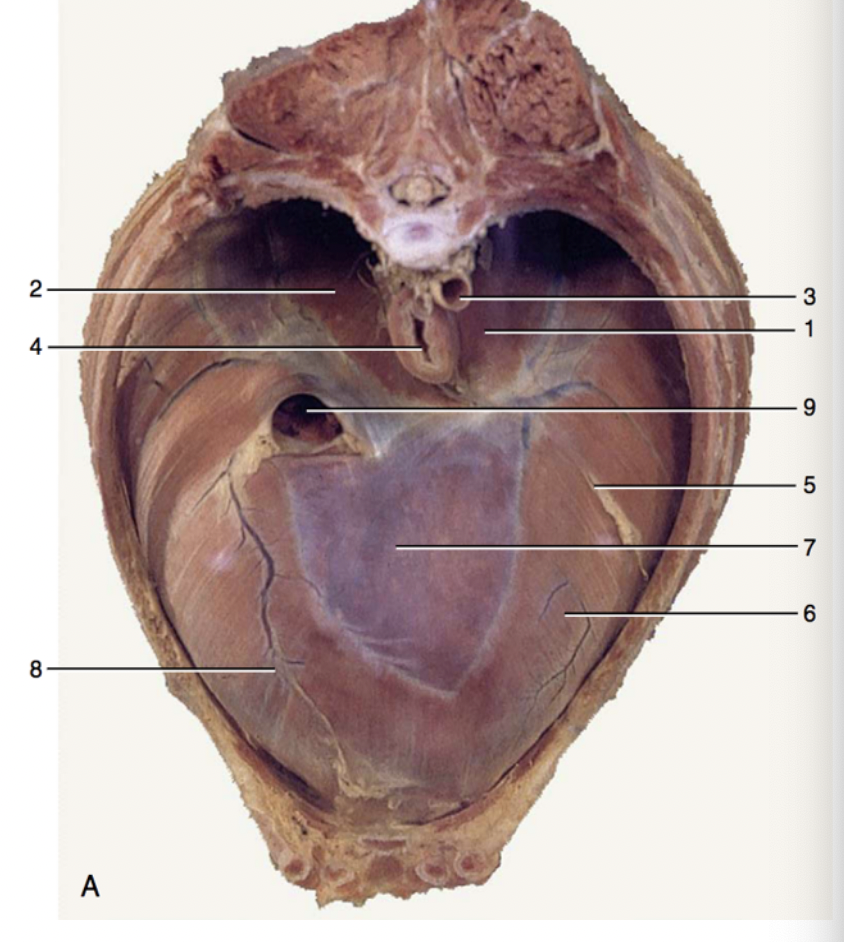
The image below is a cranial view of the respiratory diaphragm. Please match the corresponding numbers and structures - not all are matched in the question below.
3 - descending aorta passing through the aortic hiatus
4 - esophagus passing through the esophageal hiatus
9 - caudal vena cava passing through the caval foramen
7 - central tendon of the diaphram
The heart sits within the pericardium. This pericardium helps protect, anchor, and lubricate the motions of the heart.
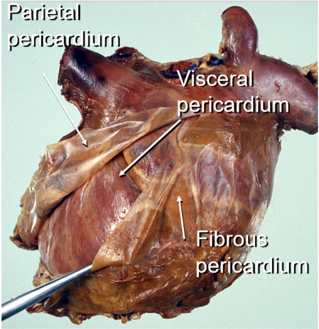
There are three layers of pericardial tissue that form the pericardial sac. The deepest/most internal layer of pericardium, known as the _____ pericardium, is directly adhered to the heart itself. There is then a fluid filled cavity, called the pericardial cavity, located between the adhered layer of pericardium and the surrounding _____ pericardium that does not directly adhere to the heart. The outermost layer of connective tissue, called the _____ pericardium, anchors the heart in place within the mediastinum.
The heart sits within the pericardium. This pericardium helps protect, anchor, and lubricate the motions of the heart.
There are three layers of pericardial tissue that form the pericardial sac. The deepest/most internal layer of pericardium, known as the visceral pericardium, is directly adhered to the heart itself. There is then a fluid filled cavity, called the pericardial cavity, located between the adhered layer of pericardium and the surrounding parietal pericardium that does not directly adhere to the heart. The outermost layer of connective tissue, called the fibrous pericardium, anchors the heart in place within the mediastinum.
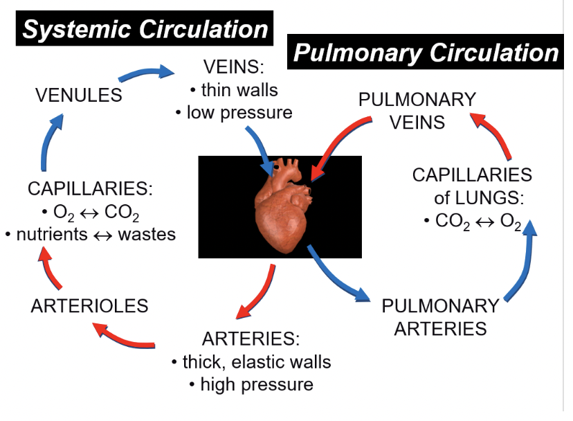
Starting in the right ventricle, imagine you are a single red blood cell in the circulatory system. Please place the following structures in the correct order in which you would pass through before returning to the right ventricle where you started.
Leaving the right ventricle
pulmonary (right semilunar) valve, pulmonary trunk, pulmonary arteries, gas exchange within the lungs, pulmonary veins, left atrium, left atrioventricular valve, left ventricle, aortic (left semilunar) valve, aortic arch, systemic arteries, systemic artierioles, gas exchange in the capillary beds of the periphery (think skeletal muscle), systemic venules, systemic veins, cranial or caudal vena cava, right atrium, right artioventricular valve
Arriving into the right ventricle
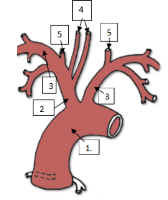
The image below is the aortic arch of a canine. Please match the names of each structure with the corresponding number.
aortic arch
brachiocephalic trunk
subclavian arteries
common carotid arteries
vertebral arteries
Moving caudally from the thoracic cavity, on the other side of the diaphragm, is the abdominal cavity. Please match the abdominal wall boundaries with the proper directional term relative to the abdominal cavity.
Dorsal - hypaxial muscles along thoracic & lumbar vertebrae
Ventral & Lateral - abdominal wall muscles
Cranial - last rib & costal arch
Caudal - bony pelvis & quadriceps femoris muscle
Please order the muscles of the abdominal wall from deep to superficial.
Deepest muscle
transversus abdominus m.
rectus abdmominus m.
internal abdominal oblique m.
external abdominal oblique m.
Superficial muscle
Within the abdominal cavity, serous membranes known as peritoneum envelope the organs to form potential spaces or boundaries. The inner abdominal wall is lined by ______ peritoneum while the abdominal organs within the cavity are covered with ______ peritoneum. Structures or organs that lie outside of this continuous peritoneal membrane, such as the kidneys, are referred to as ______ and are considered to be outside of the abdominopelvic cavity. Peritoneal folds between the visceral and parietal peritoneum are known as omenta (singular = omentum), mesenteries, or ligaments. The ______ provide a passageway for the neurovascular supply for the intestines and suspends the intestines from the dorsal abdominal wall at an anchor point called the ______.
Within the abdominal cavity, serous membranes known as peritoneum envelope the organs to form potential spaces or boundaries. The inner abdominal wall is lined by parietal peritoneum while the abdominal organs within the cavity are covered with visceral peritoneum. Structures or organs that lie outside of this continuous peritoneal membrane, such as the kidneys, are referred to as retroperitoneal. and are considered to be outside of the abdominopelvic cavity. Peritoneal folds between the visceral and parietal peritoneum are known as omenta (singular = omentum), mesenteries, or ligaments. The mesentery provide a passageway for the neurovascular supply for the intestines and suspends the intestines from the dorsal abdominal wall at an anchor point called the root of the mesentery.
We describe the PsNS and SNS as having different outflows based on their anatomy, Please match the following accordingly.
parasympathetic nervous system - craniosacral outflow
sympathetic nervous system - thoracolumbar outflow
Sort the following effects as resulting from increased parasympathetic or sympathetic tone.
Parasympathetics
airway constriction
increase salivary secretion
pupil constriction (myosis)
decrease heart rate, decrease contractile force of heart
increasing gastrointestinal motility
Sympathetics
pupil dilation (mydriasis)
increase heart rate, increase contractile force of heart
decrease gastrointestinal motility
vasoconstriction to increase blood pressure
airway dilation
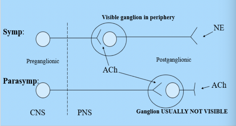
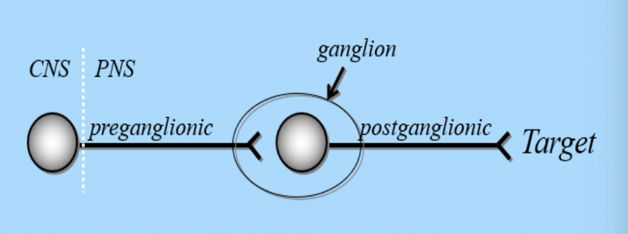
Statement In both the PsNS and SNS, the cell bodies of the preganglionic neurons are located in the _____. These cells send their axons out to communicate with the cell bodies of _____. The axons of the preganglionic neurons of the _____ are typically short compared to the longer _____ preganlionic axons. The PSNS has outflow from cranial nerves _____ as well as from sacral portions of the spinal cord.
Statement In both the PsNS and SNS, the cell bodies of the preganglionic neurons are located in the central nervous system. These cells send their axons out to communicate with the cell bodies of postganglionic neurons. The axons of the preganglionic neurons of the SNS are typically short compared to the longer PSNS preganlionic axons. The PSNS has outflow from cranial nerves III, VII, IX, X as well as from sacral portions of the spinal cord.
Please match the following innervations of the abdominal viscera with the appropriate description.
Vagus nerve (CN X), Pelvic nerves, Splanchnic nerves
Vagus nerve (CN X)
Provides the majority of the parasympathetic input to the abdominal viscera and GI tract, mostly innervates the cranial abdominal viscera
Pelvic nerves
Originating from sacral spinal cord segments, provides parasympathetic input to the caudal GI tract and the urogenital organs
Splanchnic nerves
Provides sympathetic input to the abdominal viscera, direct caudal continuation of the sympathetic chain
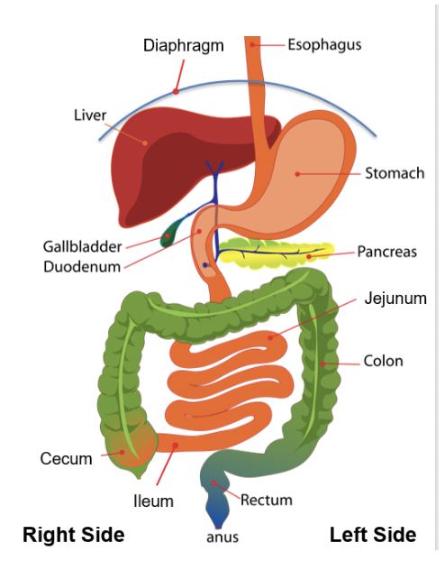
Within the thorax, the esophagus passes caudally within the mediastinum before passing through the ________ of the respiratory diaphragm to enter the abdomen. The ________ is located just caudal to the respiratory diaphragm, extending cranially into the ribcage. The gall bladder fills a fossa between the ________
and right medial lobes of the liver. The esophagus enters the cardia of the stomach at the ________. Ingesta leaves the stomach and enters the duodenum, with this outflow regulated by the ________. The duodenum receives bile produced by the liver and digestive enzymes from the ________ to aid in digestion. The ________ is located on the left side of the abdomen and is attached to the stomach via the ________ ligament.
Within the thorax, the esophagus passes caudally within the mediastinum before passing through the esophageal hiatus of the respiratory diaphragm to enter the abdomen. The liver is located just caudal to the respiratory diaphragm, extending cranially into the ribcage. The gall bladder fills a fossa between the quadrate and right medial lobes of the liver. The esophagus enters the cardia of the stomach at the cardiac sphincter. Ingesta leaves the stomach and enters the duodenum, with this outflow regulated by the pyloric sphincter. The duodenum receives bile produced by the liver and digestive enzymes from the pancreas to aid in digestion. The spleen is located on the left side of the abdomen and is attached to the stomach via the gastrosplenic ligament.
order the portions of the canine small and large intestine from oral to aboral.
oral
duodenum
jejunum
ileum
ascending colon
transverse colon
descending colon
aboral
place the regions of the equine ascending colon in order from oral to aboral.
Oral - communicating with the ileum and base of the cecum
Right ventral colon
Sternal flexure
Left ventral colon
Pelvic flexure
Left dorsal colon
Diaphragmatic flexure
Right dorsal colon
Aboral - communicating with the transverse colon
match the compartments of the ruminant stomach with the appropriate description.
Reticulum, Rumen, Omasum, Abomasum
Reticulum
Most cranial compartment which the esophagus empties into, has internal mucosa with a honeycomb-like appearance
Rumen
The largest compartment and the main site for fermentation, internal mucosa has papillae to increase surface area for nutrient absorption
Omasum
Has leaflet mucosa, commonly referred to as the "Butcher's Bible", which increases surface area for water absorption
Abomasum
The glandular 'true' stomach, secretes HCl and enzymes for digestion, can be displaced within the abdominal cavity due to it not being attached to the ventral body wall
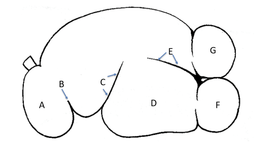
The image is a left view of a rumen drawing. Please match the letters with the correct anatomical term.
A - Reticulum
B - Ruminoreticular fold
C - Left cranial groove
D - Ventral sac
E - Left longitudinal groove
F - Caudoventral blind sac
G - caudodorsal blind sac
The primary blood supply to the abdominal viscera comes from the ________. This artery is a continuation of the descending aorta which passes caudally through the ________ of the respiratory diaphragm. This artery gives off both paired and unpaired branches before terminating caudally by dividing at the level of the seventh thoracic vertebra into the ________, internal iliac, and median sacral arteries.
The primary blood supply to the abdominal viscera comes from the abdominal aorta. This artery is a continuation of the descending aorta which passes caudally through the aortic hiatus of the respiratory diaphragm. This artery gives off both paired and unpaired branches before terminating caudally by dividing at the level of the seventh thoracic vertebra into the external iliac, internal iliac, and median sacral arteries.
Below, please sort the following arteries into paired or unpaired branches off of the abdominal aorta. Note: If there is two of a structure, for example the kidney, these typically receive blood supply from paired arteries.
Paired
Lumbar artery, Deep circumflex iliac artery, Phrenicoabdominal artery, Renal artery, Ovarian / Testicular artery
Unpaired
Caudal Mesenteric artery, Celiac artery, Cranial mesenteric artery, Median sacral artery
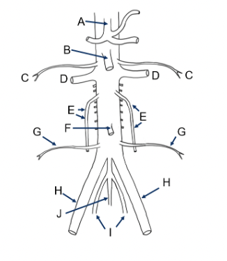
Please match the the correct artery/arteries with the indicated letter on the diagram below. As you go through this exercise, can you also describe which organs recieve blood from these vessels?
A - Celiac a.
B - Cranial mesenteric a.
C - Phrenicoabdominal a.
D - Renal a.
E - Gonadal a.
F - Caudal mesenteric a.
G -Deep circumflex a.
H - External iliac a.
I - Internal iliac a.
J - Median sacral a.
The _______ drains blood from the hind limbs, abdominal wall, kidneys, and reproductive organs. The _______ transports nutrient-rich blood from the spleen, stomach, and intestines to the liver for processing. This vein has two major branches called the _______ branch, which drains the intestines, and the the splenic vein, which drains the spleen. From the liver, the _______ bring the filtered nutrient-rich blood back to the systemic vasculature by draining into the caudal vena cava.
The caudal vena cava drains blood from the hind limbs, abdominal wall, kidneys, and reproductive organs. The hepatic portal vein transports nutrient-rich blood from the spleen, stomach, and intestines to the liver for processing. This vein has two major branches called the cranial mesenteric branch, which drains the intestines, and the the splenic vein, which drains the spleen. From the liver, the hepatic veins bring the filtered nutrient-rich blood back to the systemic vasculature by draining into the caudal vena cava.
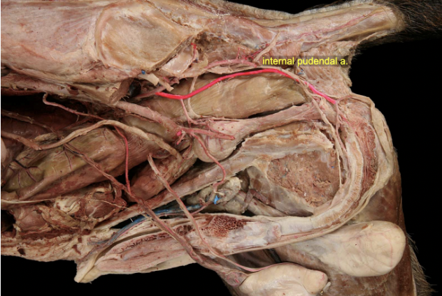
The _________ artery is a branch of the _________ artery and supplies the internal and external genitalia in the male and female with branches that occur on both the left and the right side. In the male, the major vessel is the _________ artery, which then gives off a _________ vesical artery to supply the _________. In the female, this major artery branch is called the _________ artery and gives off a branch to anastomose with the _________ artery to form the uterine artery. The urinary bladder is also supplied by the _________ vesical arteries, which branch off of the internal iliac artery.
The internal pudendal artery is a branch of the internal iliac artery and supplies the internal and external genitalia in the male and female with branches that occur on both the left and the right side. In the male, the major vessel is the prostatic artery, which then gives off a caudal vesical artery to supply the urinary bladder. In the female, this major artery branch is called the vaginal artery and gives off a branch to anastomose with the ovarian artery to form the uterine artery. The urinary bladder is also supplied by the cranial vesical arteries, which branch off of the internal iliac artery.
Sort the structures and innervation of the storage and micturition phases.
Storage phase
internal urethral sphincter tenses, sympathetic stimulation via hypogastric nerves, detrusor muscle relaxed
Micturition phase
internal urethral sphincter relaxes, detrusor muscle contracts, parasympathetic stimulation via the pelvic nerve, somatic (conscious control) via the pudendal nerve
If you have not reviewed the ANS lecture as part of this unit, then that is strongly recommended. ANS function and anatomy in the thorax, abdomen, and pelvis is also very important. For a review question, does the pelvic nerve contain pre- or post-ganglionic nerve fibers?
Preganglionic
The pelvic nerve provides parasympathetic innervation to the pelvic organs (urinary bladder, rectum, urethra, genetalia). The sympathetic innervation for these organs is supplied by the hypogastric nerve. Are the nerve fibers in the hypogastric nerve considered pre-ganglionic or post-ganglionic?
Postganglionic
In the female, the urinary bladder empties via the ________ into the vestibule. The ________ is the source of the gamete (aka oocyte or egg) and if spermatozoa are present fertilization can occur in the (aka oviduct). Implantation of the placenta and growth of the fetus occurs in the ________ in either the horns or body depending on the shape of this organ in different species. The ________ remains closed throughout gestation and only opens during _______ or when the female is in estrus (heat) and ready to be bred. When the female is bred, spermatozoa is deposited in the ________ and must then pass through the cervix to find their way to the oocyte for fertilization.
In the female, the urinary bladder empties via the urethra into the vestibule. The ovary is the source of the gamete (aka oocyte or egg) and if spermatozoa are present fertilization can occur in the uterine tube (aka oviduct). Implantation of the placenta and growth of the fetus occurs in the uterus in either the horns or body depending on the shape of this organ in different species. The cervix remains closed throughout gestation and only opens during parturition or when the female is in estrus (heat) and ready to be bred. When the female is bred, spermatozoa is deposited in the vagina and must then pass through the cervix to find their way to the oocyte for fertilization.
The male gonads are the _________. These function as
_________, producing hormones such as testosterone and germ cells (sperm). In adult life, they reside within the _________. During fetal life, they must first descend through the inguinal canal, being tethered to the body by the remnants of the _________ gubernaculum.
The male gonads are the testes. These function as endocrine and exocrine glands, producing hormones such as testosterone and germ cells (sperm). In adult life, they reside within the scrotum. During fetal life, they must first descend through the inguinal canal, being tethered to the body by the remnants of the lower gubernaculum.
A - pampiniform plexus
B - seminiferous tubules
C - head of the epididymis
D - ductus deferens
E - tail of the epididymis
Please place the following structures in order from sperm production in the seminiferous tubules to ejaculation into the external environment.
seminiferous tubule (spermatazoa production)
Head of epididymis
Body of the epididymis
Tail of the epididymis
Ductus deferens
Pelvic urethra
Penile urethra
external environment
The ductus arteriosus connects the aorta and the _______ to allow blood to bypass the lungs whereas the foramen ovale is a one-way valve allowing blood to flow from the _______ to the _______, bypassing the right ventricle and the lungs. Once the animal takes its first breath, pulmonary vascular diameter increases, causing resistance of blood flow to the lungs for gas exchange to decrease. This changes the flow of blood through the heart so that these fetal structures are no longer needed. In the adult animal, the remnant of the ductus arteriosus is the _______ and the foramen ovale is the _______.
The ductus arteriosus connects the aorta and the pulmonary trunk to allow blood to bypass the lungs whereas the foramen ovale is a one-way valve allowing blood to flow from the right atrium to the left atrium, bypassing the right ventricle and the lungs. Once the animal takes its first breath, pulmonary vascular diameter increases, causing resistance of blood flow to the lungs for gas exchange to decrease. This changes the flow of blood through the heart so that these fetal structures are no longer needed. In the adult animal, the remnant of the ductus arteriosus is the ligamentum arteriosum and the foramen ovale is the fossa ovalis.