10: Ruminant Protozoa
 Call Kai
 Learn
Learn Practice Test
 Spaced Repetition
 Match
Match Flashcards
 Knowt Play
1/83

There's no tags or description
Looks like no tags are added yet.
Name | Mastery | Learn | Test | Matching | Spaced | Call with Kai | Chat |
|---|
No analytics yet
Send a link to your students to track their progress
84 Terms
Why are Eimeria spp. (coccidia) not zoonotic
Eimeria spp. are incredibly host, site, and cell specific
T/F: Eimeria spp. are likely to cause coccidiosis
F; many species are non or mildly pathogenic
CS associated with coccidiosis
Severe diarrhea ± blood, dehydration
Patient demographic for coccidiosis
Young, old, and stressed
Ruminant species that is more likely to die from coccidiosis
Goats
Why is “winter coccidiosis” a thing
Animals in closer contact to preserve warmth spread disease more easily
Coccidiosis syndrome more common in the NW US
“Nervous coccidiosis” causing neuro signs and occasionally death
Type of life cycle in Eimeria spp.
Direct LC
Where does Eimeria spp. set up shop
Various locations in the SI
Gross lesions associated with the more pathogenic species of Eimeria spp.
Nodules in the SI
How do DH get infected with Eimeria spp.
Ingestion of sporulated oocysts
How long does it take for an oocyst to undergo sporogony
24-48 hours in environment
How does Eimeria spp. (and any apicomplexan really) cause damage in the host
Asexual replication (schizogony/merogony) is incredibly damaging
Morphology of Eimeria spp. eggs
Small but variable egg with centrally located morula or spores (depending on development), may see a micropyle cap if you are lucky
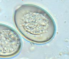
Eimeria spp. that are different than normal and are larger and brown
Eimeria macusaniensis (camelids)
Goal of Eimeria spp. treatment
Manage clinical disease
Management techniques to decrease incidence of coccidiosis
Reduce exposure to infective oocysts: clean feces, elevate feed
Prophylactic treatment
When should prophylactic coccidiostats be given
Just before or just after a period of increased stress
How do prophylactic coccidiostats allow for adaptive immunity
Knocks down the level of internal Eimeria spp. but doesn’t wipe out the entire population
Eimeria spp. treatment that is toxic to horses
Ionophores
Coccidiostats used in goats
Decoquinate, monensin
ELDU that may be warranted but cannot be used for market animals
Diclazuril or toltrazuril
Cryptosporidium life cycle
Direct life cycle
Patient demographic for cryptosporidium
Young animals
Where does cryptosporidium set up shop
Superficially associated with gut cells
T/F: crypto is zoonotic
True
Key CS associated with cryptosporidium
BAD watery diarrhea
How do cattle get cryptosporidium
Ingestion of sporulated oocysts
When do cryptosporidium oocysts become sporulated
They are infectious when excreted
How does cryptosporidium cause autoinfection
Sporozoites can bust out of sporulated oocysts while inside the host and re-infect the GIT
Primary diagnostic for cryptosporidium
Centrifugal sugar float
Morphology of cryptosporidium eggs
SMALL (almost RBC sized) eggs
Parasiticide of choice for cryptosporidium
No approved treatment :(
Treatment of choice for cryptosporidium
Supportive care and management
Babesia species in cattle
Babesia bovis and Babesia bigemina
Small babesia
Babesia bovis
Large babesia
Babesia bigemina
T/F: Babesia is reportable in cattle
True
Vector for babesia
Tick
Common name for babesia
Redwater disease
CS associated with babesia
Severe anemia and hemoglobinuria
Diagnostic for babesia
Blood smear or PCR
Blood smear findings associated with small babesia
Signet ring inclusion
Blood smear findings associated with large babesia
Teardrop or pear shaped inclusion
Treatment for babesia
Cull
Life cycle type of babesia
Apicomplexan
DH of babesia
Tick
Apicomplexan stage that causes damage in ruminants
Merozoite
Why is it so hard to control tick transmission of babesia
Female ticks can pass babesia to offspring transovarially
Tick host for bovine theileria
Longhorn tick (Haemaphysalis longicornis)
Specific type of theileria that is more pathogenic and responsible for disease in cattle
Theileria orientalis ikeda
CS associated with Theileria orientalis ikeda
Generic tick borne illness: anemia, fever
Lethargy
Abortion
Death
Top alternative differential to Theileria orientalis ikeda
Anaplasma spp.
What happens to cattle that survive a theileria infection
Carriers for life
Diagnostics for theileria
Blood smear or PCR
Blood smear findings associated with Theileria orientalis ikeda
Signet ring inclusions
How do we differentiate small babesia (reportable) from theileria
Patient history and CS: does the patient have hemoglobinuria, can you find the babesia tick (one host tick)
Management for theileria
Limit contact with vectors, reservoir cattle, and fomites
How do apicomplexans damage ruminants when they are the IH
Cysts in tissue, commonly muscle and nervous
Common zoonotic protozoa that use ruminants as IH
Toxoplasma gondii and Neospora caninum
Toxoplasma gondii and Neospora caninum route of infection for ruminants
Ingestion of sporulated oocysts or transplacental transmission
T. gondii is a major cause of abortion in which species
Small ruminants
N. caninum is a major cause of abortion in which species
Cattle
Diagnostics to confirm Toxoplasma gondii and Neospora caninum as the cause of abortion
PCR and histopath
Treatment for Toxoplasma gondii and Neospora caninum
No drugs available for the tissue stages
Prevention for Toxoplasma gondii and Neospora caninum
Prevent DH (dogs and cats) from ingesting aborted tissues or contaminated raw meat
Flagellate reproductive parasite in cattle
Tritrichomonas foetus
Life cycle of Tritrichomonas foetus
Direct life cycle with one stage
Single stage of Tritrichomonas foetus
Trophozoite
Where does Tritrichomonas foetus set up shop
Repro tract of females and prepuce of bulls
How long does Tritrichomonas foetus last outside the host
Not very long, it’s not a hardy protozoa
Patient demographic for Tritrichomonas foetus infection
Beef cattle and older bulls
CS associated with Tritrichomonas foetus in heifers
Mild vaginitis (often undiagnosed)
Breeding failure
Early abortion
CS associated with Tritrichomonas foetus in bulls
They are asymptomatic carriers
Why are beef cattle often more affected than dairy cattle
Dairy cattle are usually bred via AI
Presumptive signs of Tritrichomonas foetus infection in a herd
Widespread breeding failure
Long calving interval
Bull-sharing
Diagnostic of choice for Tritrichomonas foetus
Look at preputial wash under microscope
Mucus sample form cervix or mucus
Why do you have to culture your sample if testing for Tritrichomonas foetus
It increases the sensitivity of your microscopic and PCR tests
Oklahoma regulatory definition for a negative designation for Tritrichomonas foetus
3 consecutive negative culture tests or 1 negative PCR test
Populations that should be tested for Tritrichomonas foetus
Any bull >12 mos that is changing ownership
Potentially exposed female cattle
Any breeding bull entering OK
Parasiticide for Tritrichomonas foetus
No approved drugs
Treatment for Tritrichomonas foetus in cows
Reproductive rest for 3 most
Treatment for Tritrichomonas foetus in bulls
Cull
Purpose of Tritrichomonas foetus vaccine in cows
Minimizes disease and prevents shedding, but won’t prevent infection