Looks like no one added any tags here yet for you.
Define light.
Electromagnetic energy that is emitted in the form of waves
Define wavelength.
The distance between successive peaks or troughs
Define frequency.
The number of waves per second
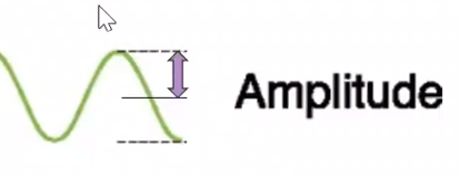
Define amplitude.
The distance between the midpoint of the wave and the peak
Define reflection.
The bouncing of light rays off a surface
Define absorption.
The transfer of light energy to a particle or surface
Define refraction.
The bending of light rays that can occur when they travel from one transparent medium to another
Define optic disk.
A pale circular region from which the retinal vessels originate
Also where the optic nerve fibers exit the retina
No photoreceptors
Define macula.
The part of the retina for central vision
Distinguished by the relative absence of large blood vessels
Define extraocular muscles.
3 pairs
Move the eyeball in the orbit
Normally not visible because they lie behind the conjunctiva
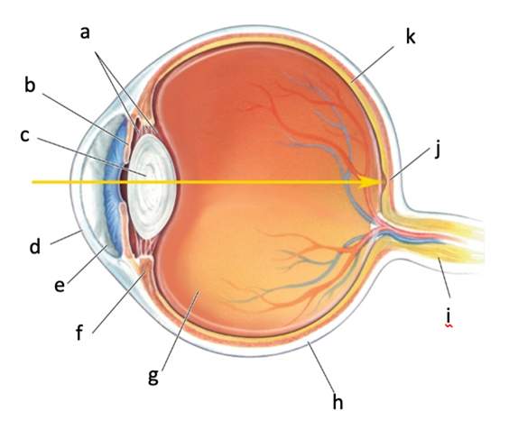
Identify and describe a.
Zonule fibers
Ligaments attached to the ciliary muscle that suspend the transparent lens behind the iris
Identify and describe b.
Iris
Surrounds the pupil
Its pigmentation provides the eye’s color
Contains two muscles that can vary the size of the pupil: one makes it smaller when it contracts, the other makes it larger
Identify and describe c.
Lens
Located behind the iris
Is suspended by ligaments (zonule fibers) attached to the ciliary muscle
Identify and describe d.
Cornea
Lacks blood vessels
Is nourished by the aqueous humor behind it
Covers both the pupil and iris
Is continuous with the sclera
Has a refractive power of 42 diopters
Identify and describe e.
Aqueous humor
Watery fluid
Located between the cornea and the lens
Nourishes the cornea
Identify and describe f.
Ciliary muscle
Forms a ring inside the eye
Zonule fibers are attached to the ciliary muscle to hold the lens in place
Identify and describe g.
Vitreous humor
Viscous, jelly-like fluid
Lies between the lens and the retina
Keeps the eyeball spherical
Identify and describe h.
Sclera
The white of the eye
Forms the touch wall of the eyeball
Identify and describe i.
Optic nerve
Carries axons from the retina → exits the back of the eye → passes through the orbit → reaches the base of the brain near the pituitary gland
Identify and describe j.
Fovea
A dark spot about 2 mm in diameter
Marks the center of the retina
Thinner portion of the retina
Identify and describe k.
Retina
Located at the back of the eye
Contains photoreceptors specialized to convert ight energy into neural activity (cones and rods)
Describe the process of light entering the eye and the role of the cornea and the lens.
Light enters the eye through the cornea, focuses on the retina, and forms an image
Light enters the eye perpendicular to the corneal surface → Light passes straight to the retina
Light enters the eye at angle other than perpendicular to the corneal surface → Light rays are bent such that they converge on the back of the retina
Cornea has a refractive power of 42 diopters
Lens shape changes to provide extra focusing power and make crisp images of objects closer than 9 meters
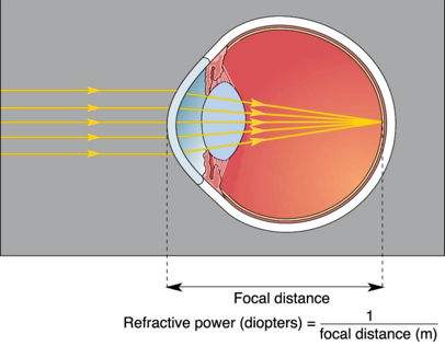
What is a diopter?
A unit of measurement that is the reciprocal of the focal distance in meters
What is focal distance and what does it depend on?
The distance from the refractive surface to the point where parallel light rays converge
Depends on the curvature of the cornea
The tighter the curve, the shorter the focal distance
Describe emmetropia.
Eye focuses parallel light rays onto the retina
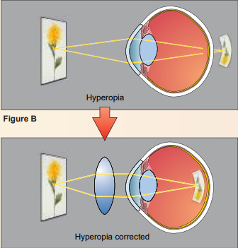
Describe hyperopia.
Farsightedness
Eye is too short → Can see far away but near objects focus behind the retina
Corrected with concave lenses
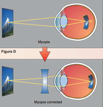
Describe myopia.
Nearsightedness
Eye is too long → Image converges in front of retina
Corrected with convex lens
Describe astigmatism.
Irregular curvature of the eye → different refraction in horizontal and vertical planes
Describe presbyopia.
“old eye”
Hardening of the lens with aging
Lens is unable to change shape and accommodate sufficiently
Corrected with bifocals
Describe radial keratotomy.
Tiny incisions in the peripheral cornea to relax central cornea
Corrects myopia
Describe PRL.
Photorefractive keratectomy
Laser reshapes outer surface of cornea by vaporizing thin layers
Describe LASIK.
Laser in situ keratomileusis
Thin flap on the outer cornea is temporarily lifted
Laser reshapes cornea from the inside
Describe strabismus.
Misalignment or lack of coordination between the eyes (cross-eyed)
Correction in early childhood
Surgery to correct extraocular muscles
Prismatic glasses
Describe esotropia.
Directions of the gaze of each eye crosses
Describe exotropia.
Direction of the gaze of each eye diverges
Describe cataracts.
Clouding of the lens
Corrected with surgical replacement with artificial lens and glasses
Describe glaucoma.
A progressive loss of vision associated with elevated intraocular pressure
Can compress the optic nerve axons
Leading cause of blindness
Describe detached retina.
The retina pulls away from the underlying wall of the eye from a blow to the head or by shrinkage of vitreous humor
Fluid from the vitreous humor flows through small tears in the retina resulting from the trauma, causing more of the retina to separate
Describe retinitis pigmentosa.
Characterized by a progressive degeneration of the photoreceptors
First sign is loss of peripheral vision and night vision
No cure but Vitamin A may slow its progression
Describe macular degeneration.
Loss of central vision
Common, affecting more than 25% of all Americans over 65 years of age
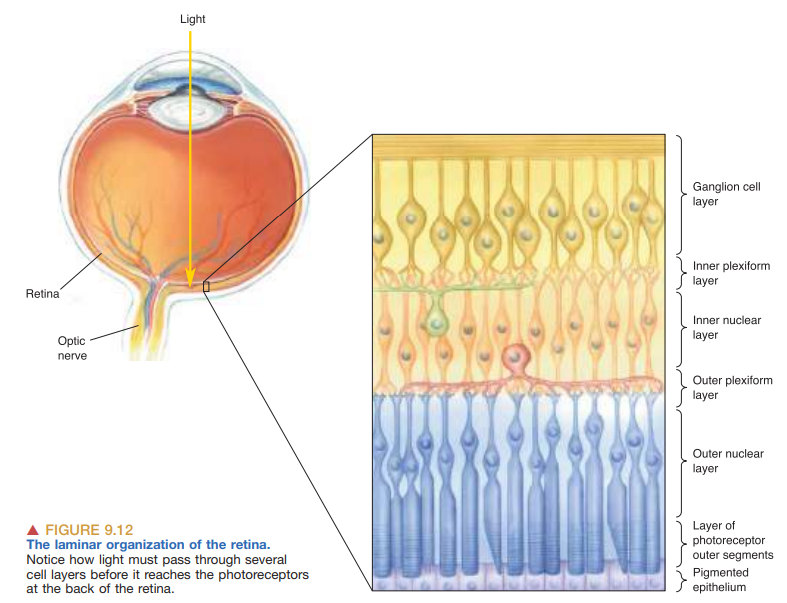
Describe the path of light when traveling through the cell layers in the retina.
Ganglion cell layer → Contain the cell bodies of ganglion cells
Inner plexiform layer → Contains the synaptic contacts between bipolar cells, amacrine cells, and ganglion cells
Inner nuclear layer → Contains the cell bodies of the bipolar cells, horizontal cells, and amacrine cells
Outer plexiform layer → Where photoreceptors make synaptic contact with the bipolar and horizontal cells
Outer nuclear layer → Contains the cell bodies of the photoreceptors
Layer of photoreceptor outer segments → Contains the light-sensitive elements of the retina
Pigmented epithelium → Absorbs any light that passes entirely through the retina
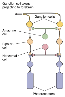
Describe the pathway of information flowing through retinal cells.
Photoreceptors → respond to light and influence the membrane potential of the bipolar cells connected to them
Horizontal cells → receive input from photoreceptors and project to other photoreceptors and bipolar cells
Bipolar cells
Amacrine cells → receive input from bipolar cell and project to ganglion cells, bipolar cells, and other amacrine cells
Ganglion cells → fire action potential in response to light → these impulses propagate along the optic nerve to the rest of the brain
Describe the role of horizontal cells.
Receive input from photoreceptors and project to other photoreceptors and bipolar cells
Describe the role of amacrine cells.
Receive input from bipolar cells and project to ganglion cells, bipolar cells, and other amacrine cells
Describe the role of pigmented epithelium.
Located behind photoreceptors (rods and cones)
Absorbs scattered light
What is tapetum lucidum and what kind of mammals possess it?
A reflective layer beneath receptors
Bounces light back toward retina → sensitive to low light levels
Nocturnal animals possess it
Describe the morphological (shape) differences between rods and cones,
Rods → longer, cylindrical outer segments containing many disks
Cones → shorter, tapering outer segment with fewer membranous disks
At the fovea, there are no…
Rods, only cones
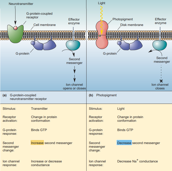
Describe dark current and how it changes in light.
Rod outer segments are depolarized (Vm = -30 mV) in the dark because of they have a steady influx of Na+
cGMP is produced in the photoreceptor by enzyme guanylyl cy → keep Na+ channels open
Light reduces cGMP → Na+ channels close → membrane potential becomes more negative → rod receptors hyperpolarize in response to light
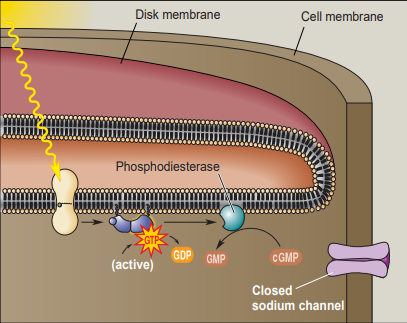
Write the phototransduction pathway in light.
Light activates (bleaches) rhodopsin by retinal undergoing a conformational change to all-trans retinal
Activates Gtransducin
Activates phosphodiesterase (PDE)
PDE activity reduces cGMP level s by converting cGMP to linear GMP.
cGMP-gated Na+ channels close
Cell membrane hyperpolarizes (becomes more negative)
Describe the Young-Helmholtz trichromacy theory of color vision.
The brain assigns colors based on signal from all 3 cone types (red, green, blue)
Equal signal from all 3 cones = white
Describe dark adaptation.
All-cone daytime vision → all-rod nighttime vision
Can take minutes to nearly an hour
Increases sensitivity to light a millionfold or more during this period
What factors contribute to dark adaptation?
Dilation of pupils → allows more light to enter the pupil
Regeneration of unbleached rhodopsin
Adjustment of functional circuitry
Why do pirates wear and eye patch over one eye?
To adapt more easily between light and dark vision
Describe light adaptation.
Cone adapts to relative changes in light
Dark → Light
Eye saturates
Reverses the dark adaptation
Cones are hyperpolarized to Ek (equilibrium potential for K+) in light → cones then gradually repolarize to -35 mV to continue sensing light
When you stare at a yellow box and then switch to a green box, the green box appears a different color. What color? Why?
Purple, it is the opponent color of green; also bleaching of the receptors
What is a receptive field?
The area of the retina where light changes neuron’s firing rate
What is the difference between ON and OFF bipolar cells?
ON bipolar cells
Hyperpolarized by light shined onto a cone
Cone releases LESS NT
Less ligand for ionotropic glutamate receptors
Vm decreases
OFF bipolar cells
Depolarized by light in receptive field center
Light hyperpolarizes the cone → Less NT
Less hyperpolarization from GPCR → depolarize ON bipolar cell
Describe center-surround receptive fields.
The receptive field of a bipolar cell consists of two parts
A circular area of retina providing direct photo receptor input (the receptive field center)
A surrounding area of retina providing input via horizontal cells (the receptive field surround)
The response of a bipolar cell’s membrane potential to light in the receptive field center is opposite to that of the light in the surround (antagonistic)
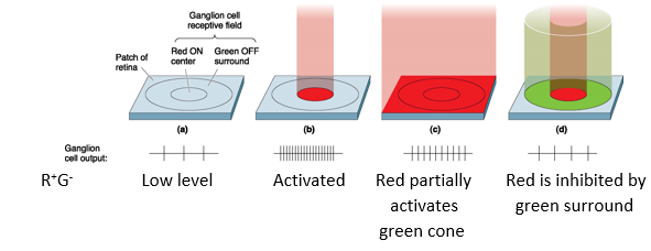
Describe color-opponent cells.
P cells and nonM-nonP are sensitive to wavelength of light (color)
Response to one color in center is cancelled by an opponent color in the surround
Red vs green; blue vs yellow
R+G- = Activated by red (center), inhibited by green (surround)
Describe ipRGCs.
Intrinsically photosensitive retinal ganglion cells
Only a few thousand ganglion cells
Contain melanopsin photopigment (first discovered in frog skin)
Depolarize in response to light
Have a large receptive field
Important for synchronizing behavior to daily changes in light level (establishing circadian rhythms)
Describe parallel processing.
Simultaneous input from two eyes
Input from eyes compared in cortex → determines depth and distance of object
Information about light and dark → ON-center and OFF-center ganglion cells
Different receptive fields and response properties of retinal ganglion cells: M and P cells, and nonM-nonP cells