Thoracic Innervation
1/39
There's no tags or description
Looks like no tags are added yet.
Name | Mastery | Learn | Test | Matching | Spaced | Call with Kai |
|---|
No analytics yet
Send a link to your students to track their progress
40 Terms
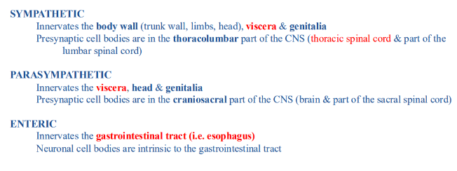
efferent innervation of thoracic viscera
-efferent innervation to viscera entirely autonomic
-ANS innervates smooth muscle, cardiac muscle, and glands
-NO skeletal muscle in viscera, therefore NO GSE neurons
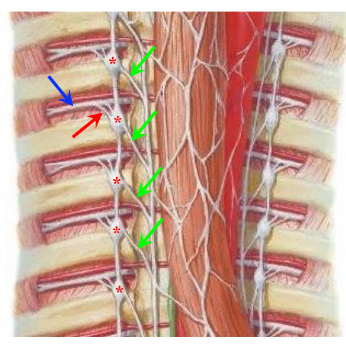
sympathetic innervation of thoracic viscera
-all the viscera in the thorax, abdomen, and pelvis are innervate by splanchnic branches of the sympathetic chain (green arrows)
-splanchnic branches: arise from the sympathetic chain ganglia at EVERY level (cervical, thoracic, lumbar, and sacral)
-splanchnic branches are separate from the rami communicantes (red arrow) and the ventral rami of spinal nerves (blue arrow)
-splanchnic branches go medially
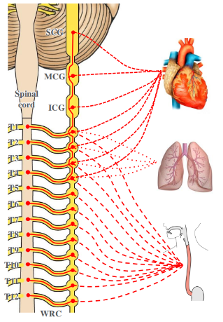
viscera in thorax innervated by ____ from the _____ parts of the sympathetic chain
-splanchnic branches
-cervical and/or thoracic
-ICG: inferior cervical ganglion
-MCG: middle cervical ganglion
-SCG: superior cervical ganglion
-WR: white rami communicantes
parasympathetic innervation of thoracic viscera
-innervated by branches of the vagus nerve (X)
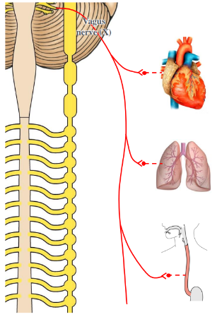
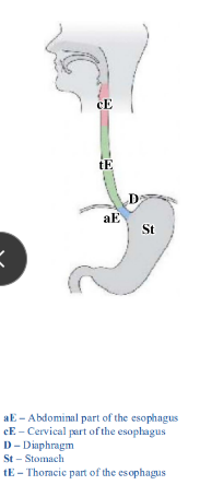
innervation of the esophagus- parts of esophagus
-cervical, thoracic, and abdominal parts of the esophagus
-cervical part: skeletal muscle, innervated by the recurrent laryngeal nerves (from X)
-thoracic and abdominal parts: smooth muscle and therefore innervated by the ANS (GVE)
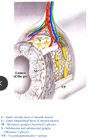
innervation of the esophagus
-gut made up of several layers: mucosa, submucosa, inner circular and outer longitudinal layer of smooth muscle, and visceral peritoneum
-postsynaptic sympathetic, presynaptic parasympathetic, and sensory axons piggyback on arteries to reach the esophagus
-ganglia within the submucosa and between layers of smooth muscle: submucosal and myenteric ganglia- contain postsynaptic parasympathetic neurons and neurons of the enteric nervous system
-enteric nervous system: part of the ANS, initiates and coordinates peristalsis
usually, the sympathetic nervous system ____ and the parasympathetic nervous system ___
-decreases the rate of peristalsis and constricts sphincters
-increases the rate of peristalsis and relaxes sphincters (vagal stimulation may improve learning, memory, and mood)
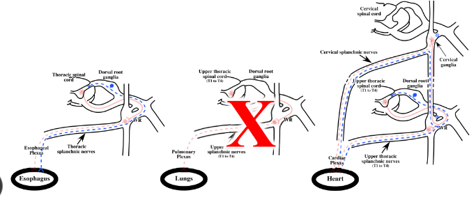
autonomic innervation of the esophagus
-sympathetic innervation: thoracic part of esophagus receives sympathetic innervation from splanchnic nerves branching off the sympathetic chain at all thoracic levels
-parasympathetic innervation: thoracic part of esophagus receives parasympathetic innervation form branches of the vagus nerve
-both sympathetic and parasympathetic axons ramify on the esophagus as the esophageal plexus
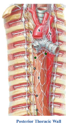
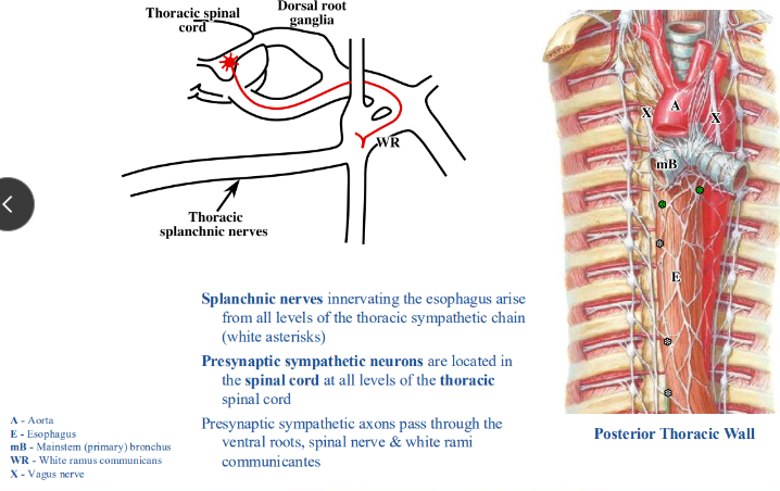
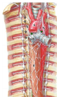
sympathetic innervation of the esophagus
-splanchnic nerves innervating esophagus arise from all levels of the thoracic sympathetic chain (white asterisks)
-presynaptic sympathetic neurons located in spinal cord at all levels of thoracic spinal cord
-presynaptic sympathetic axons pass through the ventral roots, spinal nerve, and white rami communicantes
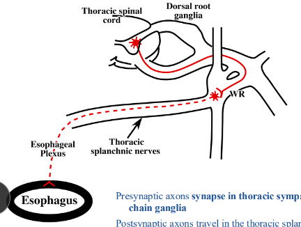
presynaptic axons synapse in _____; postsynaptic axons travel _____
-thoracic sympathetic chain ganglia
-in the thoracic splanchnic nerves, ramify within the esophageal plexus, and enter the wall of the esophagus
-sympathetic neurons decrease the rate of peristalsis
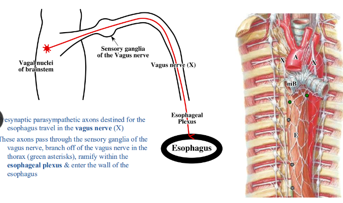
parasympathetic innervation of the esophagus
-presynaptic parasympathetic axons destined for esophagus travel in the vagus nerve (X)
-axons pass through the sensory ganglia of the vagus nerve, branch off the vagus nerve in the thorax, ramify within the esophageal plexus, and enter the wall of the esophagus
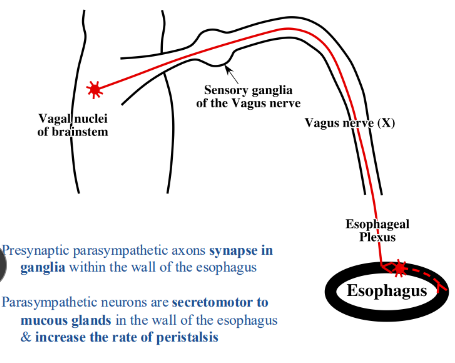
presynaptic parasympathetic axons _____
-synapse in ganglia within the wall of the esophagus
-parasympathetic neurons are secretomotor to mucous glands in the wall of the esophagus and increase the rate of peristalsis
autonomic innervation of the lungs
-sympathetic innervation: from splanchnic nerves branching off the sympathetic chain from T1 through T4
-parasympathetic innervation: lungs receive parasympathetic innervation from branches of the vagus nerve
-both sympathetic and parasympathetic axons ramify within the pulmonary plexus located anterior and posterior to the root of the lung, as well as along branches of the bronchial tree
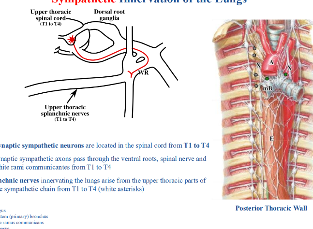
sympathetic innervation of the lungs
-presynaptic sympathetic neurons located in spinal cord from T1 to T4
-presynaptic sympathetic axons pass through the ventral roots, spinal nerve, and white rami communicantes from T1 to T4
-splanchnic nerves innervating the lungs arise from the upper thoracic parts of the sympathetic chain from T1 to T4 (white asterisks)
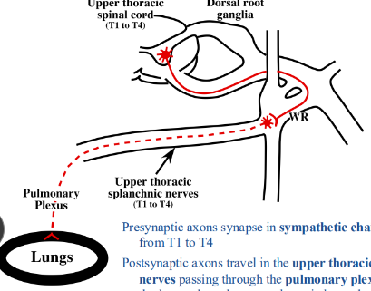
presynaptic axons synapse in _____; postsynaptic axons travel in the ____
-sympathetic chain ganglia from T1 to T4
-upper thoracic splanchnic nerves passing through the pulmonary plexus to reach the lungs; pulmonary plexus located anterior and posterior to the root of the lung and along branches of the bronchial tree
activation of sympathetic neurons results in
-inhibition of bronchial glands and smooth muscle in bronchial walls (bronchodilation)
-selective vasoconstriction of poorly oxygenated areas to allow more blood to go to the well oxygenated ones
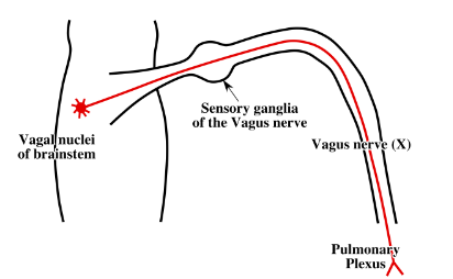
parasympathetic innervation of the lungs
-vagus nerve (X) provides parasympathetic innervation to the lungs
-presynaptic parasympathetic neuronal cell bodies located within the vagal nuclei in the brainstem
-presynaptic parasympathetic axons destined for the lungs travel in the vagus nerve (X) passing through the sensory ganglia of the vagus nerve and entering the pulmonary plexus
presynaptic parasympathetic axons synapse _____; postsynaptic parasympathetic neuronal cell bodies in pulmonary plexus have axons that ____
-in microscopic ganglia in pulmonary plexus
-continue in the plexus to reach the lungs
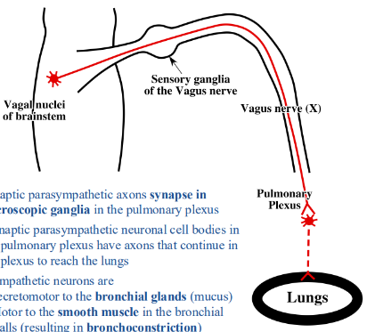
parasympathetic neuron characteristics
-secretomotor to the bronchial glands (mucus)
-motor to the smooth muscle in the bronchial walls (resulting in bronchoconstriction)
-cause vasodilation of the pulmonary vasculature
factors influencing cardiac function
-conducting system (cardiac muscle cells)
-carotid body and sinus (chemoreceptors and baroreceptors)
-adrenal glands (catecholamine releasing cells in the medulla)
-autonomic nervous system (sympathetic and parasympathetic neurons)
conducting system
-entirely specialized cardiac muscle cells
-microscopic
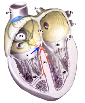
-SA node and AV node responsible for initiating and coordinating the heartbeat
-SA node (blue asterisk): at junction of superior vena cava and the right atrium
-AV node (red asterisk): located in interatrial septum near the opening of the coronary sinus
-both SA and AV nodes usually supplied by branches of the right coronary artery
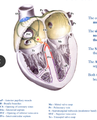
both the SA and AV node contain ______ that _____
-muscle cells; contract spontaneously
-intrinsic rate of contraction of SA node usually faster than AV node
-SA node drives rate of heartbeat- called “pacemaker”
impulses from SA node travel _____; impulses from AV node travel ____
-through specialized cardiac muscle pathways of both right and left atria and to the AV node
-in the AV bundle (blue arrow) through the fibrous skeleton to the muscular portion of the interventricular septum at which point it bifurcates into right and left bundle branches (red)
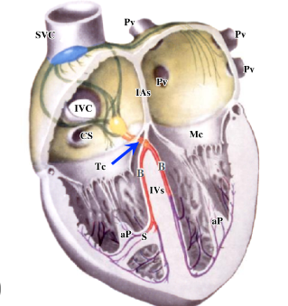
AV bundle
-ONLY way for impulses to get to the ventricles because the skeleton of the heart electrically isolates the atria from the ventricles
-AV block involves impairment of conduction through the AV bundle
subendocardial branches
-purple lines
-consist of Purkinje fibers that extend along the interventricular septum and the walls of both ventricles
-subendocardial branch to the right anterior papillary muscle contained within the septomarginal trabecula (moderator band)
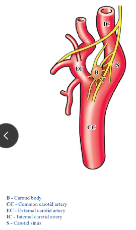
carotid body and sinus
-structures in the neck
-carotid body: small mass of tissue that lies on the medial side of the internal carotid artery close to the bifurcation of the common carotid artery
-carotid body contains chemoreceptors that respond to reduced blood gas levels (particularly O2) by stimulating the cardiac rate and respiration (increasing blood pressure)
-carotid sinus: slight dilation of initial segment of internal carotid artery
-carotid sinus contains baroreceptors that respond to increased arterial blood pressure by decreasing blood pressure
-glossopharyngeal nerve (IX) innervates the carotid body and sinus
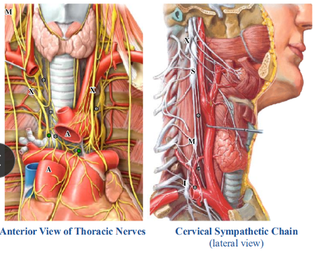
autonomic innervation of the heart
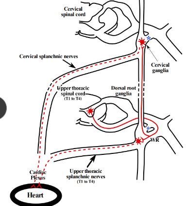
-sympathetic innervation: from splanchnic nerves branching off the sympathetic chain at CERVICAL levels and from T1 through T4
-parasympathetic innervation: from branches of the vagus nerve
-both sympathetic and parasympathetic axons ramify within the cardiac plexus located posterior to the ascending aorta and anterior to the bifurcation of the trachea
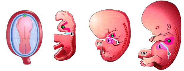
development of the heart
-changes its position during embryonic period
-after folding, the heart (green asterisk) is anterior to cervical somites
-by the end of the embryonic period, heart is within the thorax
-because of its original position, the heart receives innervation from cervical splanchnic nerves
-diaphragm also descends- explains innervation by ventral rami of C3/4/5
sympathetic innervation of the heart
-presynaptic sympathetic neurons innervating heart located in spinal cord from T1-T4
-NO presynaptic sympathetic neurons in the spinal cord superior to T1
-presynaptic sympathetic axons pass through the ventral roots, spinal nerve, and white rami communicantes from T1 to T4
-some of the presynaptic axons remain at thoracic levels while others go up the sympathetic chain to cervical levels
presynaptic axons synapse _____; postsynaptic axons _____
-in either sympathetic chain ganglia from T1 to T4, or in one of the cervical sympathetic chain ganglia
-travel in the cervical and upper thoracic splanchnic nerves passing through the cervical plexus to reach the heart
sympathetic axons distribute to
-SA node: increases heart rate (chronotropy)
-AV node: increases conduction velocity (dromotropy)
-ventricles: increases force of contraction (inotropy) and rate of myocardial relaxation (lusitropy)
-coronary arteries: vasodilation
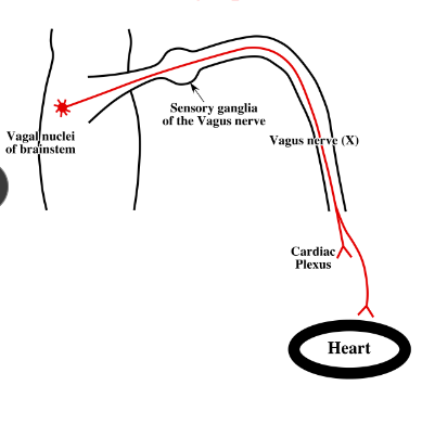
parasympathetic innervation of the heart
-vagus nerve (X) provides parasympathetic innervation to the heart
-presynaptic parasympathetic neuronal cell bodies located within the vagal nuclei in the brainstem
-presynaptic parasympathetic axons destined for the heart travel in the vagus nerve (X)
-axons pass through the sensory ganglia of the vagus nerve, branch off in the neck and thorax, and enter the cardiac plexus
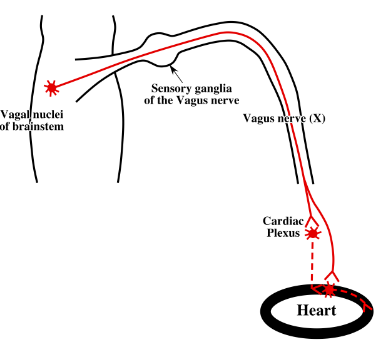
presynaptic parasympathetic axons synapse ____; postsynaptic parasympathetic neuronal cell bodies in the cardiac plexus have axons that ___
-in either tiny ganglia within the cardiac plexus or within the walls of the atria
-continue in the plexus to reach the heart
-postsynaptic parasympathetic neuronal cell bodies in walls of atria have relatively short axons
parasympathetic axons distribute to
-SA node: decreases heart rate (chronotropy)
-AV node: decreases conduction velocity (dromotropy)
-coronary arteries: vasoconstriction
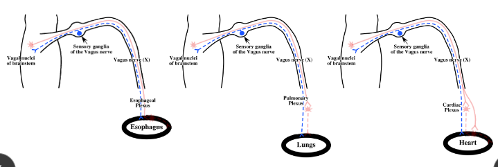
sensory innervation of thoracic viscera- reflexes
-GA axons from esophagus, lungs, and heart return with the vagus nerve
-GA neuronal cell bodies located in sensory ganglia of vagus nerve
-GA neurons mediate reflexes: swallowing (esophagus), coughing/gasping/prolonged inspiration (lungs), decreased heart rate as a result of increased intravascular pressure (heart)
sensory innervation of thoracic viscera- pain
-GA axons returning with sympathetic nerves mediate pain
-pain can be caused by distension of esophagus or insufficient blood flow to heart (myocardial ischemia)
-GA axons returning from esophagus and heart with cervical and/or upper thoracic splanchnic nerves
-there are NO GA axons returning with splanchnic nerves from the lungs, so they are insensitive to pain
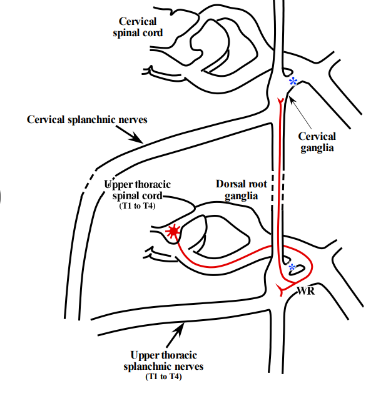
sensory axons pass through ____; sensory cell bodies located in ____
-sympathetic ganglia and white rami communicantes to reach spinal nerves and dorsal roots, since there are no white rami communicantes at cervical levels those axons must descend the sympathetic chain; dorsal root ganglia that are at the same level as the presynaptic (preganglionic) sympathetic neuronal cell bodies in the spinal cord
-all thoracic dorsal root ganglia contain sensory neurons relaying information from the esophagus, but only the dorsal root ganglia from T1 through T4 contain sensory neurons relaying information from the heart
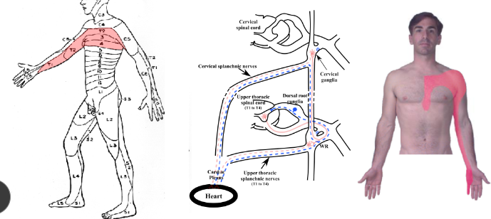
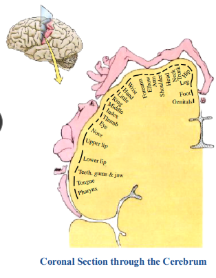
pain from viscera- referred pain
-regions of the body innervated by specific parts of the sensory cortex
-pain from viscera is perceived as referred pain
-pain felt in dermatomes where the visceral sensory neurons are located
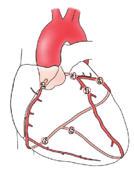
myocardial ischemia
-blockages in the coronary arteries likely to result in ischemia because they are functional end arteries; usually insufficient anastomoses with adjacent vessels to compensate for the lack of oxygen
-ischemia stimulates the sensory endings in the heart (cutting the heart or temp fluctuations do not cause cardiac pain)
-blockages may be due to transient platelet aggregation, vasospasm, a blood clot (thrombus), or rupture of a vulnerable atherosclerotic plaque
-blockages most common in anterior interventricular (LAD), right coronary, and circumflex arteries
-blockages may be asymptomatic, cause angina pectoris with transient pain, or a MI in which there are prolonged pain and permanent damage to heart muscle
cardiac pain
-pain from heart usually moderate to severe and is perceived as referred pain
-areas in which pain felt correlates (more or less) with the dermatomes innervated by sensory neurons whose cell bodies are in the same dorsal root ganglia as those of the heart (dorsal root ganglia from T1 through T4)
-cardiac pain typically felt in left arm, shoulder, or sternum
-women somewhat more likely than men to experience some of the other common symptoms, particularly shortness of breath, nausea/vomiting, and back or jaw pain as opposed to chest pain
-not known why pain may only be present in part of a dermatome