ANP1111 Midterm 2
1/279
There's no tags or description
Looks like no tags are added yet.
Name | Mastery | Learn | Test | Matching | Spaced |
|---|
No study sessions yet.
280 Terms
Structure of axial skeleton
Contains 80 bones, made up of the major regions: skull, vertebral column and rib cage.
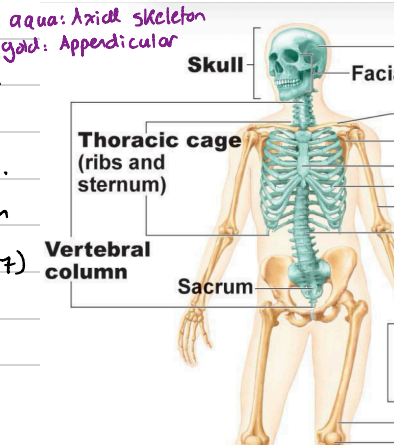
The skull
22 bones total, made up of cranial + facial bones.
Function of the skull is to protect brain and sensory organs. Has 85 openings for nerves and vessels.
Cranial bones
Protect the brain. Most skull bones are flat bones, united by sutures (interlocking joints). The cranial bones enclose and protect the brain.
8 total bones. Paired bones are parietal, temporal. Unpaired ones are frontal, occipital, sphenoid and ethmoid.
The cranium can be divided into the vault and base.
Vault
Forms superior, lateral and posterior aspects of skull + forehead.
Base
Forms the inferior aspects of the skull
Facial bones
Form the framework of the face, anchor muscles, hold teeth and house sensory organs.
There are 14 total.
Unpaired ones are mandible and vomer.
Paired ones are maxillae, zygomatic, nasal, lacrimal, palatine and inferior conchae.
Vertebral column
'Made up of 33 bones
Its structure is 24 movable vertebrae + 9 fused (sacrum and coccyx) which are roughly 70 cm long.
Regions are 7 cervical bones, 12 thoracic bones, 5 lumbar bones, 4 fused sacral and 3-4 fused coccygeal.
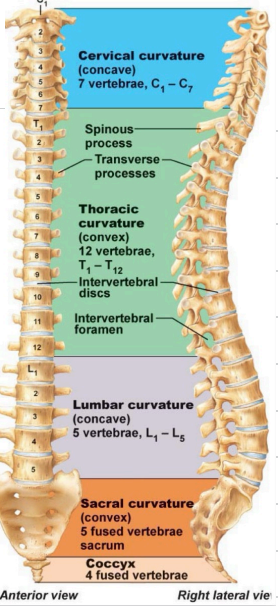
Cervical and lumbar bones are concave, thoratic and sacral are convex.
Function of vertebral column bones are bearing weight, attaching muscles and protecting spine.
Intervertebral discs
Cushion the vertebrae, make up 25% of vertebral column length. (explains why you’re taller in the morning compared to evening.)
Nucleus pulposus
An elastic, annulus (ring shaped) fibrosus which holds vertebrae together.
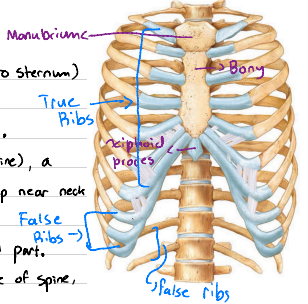
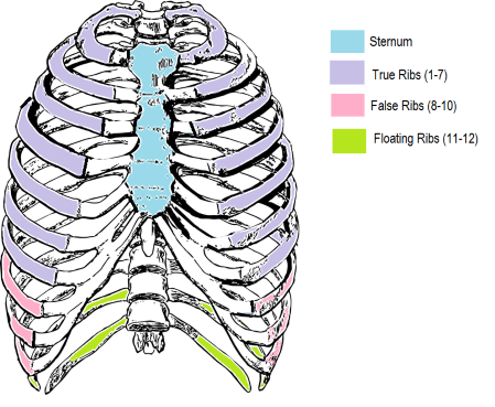
Bony thorax (rib cage)
Made up of thoracic vertebrae, 12 pairs of ribs, sternum and costal cartilage.
Function is to protect the heart and lungs, support shoulder + muscles.
Sternum
Composed of 3 parts. The manubrium which articulates with clavicles and 1st rib. The body ribs (ribs 2-6) and xiphoid which are responsible for muscle attachment.
Ribs
Ribs 1-7 are true ribs (attach directly to sternum)
False ribs (8-10) attach indirectly.
Floating ribs (11-12) have no anterior (front) attachment.
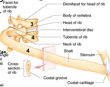
A typical rib has a head (back end connecting to spine), a neck (short section right after head), a tubercle (small bump near neck connecting to a side part of vertebrae called transverse process), and a shaft (body) which is the long, curved part.
Basically…
Head: Connects to spine
Tubercle: Connects to side of spine
Shaft: Main curved part around chest
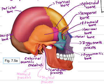
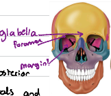
Frontal bone
A dome shaped cranial bone. Forms forehead and roof of orbits. It features supraorbital foramen (small openings located on upper edge of eye socket), supraorbital margin (prominent bony ridge in upper edge of eye socket) and glabella (smooth, bony area between eyebrows, just above bridge of nose)
Parietal bone
Pair of bones. They form superior and lateral aspects of the skull. (Form the bulk of the cranial vault.)
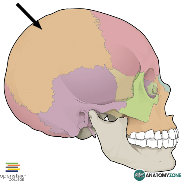
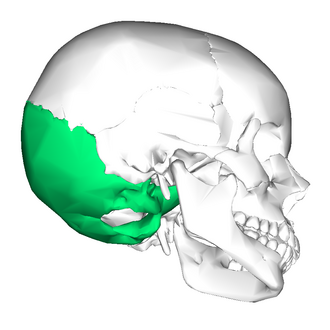
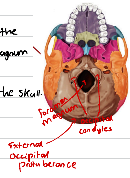
Occipital bone
Single bone at the base of the skull. It helps form posterior aspect of the skull (wall and base). It attaches anteriorly to the 2 parietals and 2 temporals and attaches to sphenoid.
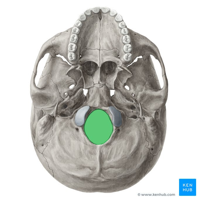
Foramen magnum
Large hole at the base of the skull. Its the passage for the spinal cord.
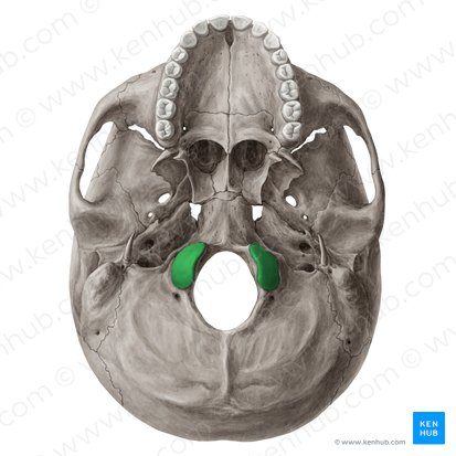
Occipital condyle
Found on each side of the foramen magnum and are the site of articulation with first cervical vetebra.
External occipital protuberance
A projection at the back of the skull. More prominent in men compared to women.
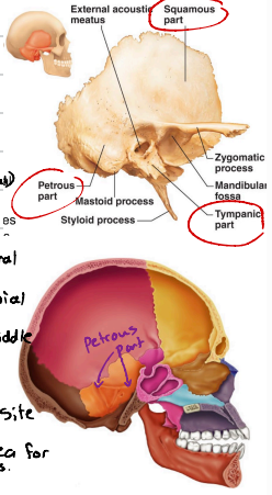
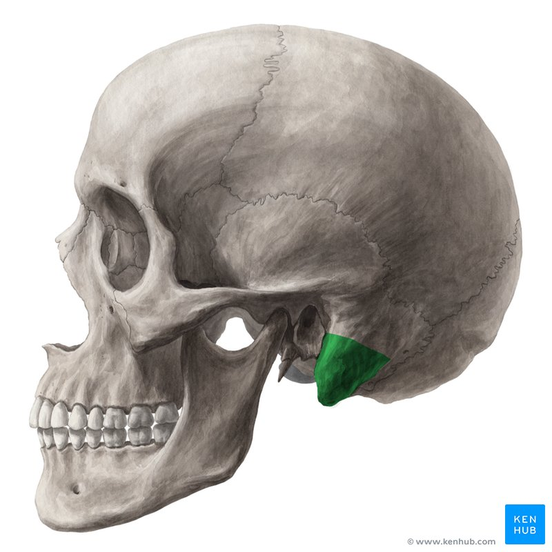
Temporal bones
Paired bones, they form the inferior and lateral aspects of the skull and parts of the cranial floor. They are located just below the 2 parietal bones and have 3 very different areas. Squamous part, tympanic part and petrous part.
Squamous part
A flattened part which forms zygomatic process to the cheekbone (aka zygomatic bone.) The mandibular fossa receives condyle of mandible.
One of three areas of temporal bone.

Tympanic part
Surrounds external acoustic meatus (external ear canal)
One of three areas of temporal bone.
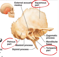
Petrous part
Found in the internal aspect of the temporal bone. It contributes to cranial base, and houses inner and middle ear cavities.
Mastoid process
Attachment site for some neck muscles. Forms a prominent part of temporal bone at the base of the skull.
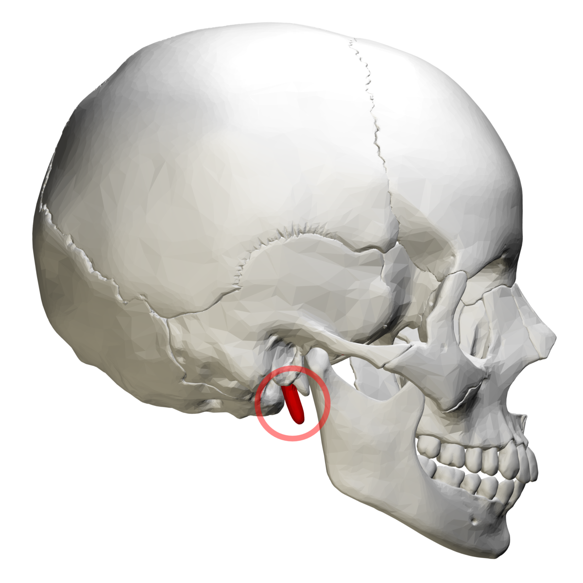
Styloid process
Attachment area for muscles of tongue and neck muscles.
Extends from temporal bone.
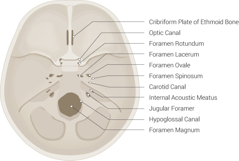
Important foramina (openings) associated with petrous part of temporal bone
Jugular foramen: Allows passage of the internal jugular vein and three cranial nerves (IX, X, XI)
Carotid canal: Allows passage of the internal carotid artery into the cranial cavity.
Internal acoustic meatus: Transmits cranial nerves VII and VIII
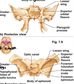
Sphenoid bones
A “bat shaped” complex bone, articulates with all other cranial bones. It forms the base of middle cranial fossa, and contributes to the base of anterior cranial fossa central body which contains sphenoid sinuses.
It has three projections. Greater and lesser wings + pterygoid processes.
Order is greater wings → middle cranial fossa → lesser wings → anterior cranial fossa
Greater wings
Project laterally (towards sides) from sphenoid body, forming parts of the middle cranial fossa, posterior walls of orbits, and external walls of the skull.
One of three projections of sphenoid bone.

Lesser wings
Form part of the floor of the anterior cranial fossa and part of the medial walls of the orbits.
One of three projections of sphenoid bone.
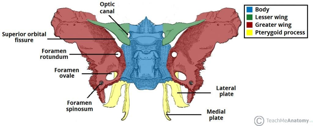
Pterygoid processes
They project inferiorly from the junction of the body and greater wings. They anchor the pterygoid muscles which are important for chewing.
One of three projections of sphenoid bone.

Optic foramina (canals)
Lie anterior to the sella turcia and allow optic nerves to pass.
Sphenoid bone.
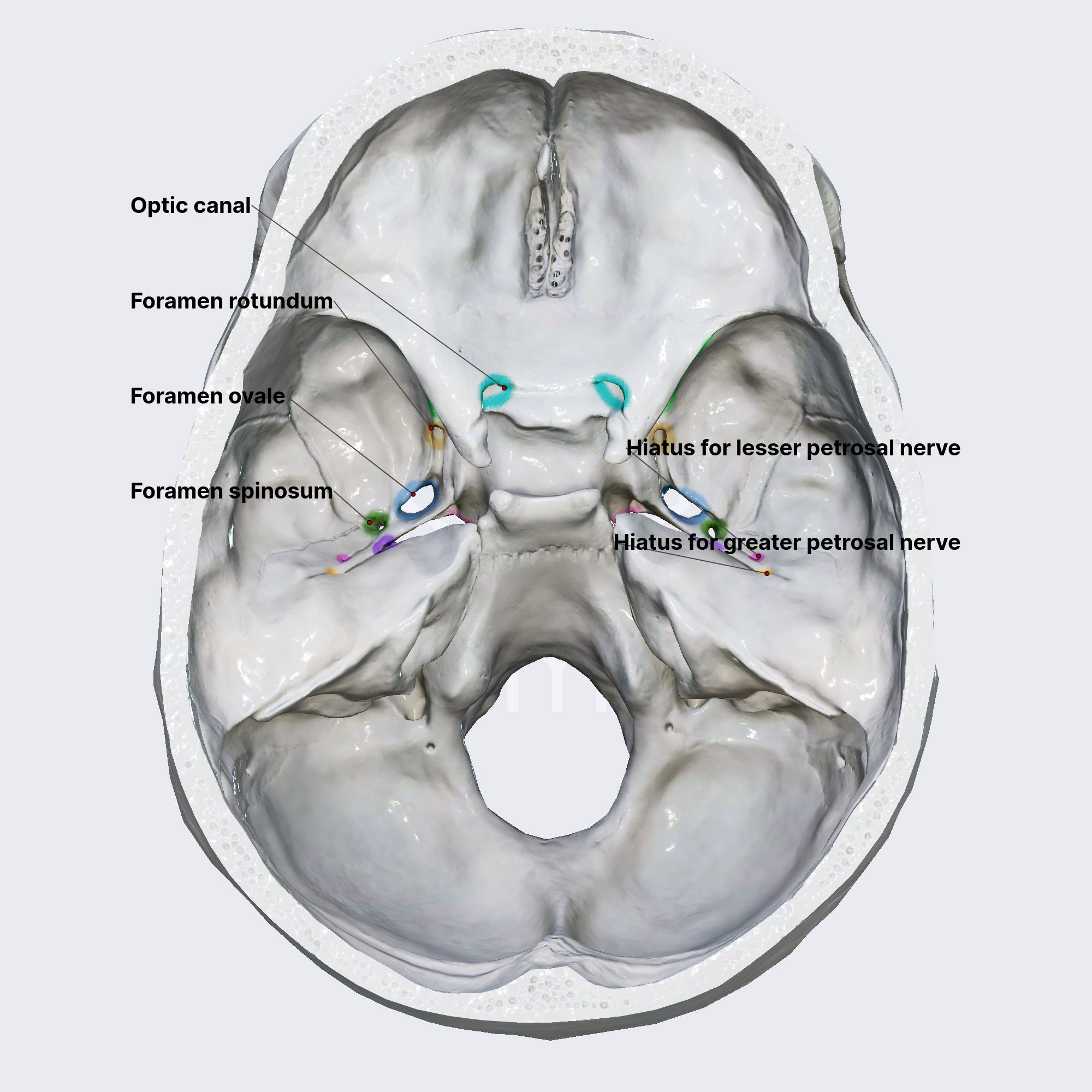
Superior orbital fissure
Found between greater and lesser wings. Allows for passage of cranial nerves that control eye movement.
Sphenoid bone.
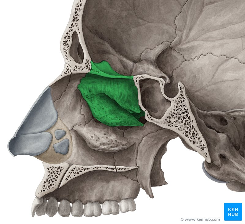
Ethmoid bone
Lies between sphenoid and nasal bones of the face. It forms most of the bony area between nasal cavity and the orbits.
The lateral masses on both sides contain ethmoid sinuses.
Extending medially from the lateral masses, there are superior and middle nasal conchae.
The lateral surfaces of the ethmoid’s lateral masses are called orbital plates which contribute to medial walls of the orbits.
Cribiform plate
Forms roof of nasal cavity and floor of anterior cranial fossa. Tiny holes (olfactory foramina) allow passage of olfactory nerves.
Ethmoid bone.

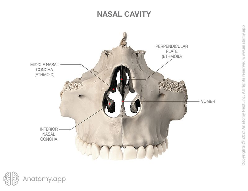
Perpendicular plate
Projects inferiorly to contribute to nasal septum.
Ethmoid bone.
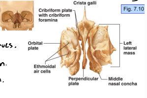
Crista galli
Projects superiorly to attach to dura mater of brain.
Ethmoid bone.

4 key sutures connecting cranial bones
Coronal, squamous, lambdoid, sagittal
Sutural bones are tiny irregular bones which occur within sutures, most often in lambdoid suture.
Coronal suture
Where the 2 parietal bones meet the frontal bone anteriorly.
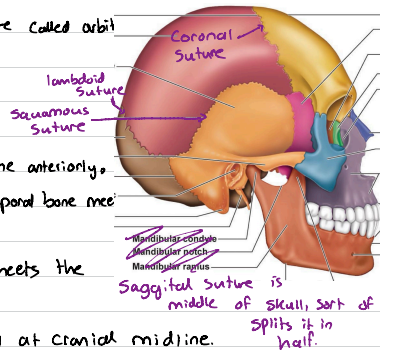
Squamous suture
One on each side, where parietal bone and temporal bone meet on lateral aspect of the skull.

Lambdoid suture
Where the occipital bone posteriorly meets the parietal bones

Sagittal suture
Where parietal bones meet superiorly at cranial midline.

Mandible (lower jawbone)
The largest, strongest facial bone.
It has a body which forms the chin, and 2 upright rami (branches) which meet the body posteriorly at the mandibular angle.
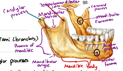
Mandibular notch
Separates the coronoid and condylar processes

Coronoid process
An insertion point for the large temporalis muscle that elevates lower jaw during chewing.

Mandibular condyle
Upper part of condylar process of mandible. It articulates with the mandibular fossa of the temporal bone, forming the temporomandibular joint (acts as a ball and socket joint)

Aveolar margin
The top tip of alveolar process which contains tooth sockets.

Mandibular foramina
Found on the medial surface of each ramus. Permit the nerves responsible for tooth sensation to pass to the teeth in lower jaw.

Mental foramina
Openings on the lateral aspects of mandibular body, allowing blood vessels and nerves to pass to the skin of the chin and lower lip.

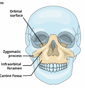
Maxillary bones
Are fused medially. Alveolar margins hold teeth of upper jaw.
Palatine processes
Project posteriorly from alveolar process, forming the anterior 2/3 of the hard palate/bony roof of mouth.
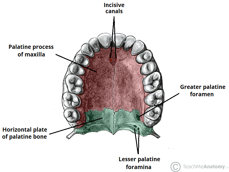
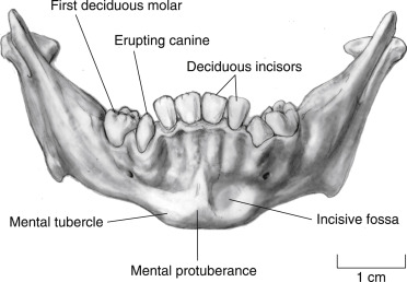
Incisive fossa
A midline foramen which leads into incisive canal which is a passageway for blood vessels and nerves
Frontal processes
Extend superiorly to the frontal bone and forms part of the lateral aspects of thenose bridge.
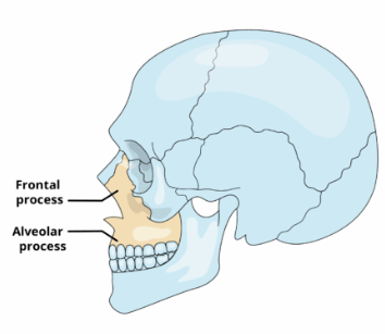
Zygomatic processes
An articulation site where the maxillae laterally articulate with bones.
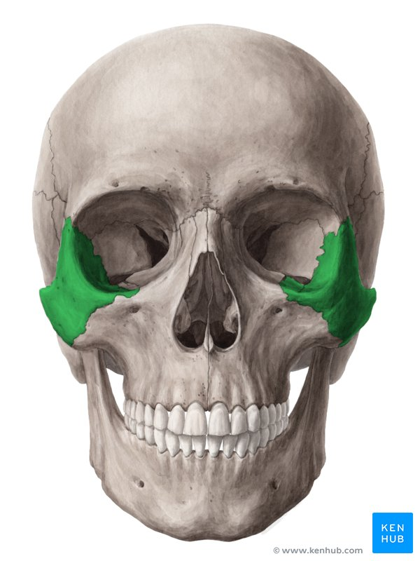
Zygomatic bones
Also known as cheekbones. They articulate with zygoimatic processes of maxilla, and frontal and temporal bones.
They also contribute to inferolateral margins of the orbit
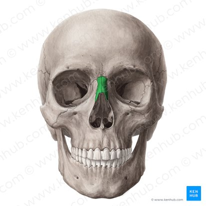
Nasal bones
2 tiny, rectangular bones that fuse medially to form nose bridge.
They articulate superiorly with frontal bone, and laterally with maxillary bones.
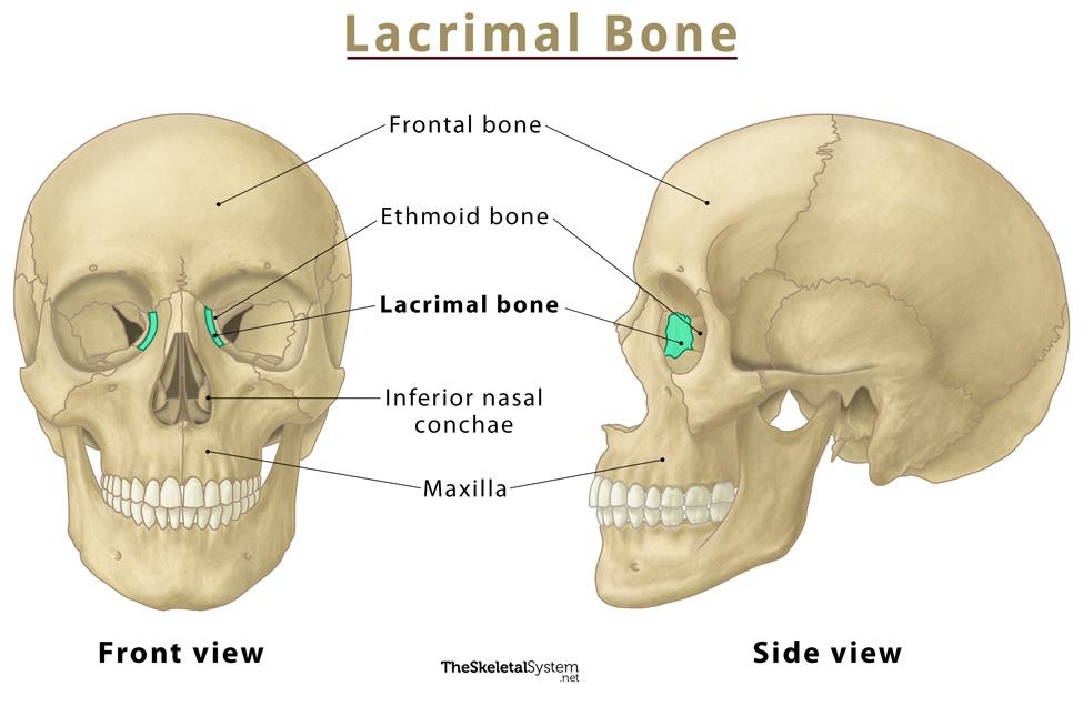
Lacrimal bones
Pair of fingernail shaped bones which contribute to medial walls of each orbit. They articulate with frontal bone, ethmoid bone and maxillae.
Each lacrimal bone has a depression (lacrimal fossa) for lacrimal sac.
Orbits
They are bony cavities in which eyes are firmly encased and cushioned by fatty tissue. There are 7 of them.
Zygomatic, frontal, maxilla, ethmoid, lacrimal, sphenoid and palatine (orbital?) process.
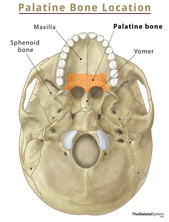
Palatine bones
2 L-shaped bones which form two bony plates.
Horizontal plate: Joined at the medial palatine suture and form part of the hard palate.
Vertical plate: Forms part of the nasal septum (cavity and orbit
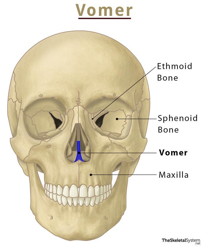
Vomer
Plow-shaped facial bone that lies in the nasal cavity which forms the nasal septum
Inferior nasal conchae
Pair of bones, they are thin and curved and project medially from lateral walls of nasal cavity
They are the largest of the three pairs of nasal conchae. The other two (superior and middle) are simply part of ethmoid bone.
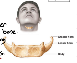
Hyoid bone
Only bone in body that doesn’t articulate with any other bone. it supports the tongue and gives attachment to muscles for swallowing and speech. It has a horseshoe shape with a body + 2 pairs of horns.
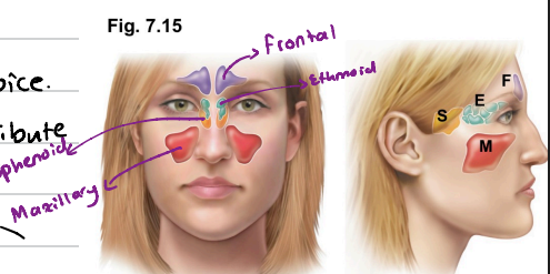
Paranasal sinuses
Mucosa-lined, air-filled sinuses found in the frontal, sphenoid, ethmoid and paired maxillary bons.
They lighten the skull and enhance resonance of voice.
They connect to the nasal cavity so they also contribute in warming and humidifying incoming air.
Vertebral column
33 bones in total. 24 of them remain separate to allow flexibility. The remaining 9 fuse to form composite bones sacrum and coccyx.
Roughly 70cm long in average height adult
Regions of vertebral column
Cervical, thoracic, lumbar, sacral and coccygeal.
7 cervical, 12 thoracic, 5 lumbar, 5 sacral, 3-4 coccygeal.
Ligaments of vertebral column
Strap-like and support the columns so the bones stay upright. The major ones are the anterior longitudinal ligament and the posterior longitudinal ligament.
Anterior longitudinal ligament
Strongly attached to the bony vertebrae and the discs. It provides support and prevents hyperextension (too far backwards) of the spine.
Posterior longitudinal ligament
Resists hyperflexion of the spine (too far forward). It’s narrow, weaker and attached only to the discs.
Intervertebral discs of vertebral column
Cushion like pad between bony vertebral bodies.
They act as shock absorbers. Each is circular with nucleus pulposus in the center (gel-like, gives elasticity and compressibility) and annulus fibrosus around periphery which holds together successive vertebrae and resist tension in spine.
The discs are thickest in the lumbar/cervical regions → provides flexibility.
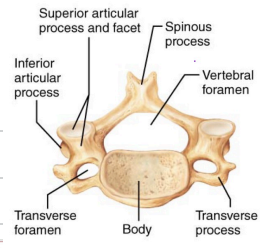
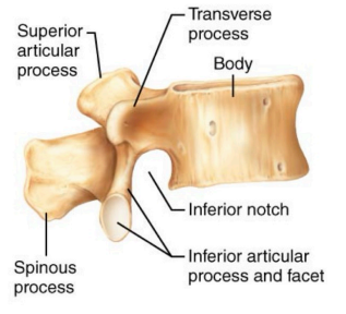
General structure of a vertebra
They get longer as one descends the column. They are made up of a weight-bearing body (anteriorly) and a vertebral arch posteriorly.
The arch and body enclose the vertebral foramen. The vertebral canal is formed by successive vertebral foramina of the articulated vertebrae.
Pedicles have notches on the superior and inferior surfaces, providing lateral openings between adjacent vertebrae called intervertebral foramina.
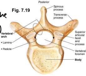
Vertebral arch
Composite structure formed by 2 pedicles and 2 laminae
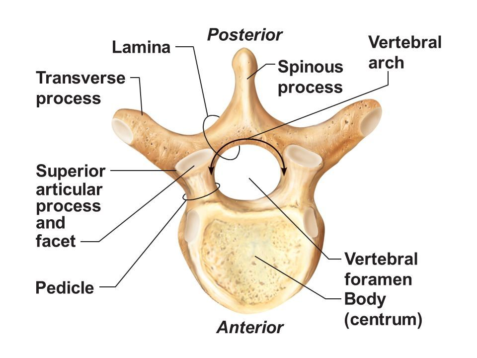
Processes of each vertebral arch
1 spinous process for muscle attachment, 2 transverse processes. One per side for muscle attachment, aired superior and inferior articular processes which link vertebrae above and below smooth, collagen-coated facets for articulation.
7 total at each vertebral arch.
Cervical vertebrae
7 total, 1 (atlas) and 2 (axis) have unusual structures + no disc.
3-7 are typical. They have an oval body, are broader side to side than front to back.
Have short spinous processes that split at the end (except 7 which sticks out and can be palpated/not split.
Vertebral foramen (opening) is a large triangle.
Each transverse process contains a transverse foramen for passage of vertebral artery to brain.
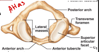
Atlas
Cervical vertebrae 1. Has no body and no spinous processes. Has posterior and anterior neural arches, and lateral masses with superior and inferior articular facets.
The articulation of superior articular facets with occipital condyles basically allowing nodding.
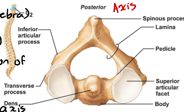
Axis
Cervical vertebrae 2. Resemble more like c3-c7 (typical vertebrae) except for the fact that it has dens or odontoid process projecting superiorly from its body.
The dens act as a pivot for the rotation of the atlas, as a result allowing you to move head side to side.
Dens are the “missing” body from the atlas, which fuses with the axis during embryonic development.
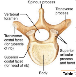
Thoracic vertebrae
12 total, they all have ribs attached and increase in size as you go down.
Their body is roughly heart shaped and bear facets for ribs (commonly called demi-facets).
The vertebral foramen is circular, spinous process is long and points down, transverse processes have facets for articulation with tubercles of ribs.
Lumbar vertebrae
This region receives the most stress. The bodies are kidney shaped and increase in size from top to bottom.
Characteristics are as follows:
Pedicles and laminae are shorter and thicker than those of other vertebrae
Spinous processes are flat and short and project directly backwards
The vertebral foramen is triangular shaped
The orientation of inferior and superior facets are unique compared to others.
(superior facet faces in, inferior facet faces out.)
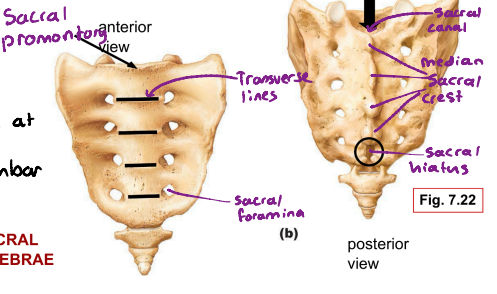
Sacral vertebrae
Starts off as 5 separate vertebrae but fuse at adolescence into the sacrum. it articulates with the 5th lumbar and laterally with hip bones via sacroiliac joint.
Sacral promontory: Basically the high point of sacral vertebrae
Transverse lines: They mark the lines of fusion of the sacral vertebrae
Sacral foramina: They transmit blood vessels and anterior rami of sacral spinal nerves.
Median sacral crest: The fused spinous processes of sacral vertebrae
Sacral canal: The area where the vertebral canal continues inside the sacrum
Sacral hiatus: An enlarged external opening.
Coccygeal vertebrae
The tailbone is made up of 3 or 4 fused coccygeal vertebrae.
It’s the attachment area for some pelvic ligaments. Otherwise it’s useless.
Bony thorax
Refers to the thoracic vertebrae + ribs + costal cartilages + sternum
It forms a protective cage around the heart, lungs and major blood vessels.
It supports the shoulder girdle and upper limbs
It provides an area of muscle attachment for the back, chest and shoulders.
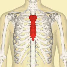
Sternum
It lies in the anterior midline of the thorax. It’s a fusion of three bones. Manubrium, body and xiphoid process.
Manubrium
Superior portion of the sternum.
It articulates via its clavicular notches with the clavicle. It also articulates with the 1st pair of ribs
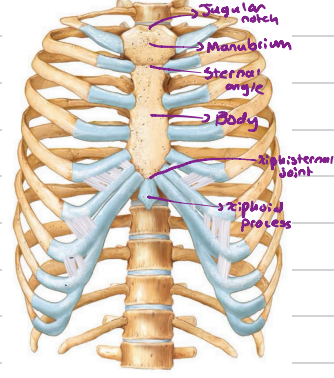
The body
The midportion.
It forms the bulk of the sternum.
It has notches for articulation with 2nd to 7th ribs.

Xiphoid process
Forms inferior end of sternum. It’s a plate of hyaline cartilage during youth but ossifies in adulthood.
It articulates only with sternal body and serves as an attachment point for some abdominal muscles.

Jugular notch
Indentation you can palpate. Basically a visible dip.

Sternal angle
It’s a cartilaginous hinge between the manubrium and body of sternum.

Xiphisternal joint
A fusion of the sternal body and xiphoid process

Ribs
There are 12 on each side and they all attach at the back of the vertebral column. They curve inferiorly and anteriorly.
There are 7 true ones which attach to the sternum directly. The remaining 5 are false ribs.
Ribs 8-10 attach to the sternum indirectly via costal cartilages and rib 7.
Ribs 11 and 12 are not attached anteriorly (front). They are floating ribs.
Anatomy of a typical rib
Ribs are typically a bowed, flat bone.
Made up of a shaft, head, neck and tubercle. The shaft is the main portion.
The costal groove is an indented line found on inner surface of ribs and it lodges the intercostal nerves and blood vessels.
The head of rib articulates with vertebral body via 2 facets.
One articulates with the demi-facet on the body of the same numbered thoracic vertebra.
The other one on the body of superior vertebra.
The tubercle articulates with the transverse process of the same numbered thoracic vertebra.
The appendicular skeleton
Refers to bones of the limbs and their girdles (pectoral girdle, pelvic girdle plus upper and lower limbs.
Bones in the pectoral girdle are light and very movable. They are attachment points of muscles to move to upper limbs.
The scapulae are attached to other bones only laterally, and the socket of glenoid cavity (shoulder joint) is shallow and poorly reinforced.
30 separate bones, arm, forearm, wrist and hand in upper limbs.
Bones of lower limbs involves bones of thighs, leg and foot.

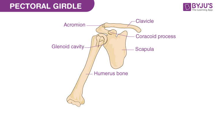
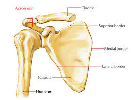
Pectoral girdle
Consists of 2 pairs of bones. Clavicles and scapulae which almost form a complete circle around upper trunk to make shoulders.
Anteriorly the medial end of each clavicle joins the sternum.
Laterally, the distal ends of the clavicles meet the scapulae.
The scapulae fail to complete the ring posteriorly, though, since the medial borders don’t meet at the axial skeleton.
Bones in the pectoral girdle are light and very movable. They are attachment points of muscles to move to upper limbs
Clavicles
Also known as collarbones. They are mildly S-shaped.
They act as insertion points for muscles, and also a brace too push arms laterally.
The curvature ensures that if there’s a fracture, it’s outward → away from the subclavian artery.
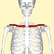
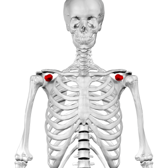
Scapulae
Also known as the shoulder blade.
They are thin, triangular, flat bones. They lie on the dorsal surface of the rib cage, between ribs 2 and 7. Each scapula has three borders.
Superior border: Shortest, sharpest border
Medial border: parallels the vertebral column.
Lateral border: Thick, lies next to the armpit and ends superiorly in glenoid cavity.
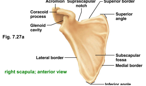
Spine acromion
Articulates with acromial end of clavicle.
Coracoid process
Helps anchor the biceps muscle of the arm.
Humerus
The longest bone of the upper limb and the only “arm” bone. It articulates with scalpula proximally and with the radius and ulna distally.
Note: The head, anatomical neck, greater and lesser tubercle, intertubercular sulcus, surgical neck, deltoid tuberosity.
Also the 2 condyles and epicondyles which form ends of joints.

The head of humeurs
Region in the humerus.
Inserts into glenoid cavity in a manner that allows the arm to hang freely at ones side.
Anatomical neck of humerus
A slight constriction immediately inferior to the head.

Greater and lesser tubercle
Sites of attachment of rotator cuff muscles
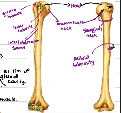
Intertubercular sulcus
Guides a tendon of bicep muscle to it's attachment point at rim of glenoid cavity.

Surgical neck
Just distal to tubercles, it’s most frequently fractured part of the humerus.

Deltoid tuberosity
It’s v-shaped. It’s the roughened attachemnt site for deltoid muscle.
