A&P Lab Midterm
1/135
There's no tags or description
Looks like no tags are added yet.
Name | Mastery | Learn | Test | Matching | Spaced | Call with Kai |
|---|
No analytics yet
Send a link to your students to track their progress
136 Terms
tapetum lucidum (cow eye)
Iridescent layer found in nocturnal animals for maximizing vision under low intensity light (What makes their eyes shine at night)
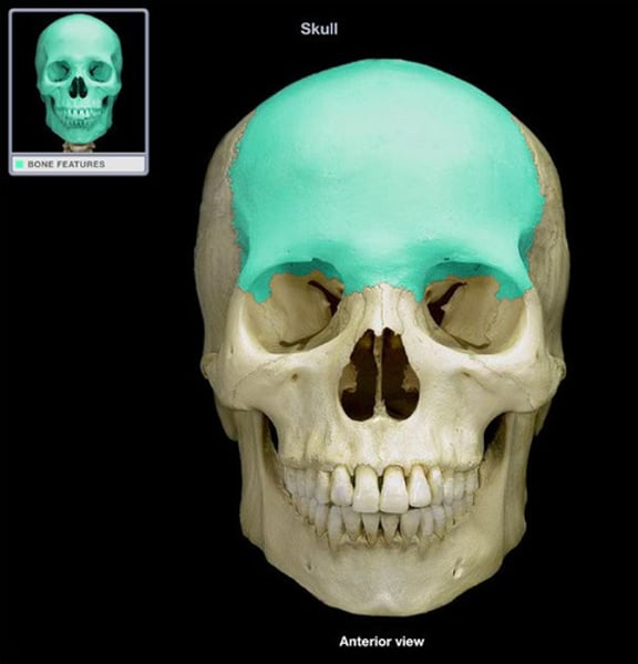
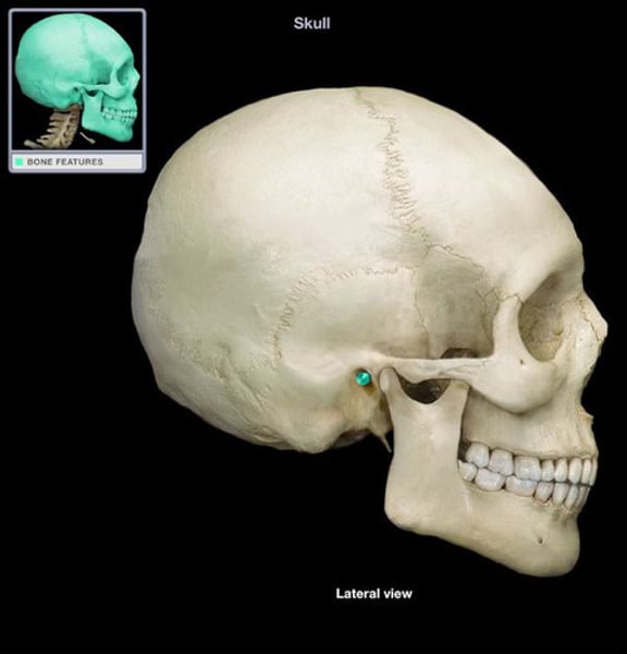
Frontal bone
bone that forms the forehead
supraorbital notch
paired opening or notch superior to the orbit
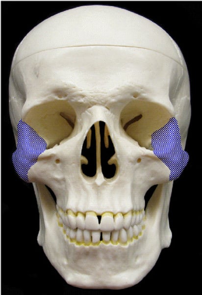
Zygomatic bones
known as the cheeckbones, articulate with the frontal bone (forehead)
zygomaticofacial foramen
an opening in the zygomatic bone
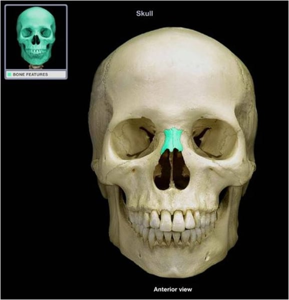
Nasal bones
form the bridge of the nose
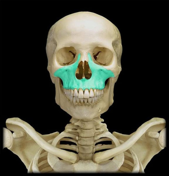
Maxillary bones
form most of the upper jaw
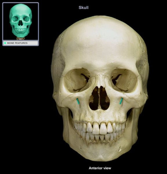
Infraorbital foramen
opening under the orbit carrying the infraorbital nerves and blood vessels the the nasal region
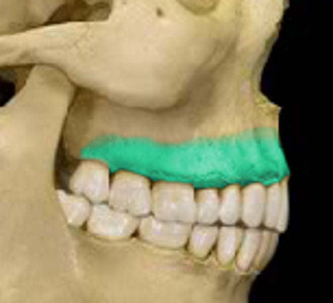
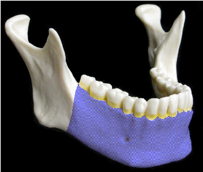
Alveolar processes
A bony ridge found on the inferior surface of the Maxilla and the superior surface of the Mandible which contains the sockets for the teeth.
Mandible
lower jaw
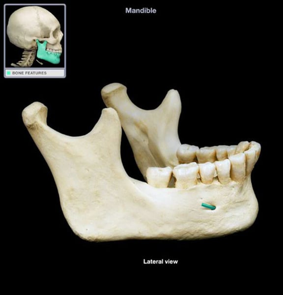
Mental foramen
Name this foramen.
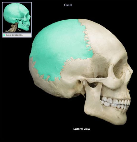
Parietal bones
Bones that form the sides and top of the cranium.
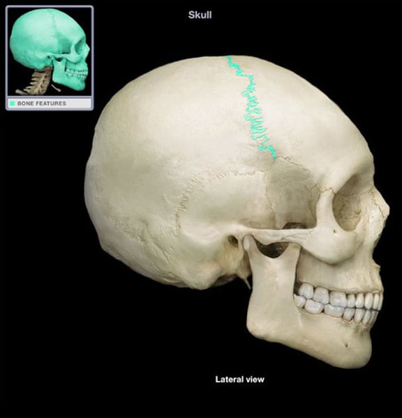
Coronal suture
the suture between the parietal and frontal bones of the skull
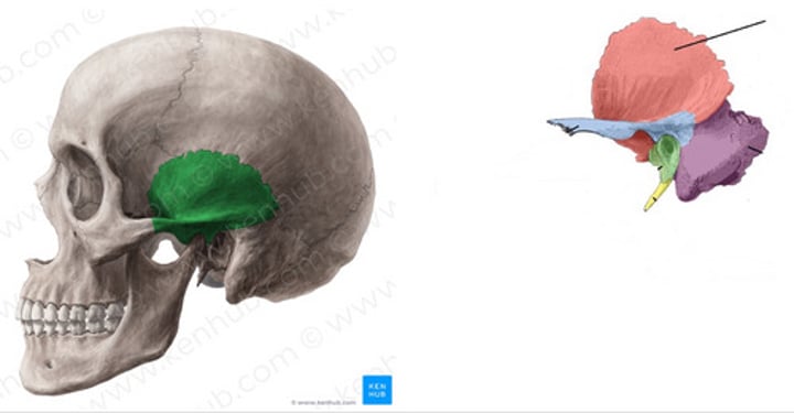
Greater wing of the sphenoid
anterior to the temporal bone

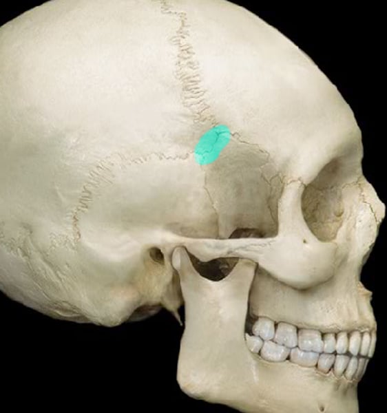
Pterion (pteri=wing)
Where the bones meet at the temple
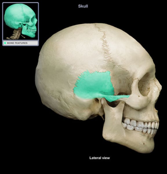
Temporal fossa
area superior and deep to the zygomatic arch
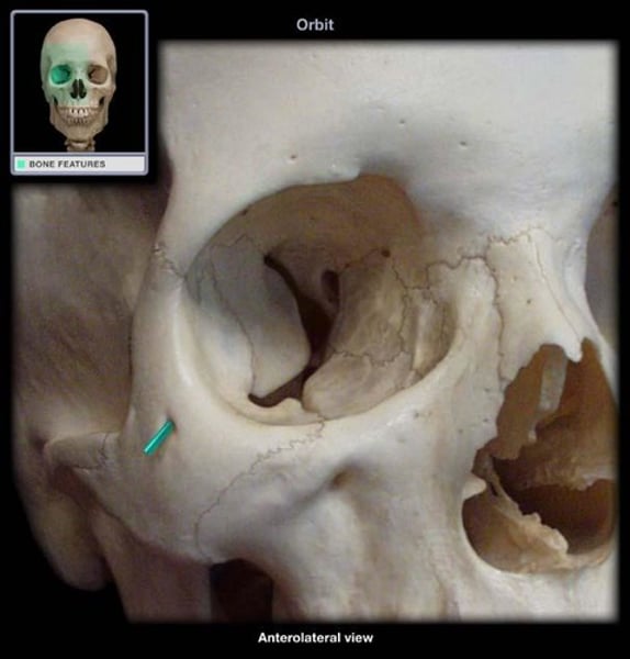
Zygomatic process
Name this part of the temporal bone.

Infratemporal crest of the greater wing of the sphenoid bone.
Name this feature
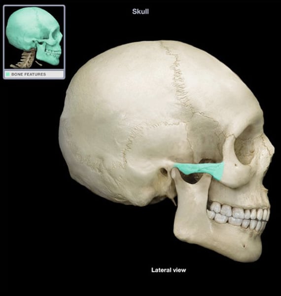
Zygomatic arch
Name the structure.
Squamous part of temporal
anterior and upper part, contributes to temporal fossa
Mastoid part of temporal
Mastoid process
Mastoid notch
Mastoid foramen
Stylomastoid foramen
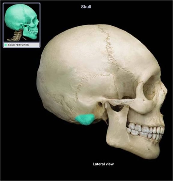
Mastoid process
round projection on the temporal bone behind the ear

Tympanic part of temporal
ring of bone around external acoustic meatus
External acoustic meatus
ear canal

Infratemporal fossa
area inferior and deep to the zygomatic arch
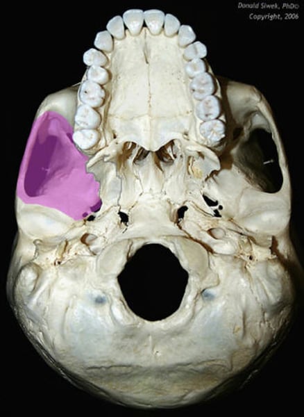
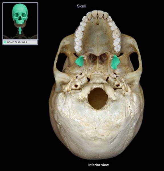
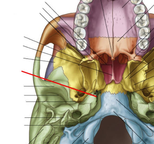
Pterygopalatine fossa
fossa deep to the infratemporal fossa and between the pterygoid process and maxillary tuberosity
Palatine bone
Name this bone.
Medial part of the greater wing of sphenoid
Name this feature

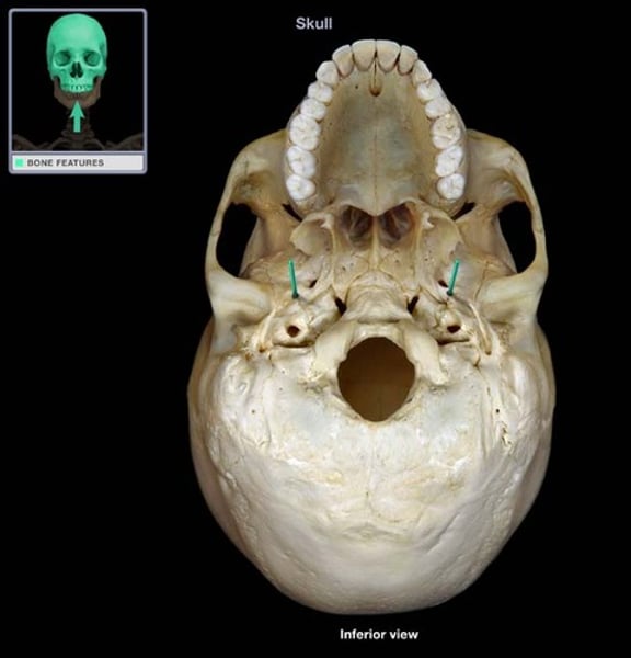
Foramen spinosum
Name this foramen.
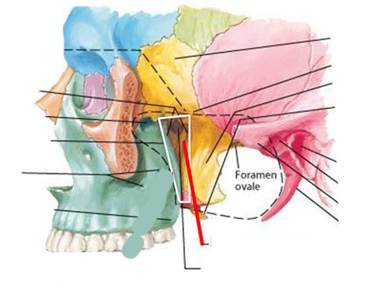
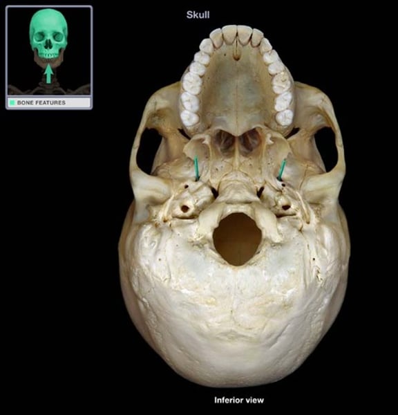
Foramen ovale
Name this foramen.
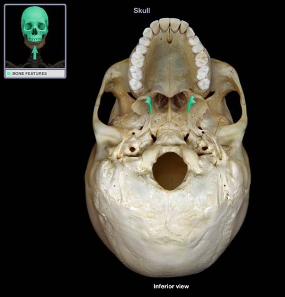
Pterygoid process of the sphenoid
Process of the sphenoid bone, consisting of two plates
Lateral and medial pterygoid plates
origin of lateral and medial pterygoids
Foramen lacerum
Name this foramen.

Pterygoid canal
Name this canal
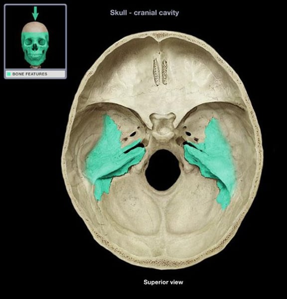
Petrous part of temporal bone
raised area on internal surface of cranial vault which encloses structures of middle and inner ear
Apex of petrous
What is the highlighted section?
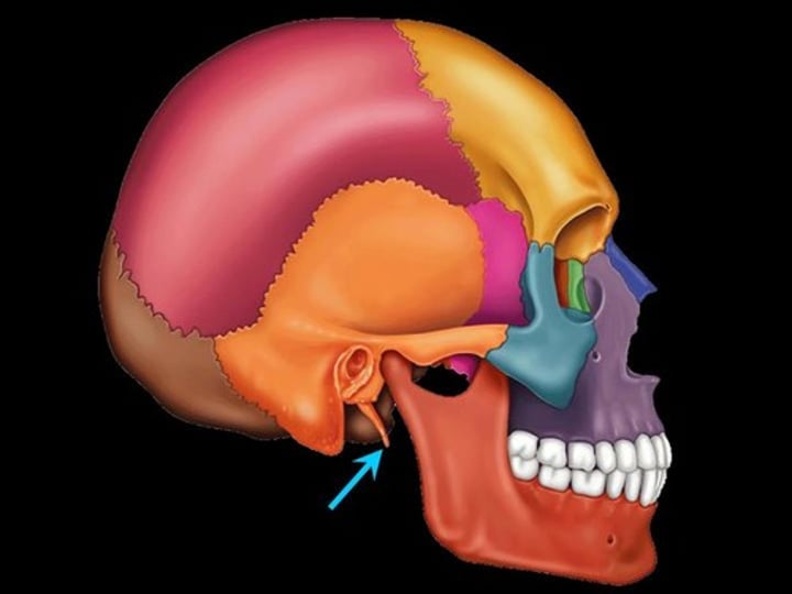
Styloid process
pole-like process extending downward from the temporal bone on each side of the skull
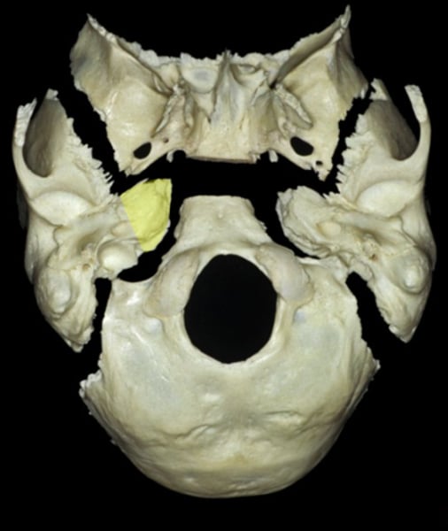
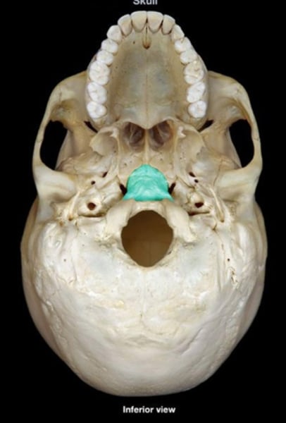
Basilar bone or basilar part of occipital
Name the highlighted feature
Occipital condyles
Rounded projections lateral to the foramen magnum that articulate with the first cervical vertebra (atlas)

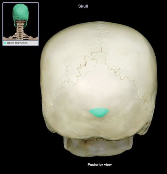
External occipital protuberance
Name this bony landmark.
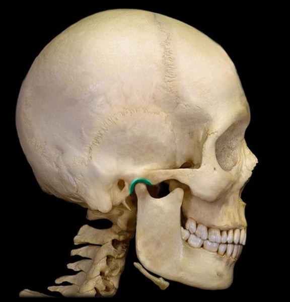
Mandibular fossa
the depression in the temporal bone into which the condyle of the mandible fits
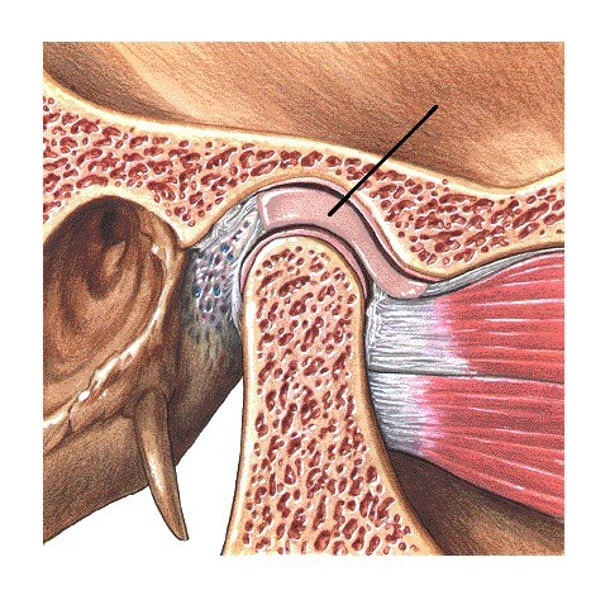
Articular fibrocartilage
tissue shaped like a disc or partial disc called a meniscus that provides cushioning at a joint
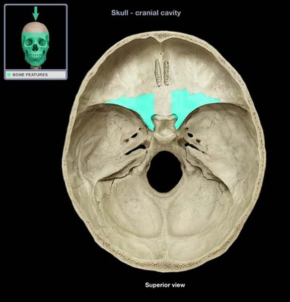
Orbital plates of frontal bone
Comprise the floor of the anterior cranial fossa, and the roof of the orbital cavity
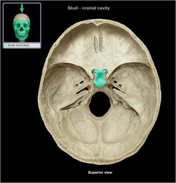
Crista galli
ethmoid bone

Cribriform plate of ethomoid bone
within the anterior region
Body of sphenoid
hollow, cube like central portion
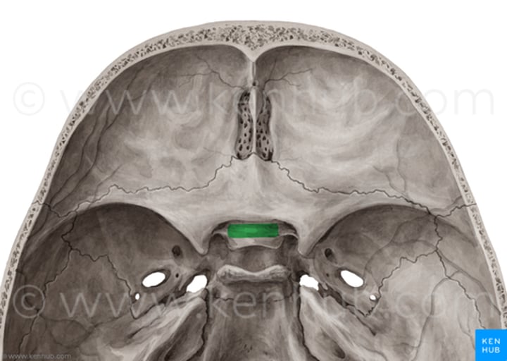
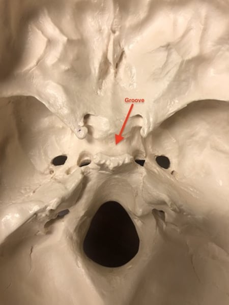
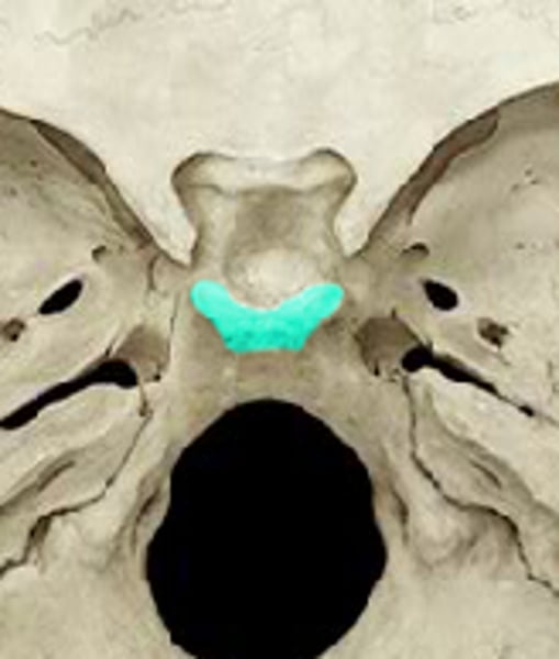
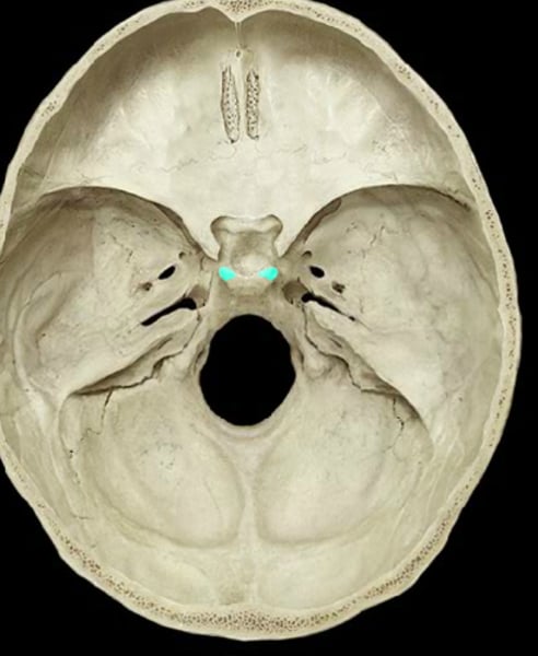
Prechiasmatic groove or sulcus of sphenoid body
name the structure
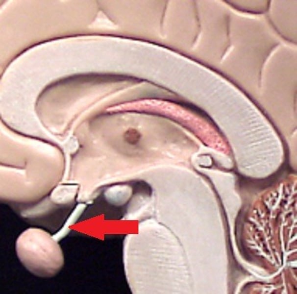
Optic nerves
the nerve that carries neural impulses from the eye to the brain
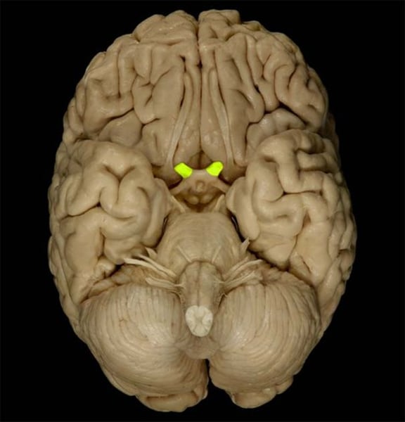
Optic chiasm
superior to pituitary gland

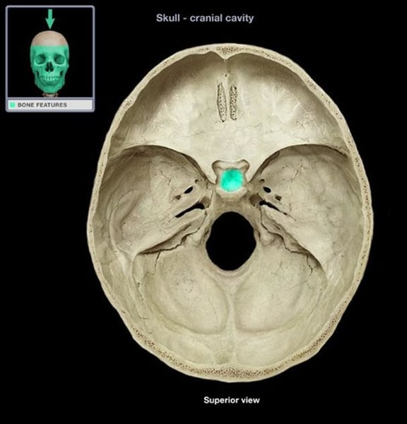
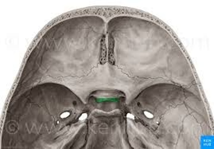
Sella turcica
cavity in the skull that contains the pituitary gland
Pituitary gland
housed in the sella turcica
Tuberculum sellae
forms the anterior border of the sella turcica
Hypophyseal fossa
a depression within the sella turcica, holds the pituitary gland
Dorsum sellae
ridge of bone at posterior edge of sella turcica
Posterior clinoids
Name the highlighted region
Internal carotid
name the canal

Petrous temporal bone
Contains the inner ear

Middle meningeal vessels
What are the arrows pointing at?
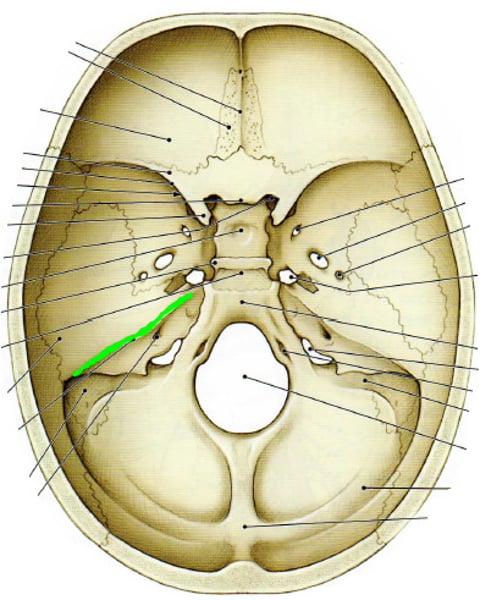
Petrous ridges
temporal
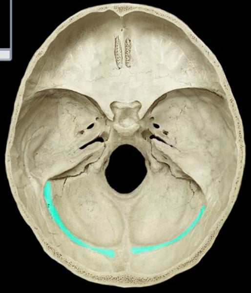
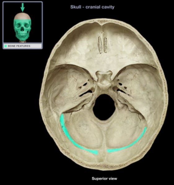
Transverse sinus grooves
What is the highlighted section?
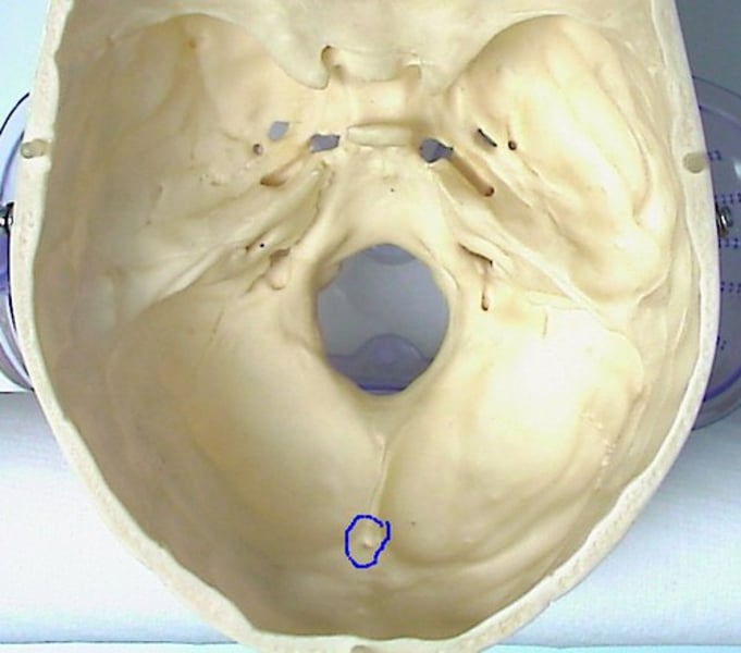
Internal occipital protuberance
internal landmark of the occipital bone
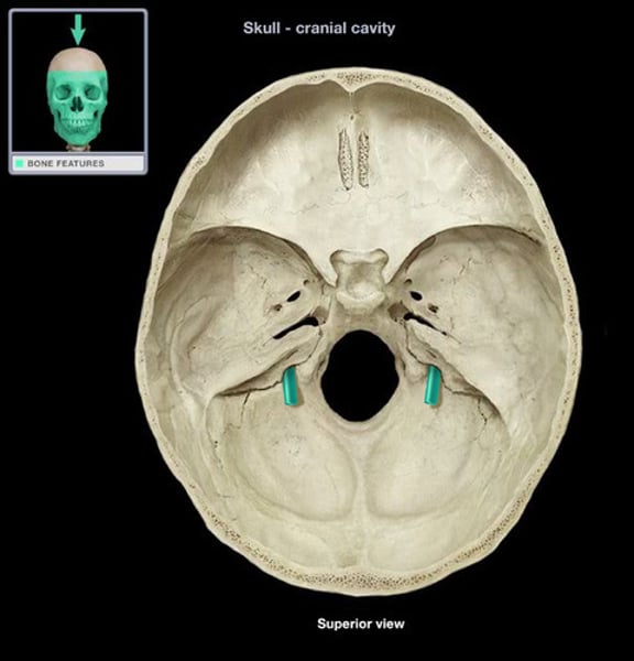
Internal acoustic meatus
A passage for CN VIII from the inner ear to the brain.

Jugular foramen
Name this foramen.
Hypoglossal canal
CN XII

Basilar portion of occipital bone
Name the green region
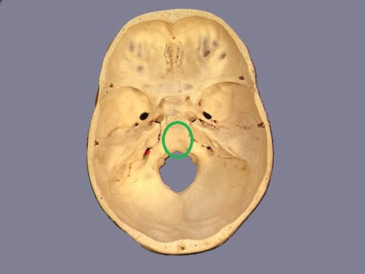
Clivus
slope
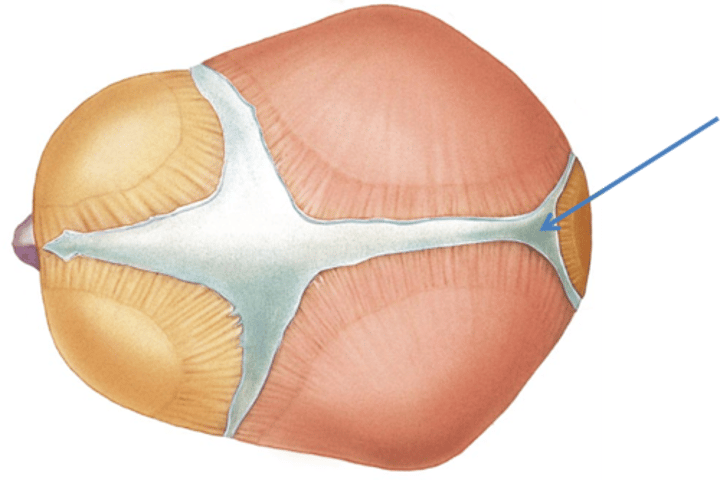
Fontanelles
soft spots normally present on the skull of a newborn
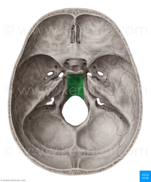
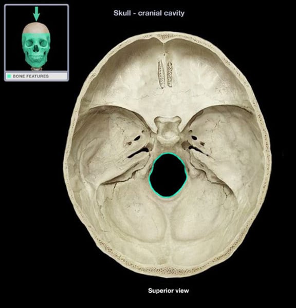
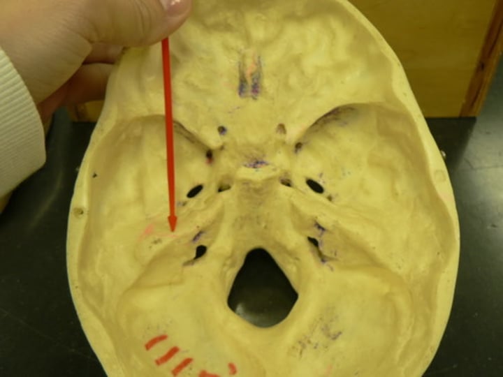
Foramen magnum
A large opening at the base of the skull through which the brain connects to the spinal cord.
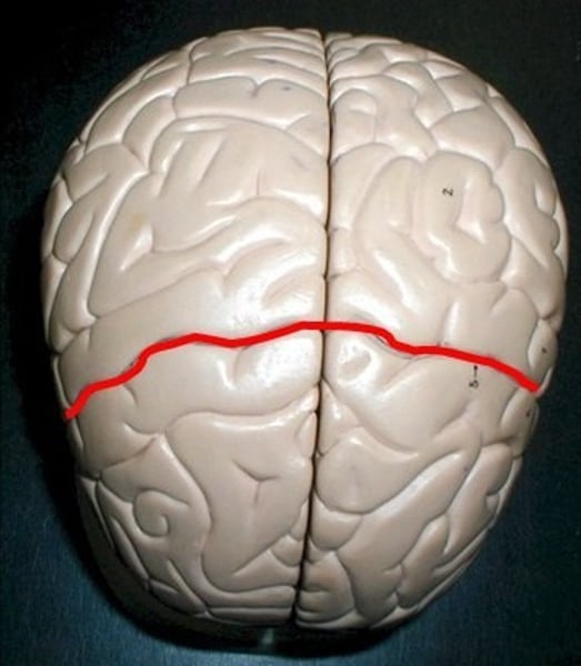
Central sulcus (inside)
anterior portion of scull
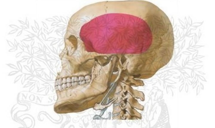
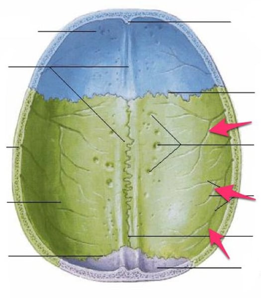
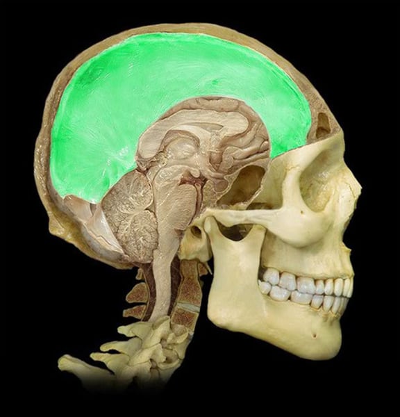
Calvaria
Top part of the skull
Parietal foramina
parietal bone near sagittal suture
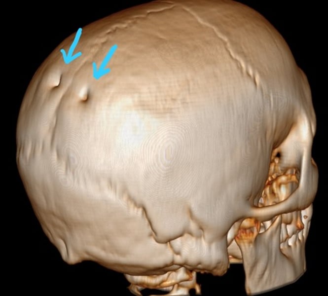
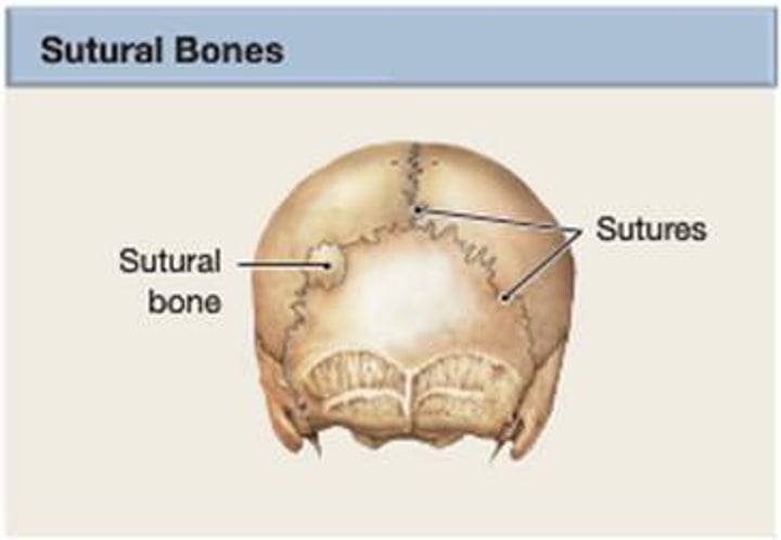
Sutural bone
tiny bones between cranial bones
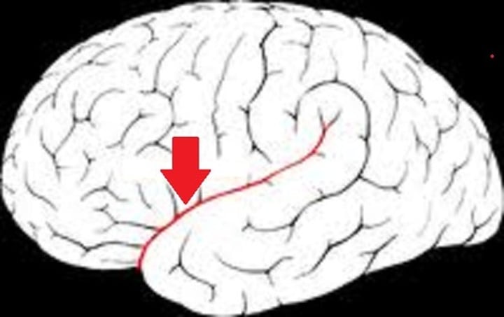
Lateral sulcus
Separates temporal lobe from parietal and frontal lobes
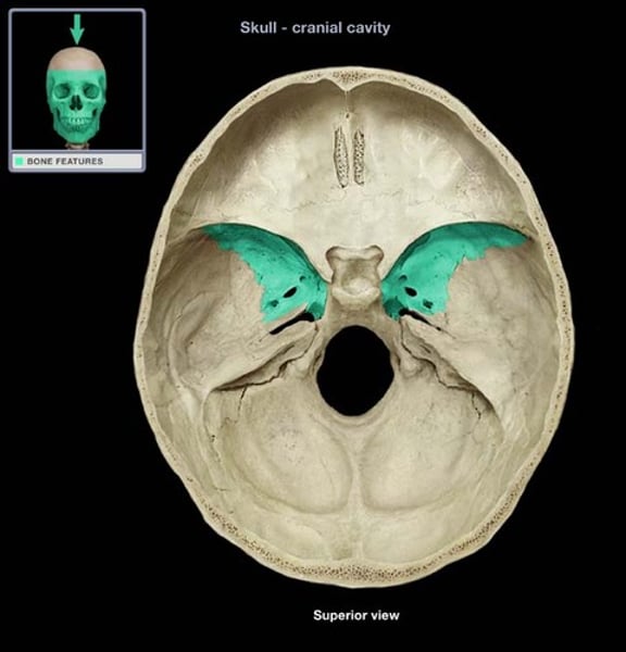
lesser wing (sphenoidal crest) of the sphenoid bone
Name the highlighted region
Greater wing of sphenoid
Name this structure
Squamous portion of the temporal bone
Thin portion of temporal bone
Petrous portion of temporal
houses structures of inner ear
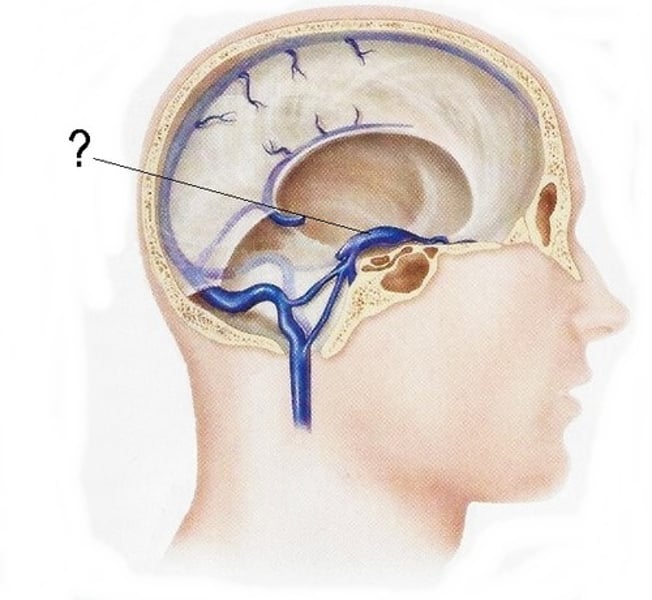
Cavernous dural sinus vein
What is the arrow pointing at?
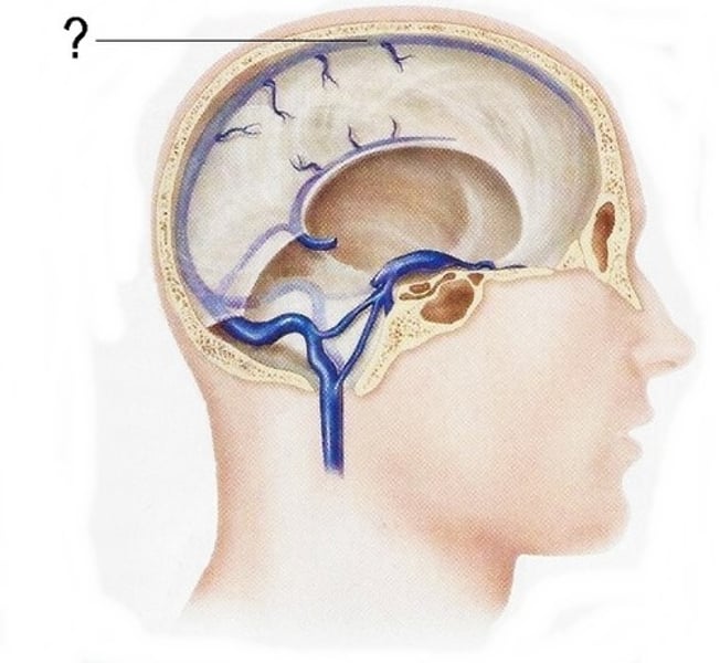
Superior sagittal sinus
Name this vein.
Granular foveolae
Shallow depressions caused by the arachnoid granulations
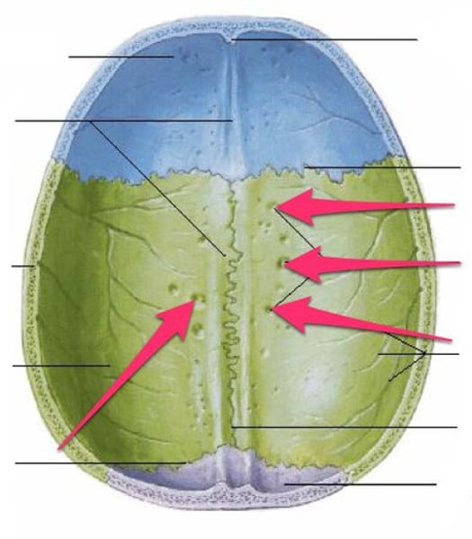
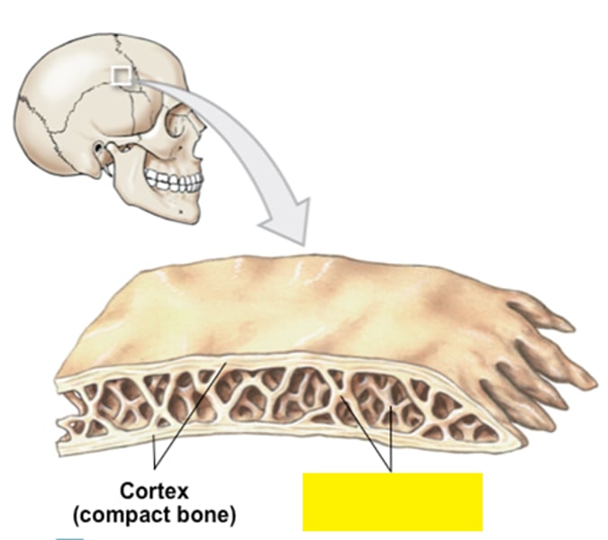
Diploë
layer of spongy bone, that is sandwiched between two the layers of compact bone found in flat bones
Dura
What is this layer?
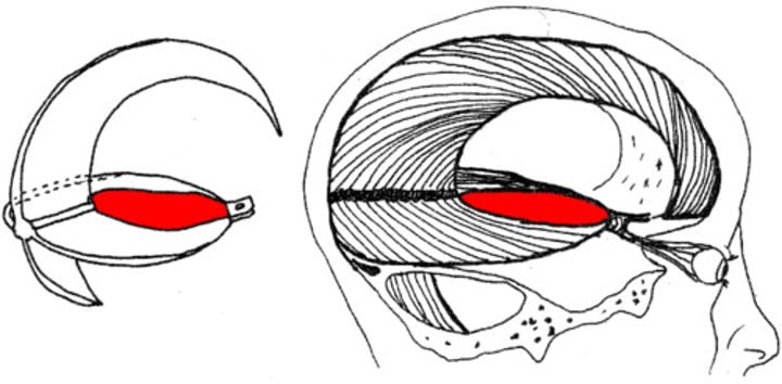
Cerebral falx or falx cerebri
What is this sheath?
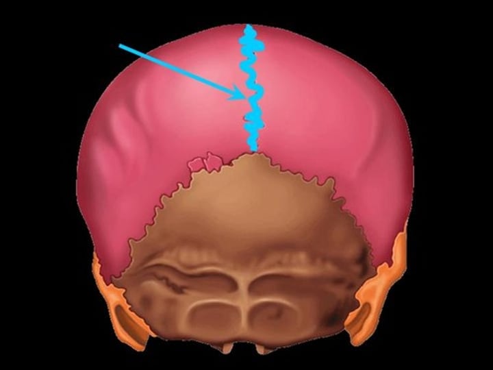
Sagittal suture
between parietal bones
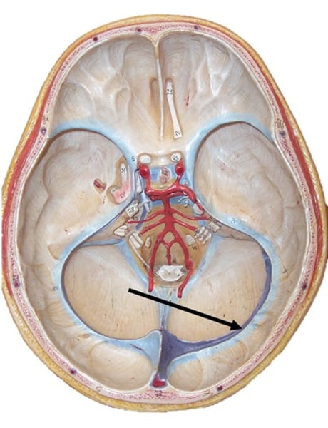
Right transverse sinus
Continuation of the superior sagittal sinus
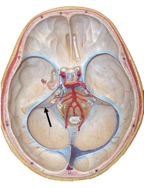
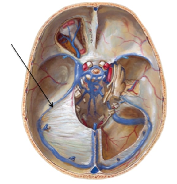
Tentorial notch
the space in the tentorium through which the brainstem passes
Superior petrosal sinuses
Stretch between cavernous and transverse sinuses
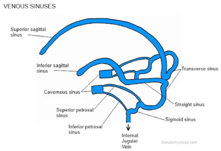
Transverse dural sinuses
What would run in this groove?
Sigmoid sinus
drains into internal jugular vein
Inferior petrosal sinus
carries blood from the cavernous sinus to the internal jugular vein
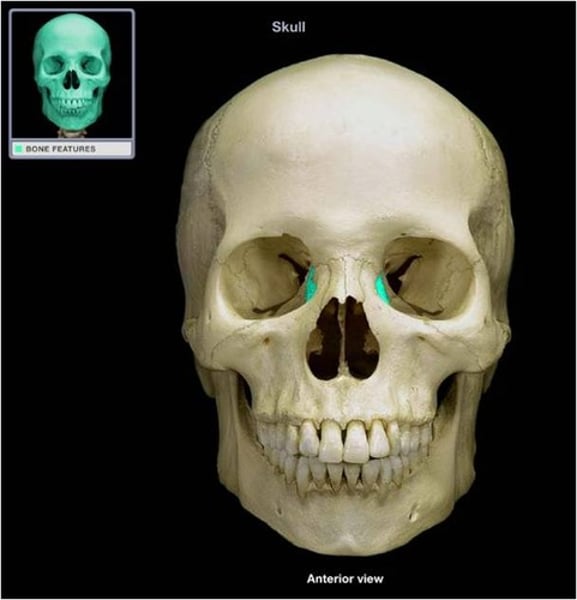
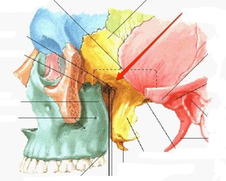
Superior orbital fissure (SOF)
Largest communication between the orbit and middle Cranial fossa
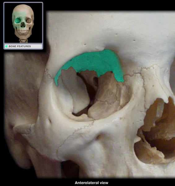
Orbital plate of frontal bone
This section of the Frontal bone forms the upper portion of the eye socket.
Lesser wing of sphenoid bone
Name this structure
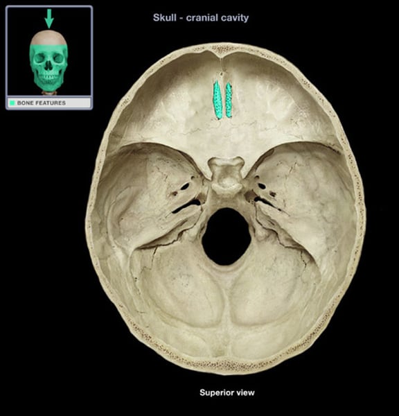
Posterior ethmoid foramen
Posterior ethmoid nerve
Anterior ethmoid foramen
anterior ethmoid nerve
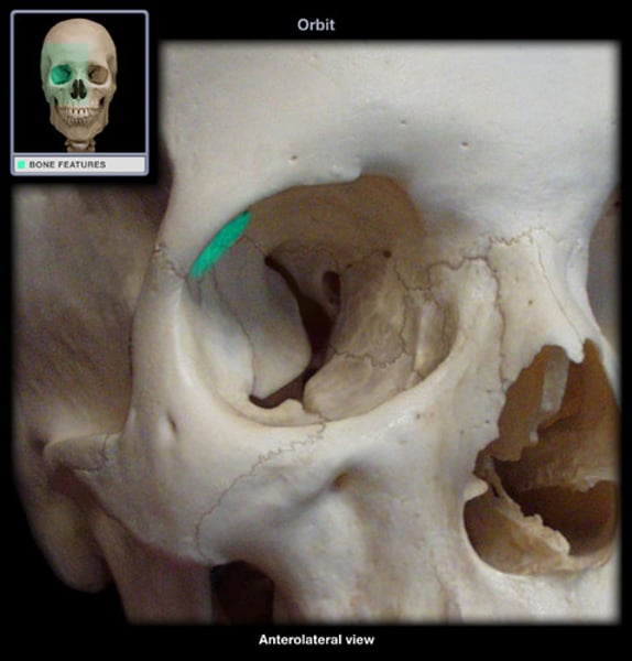
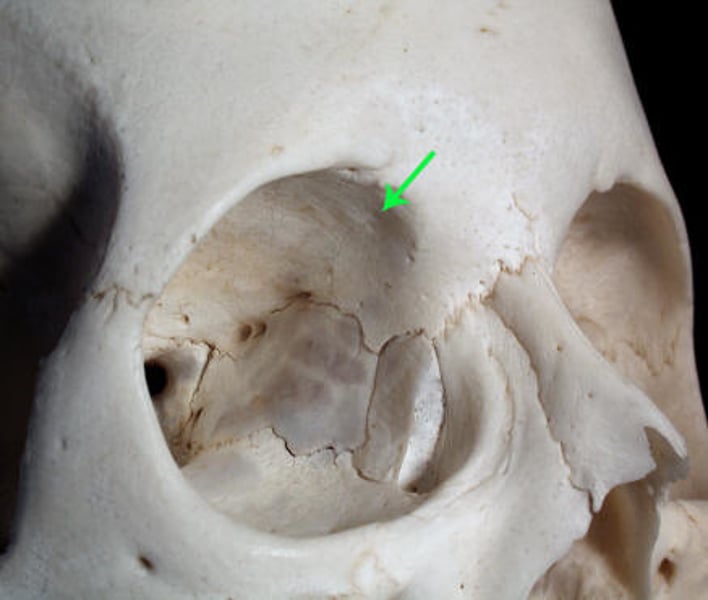
Fossa for lacrimal gland on orbital plate of frontal
Where does the lacrimal gland rest?
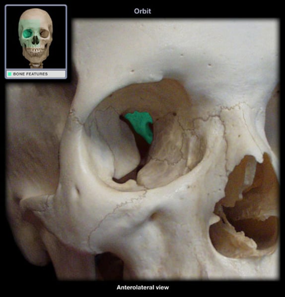
Fovea for trochlea of superior oblique muscle
The anchoring site for the pulley of the superior oblique muscle
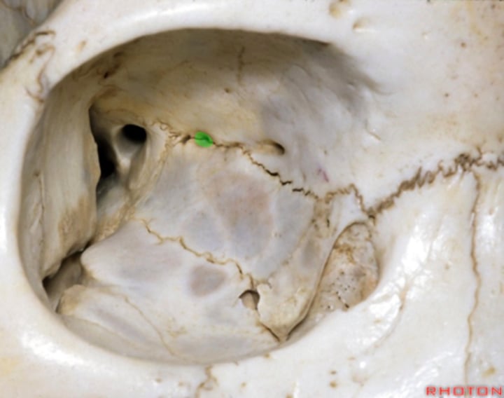
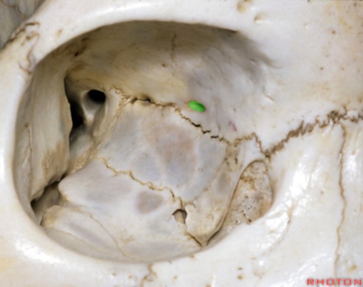
trochlear spine
Indicates that ligaments of the cartilaginous pulley were ossified. This is not always present.

Frontal process of maxilla
the ascending part of the upper jaw which gradually protrudes as it rises beside the nasal bone to meet the frontal bone; the ascending process of the upper jaw.
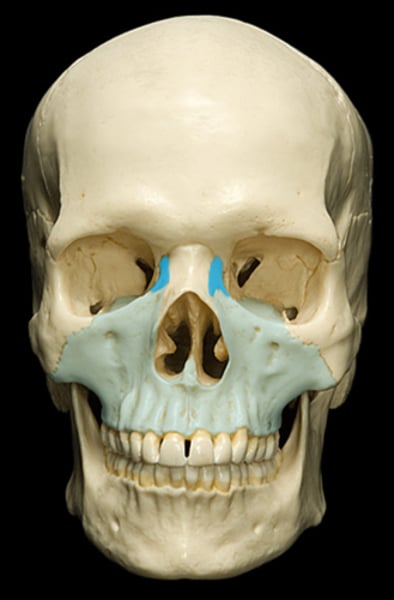
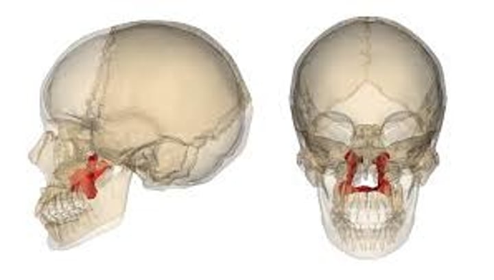
Lacrimal bone
small fragile bone making up part of the front inner walls of each eye socket and providing room for the passage of the lacrimal ducts