Regulation of Motor Control and the Limbic System
1/37
There's no tags or description
Looks like no tags are added yet.
Name | Mastery | Learn | Test | Matching | Spaced | Call with Kai |
|---|
No analytics yet
Send a link to your students to track their progress
38 Terms
Reminder**
What is contained within the telencephalon?
What is contained within the diencephalon?
Telencephalon: cerebral cortices, limbic system and basal ganglia
Diencephalon: thalamus, hypothalamus, pituitary gland and mammillary bodies
What four regions can the CNS be divided into?
Forebrain
Midbrain
Hindbrain
Spinal Cord
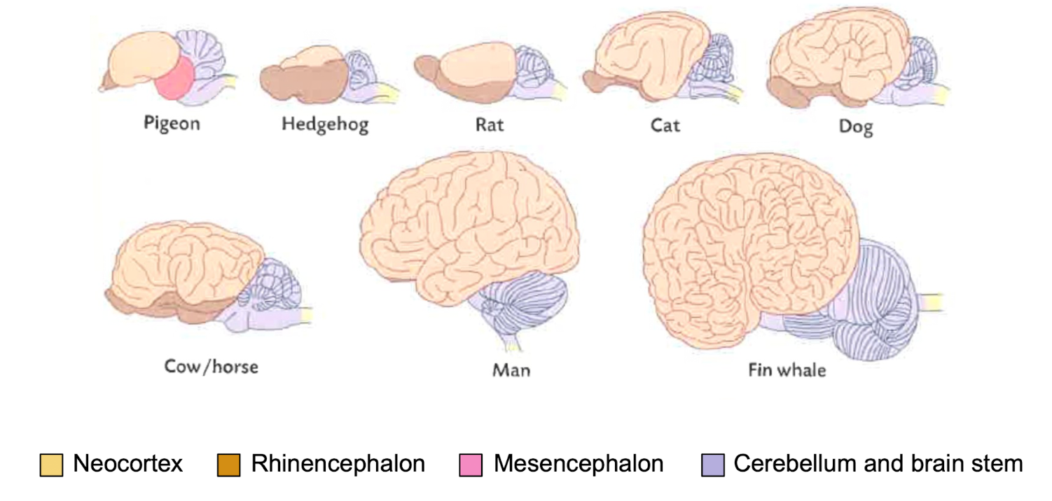
Compare the brain structure between various mammals.
Telencephalon → Region modified for that species
Rhinencephalon
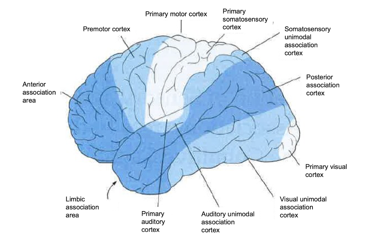
Describe the various regions of the cerebral cortices.
Cerebrum = species specific coding
Anterior Association Area - Integration of sensory and motor = personality
Primary visual cortex - visual scene → broken down here, colors encoded, eyes essentially respond to light
What are the different divisions of the limbic system?
Limbic system (i.e. 'ring') of structures that surround the thalamus.
Structures provide a useful means of conceptualizing the functional role and organization of the subcortex.
Primarily involved in the control of emotion, learning and memory.
The limbic system includes structures such as the amygdala, hippocampus, hypothalamus, and cingulate gyrus.
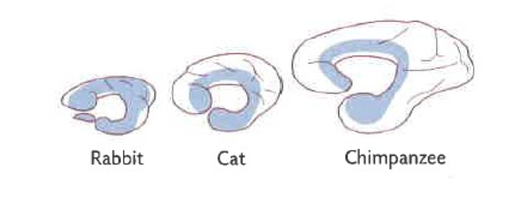
Why is the limbic system considered a primitive brain structure?
The forebrain of 'higher' order organisms has expanded over and around the limbic system to permit greater control of complex sensory-association-motor representation
Higher = more greatly developed since cortex has been used as a measure of intelligence
Describe the structure and function of the hippocampus.
The hippocampus is responsible for spatial memory and the transfer of some types of information for long-term memories.
Hippocampus - three layer cortical structure that plays a role in certain forms of memory and spatial navigation
Good for spatial orientation
Spatial memory is pivotal for survival. Hippocampal cells are: 'grid' cells that provide a universal neuronal coordinate system for spatial navigation. Other cells 'place' cells are activated only when the animal is in a specific place.
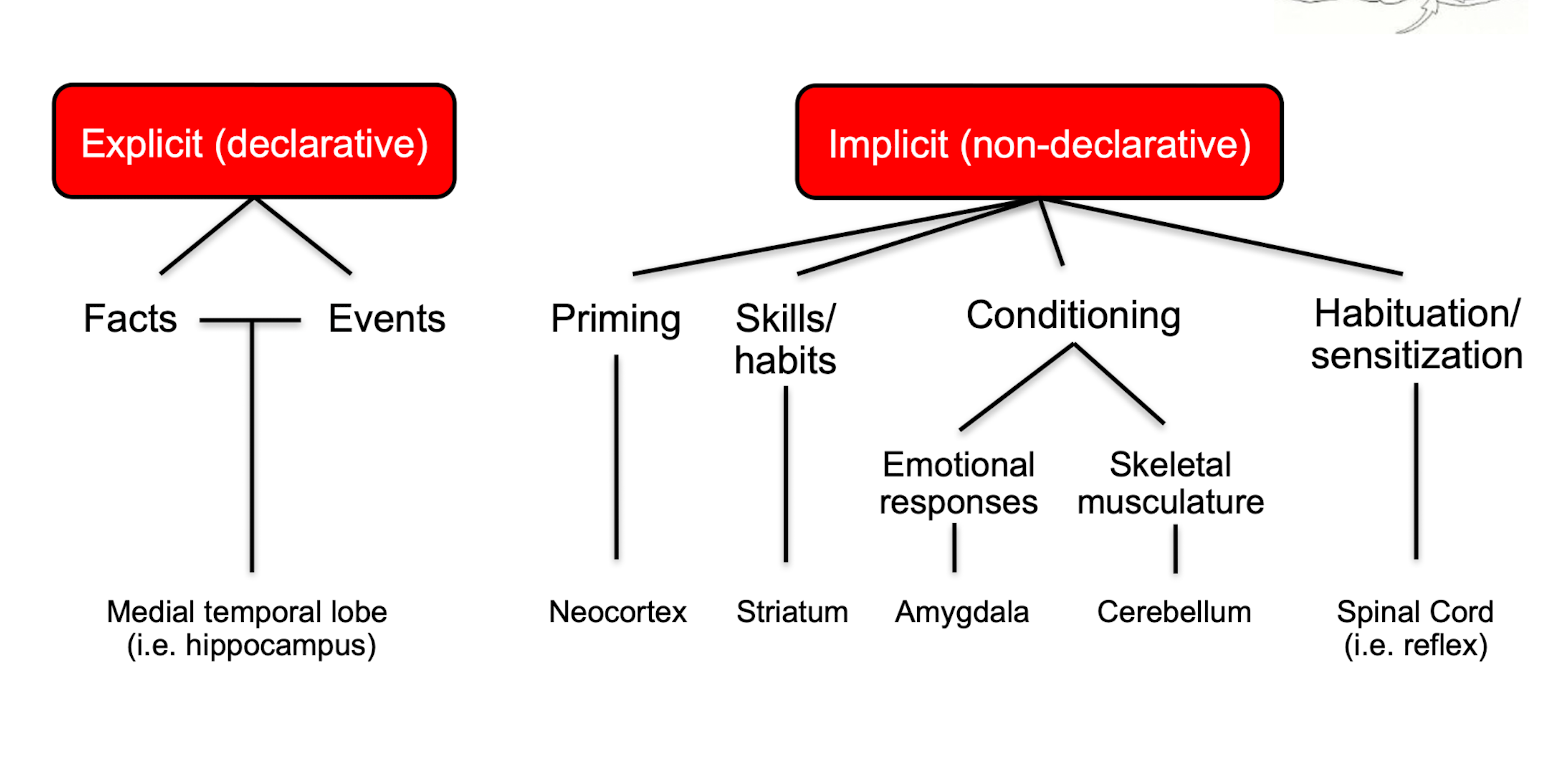
What is the hippocampus role in explicit and implicit memories?
Explicit Memories - Explicit memory is the conscious, intentional recall of facts and personal experiences. It is also known as declarative memory because it involves facts and concepts that can be voluntarily articulated.
For facts and events coded in the hippocampus
Implicit Memories - Implicit memory is a type of unconscious, long-term memory that affects our thoughts and behaviors without conscious recall.
Priming: example: repeating something until memorized
Habituation: learned or unlearned reflexes
Conditioning
Skills/Habits
How do hippocampal lesions impact explicit memories?
Discuss case of H.M.
The case of H.M. Had severe epilepsy and removed medial temporal regions (i.e. hippocampus).
Results in anterograde amnesia, unable to recall anything that had happened since his surgery. Implicit memories and working memory was intact.
Demonstration - meal times
He perceived his environment the exact same, but cannot comprehend the situation.
e.g. could learn certain forms of motor memory (like drawing the star)
Describe the structure and function of the amygdala?
A collection of nuclei in the medial temporal lobe involved in emotional (affective) behavior, motivation, and species-typical responses.
Amygdala neurons are multimodal, and respond to more than one sensory modality.
The amygdala has rich reciprocal connections with multiple brain regions, allowing it to integrate emotion with perception, memory, and physiology:
Hypothalamus → autonomic & endocrine emotion responses
Hippocampus → emotion–memory link
Cortices (esp. prefrontal) → emotion evaluation & decision-making
Brainstem → physiological & reflex responses
Basolateral nuclei – receive sensory input and link it to emotional meaning.
Central nucleus – main output; controls autonomic and behavioral emotional responses.
Which Video
What may happen if animals experience damage or lesions of the amygdala?
Describe Kluver-Bucy Syndrome
Lesions in monkeys result in:
1) Tameness and loss of fear
2) Indiscriminate dietary behaviour (eat previously rejected foods)
3) Greatly increased autoerotic, homosexual and heterosexual activity
4) A tendency to attend to and react to every visual stimulus
5) A tendency to examine all objects by mouth
In cats:
Lesions of the amygdala will result in animals wandering through a colony of monkeys completely undisturbed by their hooting and threats.
→ Results in a reduction in fear
THEREFORE Amygdala is very important for species specific behaviors and understanding what is a threat for that specific animal.
The fear conditioning methods (Pavlovian methods) are mediated by what regions of the brain?
Also learned responses such as to avoid specific animals, places and objects that are associated with danger. Fear conditioning (Pavlovian methods) of a tone-shock pairing are mediated by the amygdala.
Does not take a long time for animals to associate stimulus with pain.
→ Association learning, driven by the amygdala.
What are the main structures and functions of the fornix?
Fornix is a bundle of fibres along the medial aspect of the hemispheres.
The fornix has inter-connections with the hippocampi on both sides of the two cerebral hemispheres.
Fornix primarily connects the hippocampus to the mammillary body of the hypothalamus. Other fibres connect directly to the anterior nucleus of the thalamus.
Damage leads to deficits in learning.
What is the role of the Fornix for emotional processing?
What malfunctions are associated with this region of the brain?
Malfunctions of fornix signalling have been associated with Multiple sclerosis.
The demyelination of fibre bundles of the fornix has been identified as a functional consequence and symptoms of cognitive dysfunction such as dementia, short term memory impairments and long-term learning impairments
Damage to the fornix has also be implicated in Alzheimer's disease, schizophrenia
Describe the structure and function of the mammillary body.
The mammillary body is intricately connected with the hippocampal formation, fornix, amygdala and midbrain.
The primary function is associated with recollective memory, but also involved with emotion and goal-directed behaviours.
Trauma, stroke, tumors and alcoholism cause significant damage to mammillary bodies. The result is anterograde amnesia and reduced motivation.
What is the structure and function of the septum in the brain?
The septum is involved in emotional behaviours, sexual behaviour, aggressive behaviour, modulation of autonomic functions, and attention and memory functions
Modulator not a driver
The septum received input from the hippocampus, amygdala, hypothalamus and midbrain. The septum projects to the hippocampus and dentate gyrus (via the fornix), the thalamus and several hypothalamic nuclei.
What do lesions of the septum cause?
Lesions of the septum induce rage. In rodents, damage resulted in exaggerated reactivity to both appropriate and innocuous stimuli (sham rage).
Inability to regulate rage emotions.
In contrast, implanted electrodes in the septal nuclei for electrical self-stimulation studies resulted in prolonged and repeated stimulation, indicative of pleasurable responses.
What is the structure and function of the cingulate cortex?
The cingulate cortex is a neural interface between emotion, sensation, and action.
It has reciprocal connections with the limbic (medial dorsal and anterior) thalamic nuclei and with other limbic areas including the subiculum and entorhinal cortex (i.e. hippocampal formation). Cingulate cortical neurons also send axons to motor and premotor areas including the striatum, motor and premotor cortex.
The cingulate cortex is involved in planned motor movements.
What do lesions of the cingulate cortex cause?
Lesions in the cingulate cortex result in indifference to pain and other sensations that have strong emotional connotations; they produce social indifference and apathy, eliminate emotional intonation in speech, and cause personality changes.
Bilateral anterior cingulate lesions, or cingulotomies, have been done as "psychosurgery" to alleviate intractable pain and to incapacitate anxiety, obsessive-compulsive behaviour, and intractable depression.
Lesions in the posterior cingulate cortex result in diminished ability to perform spatial navigation.
What are the structures and functions of the basal ganglia?
The basal ganglia has four nuclei that are involved in voluntary movement: caudate nucleus, putamen, globus pallidus and substantia nigra.
Do not have direct input or output with the spinal cord.
Instead, there is input from the cortex and output to the midbrain. Another output pathways via the thalamus projects to the frontal, premotor and motor cortices.
Dopamine drives a direct motor program and can inhibit behaviors
Red: excitatory pathway
Gray: inhibitory pathway
What is the neural basis of parkinson’s disease
Loss of the dopamine in Parkinson's disease leads to increased output of the basal ganglia. Leads to paralysis
The overactivity of the indirect pathway results in hypokinetic disorders (i.e. impaired initiation of movement and reduced voluntary movement.
Case: LDOPA restores behavioural control. Temporarily.
Why does dopamine have both inhibitory and excitatory?
Binds to D2 → initiates inhibition
Binds to __ → Initiates excitation
What surgical intervention can improve the outcome of parkinson’s disease?
Can give dopamine BUT body becomes sensitised over time
A more permanent effect: destroying parts of the brain, two ways:
Globus Pallidus
Lesions of the internal segment of the Globus pallidus prevents abnormal output and partially restores voluntary movement.
Subthalamic nucleus
Lesions of the subthalamic nucleus reduces Parkinson's symptom's by eliminating excessive output from the internal segment of the Globus pallidus
What is the globus pallidus?
Inhibitory output
What happens if the thalamus is damaged?
COMA - inability to process information
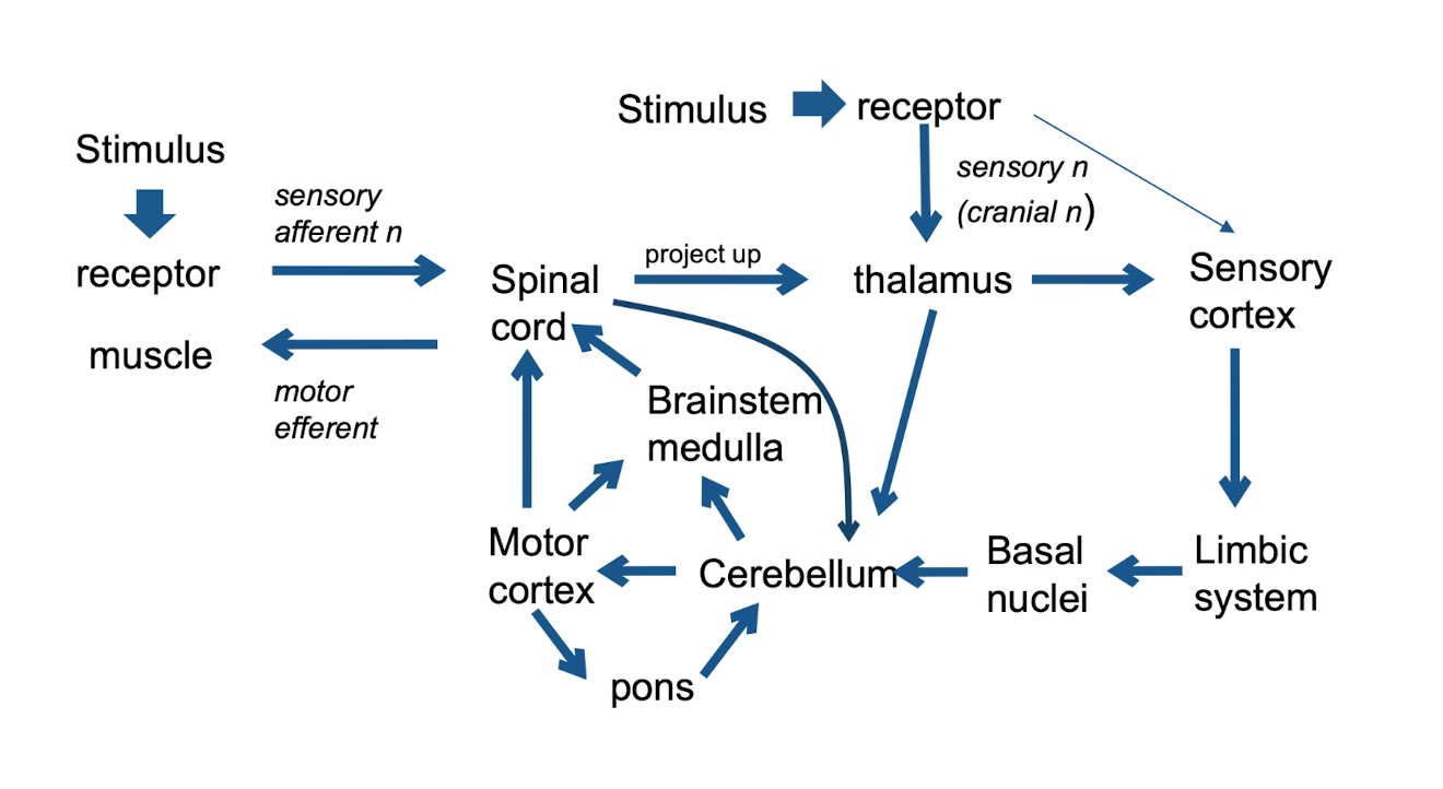
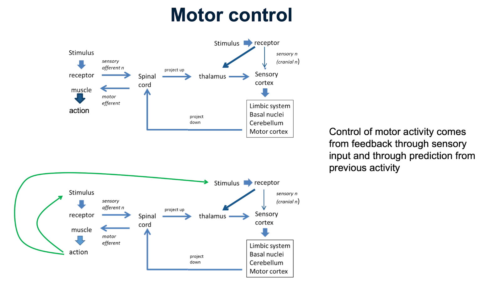
Outline how motor control is processed.
Note negative + positive feedback posibility
Describe the difference between simple and complex organisms regarding the hierarchy of motor control.
Simple organisms - local segmental control
Complex - Higher centers between spinal cord
"Higher" animals have additional control from higher centres in brain (encephalization)
In what case can spinal shock occur?
Spinal cord expects input from higher centers → if it is cut, the spinal shock occurs
Frequently experienced in car crashes
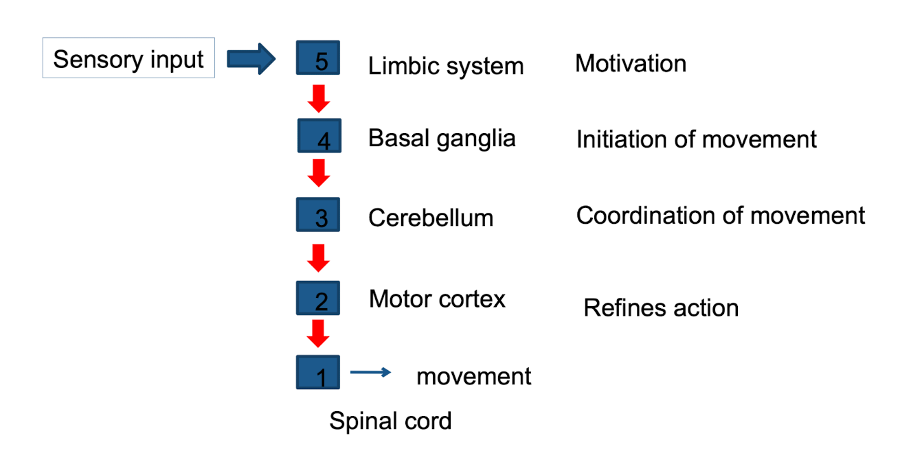
Describe the hierarchy of motor control from limbic system to the spinal cord.
What are the pyramidal tracts?
What are the extrapyramidal tracts?
Both are descending pathways → in control of the motor responses
Pyramidal tracts are the motor neurons that originate in the frontal cortex and terminate in the spinal cord (corticospinal) or brain stem (corticobulbar).
Extrapyramidal tracts are located in the pons and medulla of the midbrain and involved in involuntary movements.
What is pyramidal tract syndome?
characterized by spasticity and paralysis
What is extrapyramidal tract syndrome?
Extrapyramidal tract syndrome: characterized by involuntary movements, muscular rigidity and immobility without paralysis.
What are the features and purpose of the corticospinal tract?
Is a Pyramidal Tract
Required for fine, skilled movement.
Degree of cross-over varies between species - in dogs it is 100%, in ungulates 50%.
Fibres terminate on inter-neurones in the spinal cord.
Importance varies between species
What are the four types of extra-pyramidal tracts?
Reticulospinal tract
"old" tract, basic instincts (startle reaction): postural, initiates locomotion
Vestibulospinal tract
Input from vestibular apparatus and cerebellum. Mainly postural acting on extensors
Rubrospinal tract
Acts mainly on flexors, postural
Tectospinal tract
Input from vision and hearing, acts on cervical vertebrae, orientates head
What are the main effects of lesions in the descending tracts?
Lesions lead to hyperactivity in extensor muscles.
This is because many of the descending tracts are inhibitory.
Decorticate - Spastic
Decerebrate - Rigid
Spinal - Flaccid
Summarize the structures in the CNS.