sports science - topic 2
1/48
There's no tags or description
Looks like no tags are added yet.
Name | Mastery | Learn | Test | Matching | Spaced |
|---|
No study sessions yet.
49 Terms
2.1.1: List the principal structures of the ventilatory system
Nose, mouth, pharynx, larynx, trachea, bronchi, bronchioles, lungs and alveoli
2.1.2: Outline the functions of the conducting pathways
To provide a low resistance pathway for airflow
Defence against chemicals and other harmful substances
To warm and moisten air
2.1.3: Pulmonary ventilation
Inflow and outflow of air between the atmosphere and the lungs (breathing)
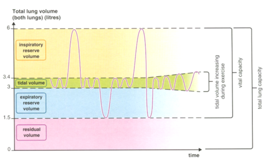
2.1.3: Tidal Volume
Volume of air inspired or expired per breath
Tidal volume increases during exercises
2.1.3: Inspiratory Reserve Volume
Additional inspired air over and above tidal volume (VT)
2.1.3: Relationship between Inspiratory Reserve Volume and Tidal Volume
when VT (Tidal volume) increases, IRV (Inspiratory reserve volume) decreases by the same volume and vice versa.
e.g. IRV decreases during exercise while VT increases
2.1.3: Expiratory Reserve Volume
Volume of air in excess of tidal volume that can be exhaled forcibly
2.1.3: Total Lung Capacity
Volume of air in the lungs after a maximum inhalation and includes the residual volume (average adult = 4-8 litres)
2.1.3: Vital Capacity
The maximal volume of air that can forcefully be expired following a maximal inspiration - the functional capacity of the lungs
2.1.3: Residual Volume
Volume of air still contained in the lungs AFTER a maximal inhalation
2.1.3: What happens to VT during exercise?
VT increases
2.1.3: Diagram of total lung volume

2.1.3: What does the red arrow represent?
Tidal volume

2.1.3: What does the blue arrow represent?
Inspiratory reserve volume

2.1.3: What does the green arrow represent?
Expiratory reserve volume

2.1.3: What does the yellow arrow represent?
Vital capacity

2.1.3: What does the purple/black arrow represent?
Total lung capacity
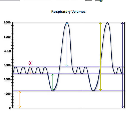
2.1.3: What does the orange arrow represent?
Residual volume
2.1.4: Explain the mechanics of ventilation in the human lung - Inhalation
Volume of thorax: increases
Diaphragm muscle: contracts
Diaphragm: flattens and pushes down digestive organs
External intercostal muscles: contracts/expands
Rib cage: moves upward and outward
Pressure in chest cavity: decreases below atmospheric pressure
Movement of air: into the lungs down the pressure gradient
2.1.4: What happens to diaphragm during inhalation?
Flattens and pushes down digestive organs
2.1.4: What happens to external intercostal muscles during inhalation?
Contract/expand
2.1.4: What happens to the volume of the thorax during inhalation?
Increases
2.1.4: What happens to pressure in chest cavity during inhalation?
Decreases (because lungs open up (volume increases) molecules inside the lungs have more space to move around)
2.1.4: Explain the mechanics of ventilation in the human lung - Exhalation
Volume of thorax: decreases
Diaphragm muscle: contracts
Diaphragm: relaxes and resumes to dome shape
External intercostal muscles: relaxes
Rib cage: inward and downward
Pressure in chest cavity: increases below atmospheric pressure
Movement of air: air forced out of lungs
2.1.4: Explain the mechanics of inhalation for a swimmer
As the head is lifted from the water:
external intercostal contract
rib cage moves upwards and outwards
diaphragm flattens/contracts
thoracic cavity volume increases in size/capacity
thoracic cavity pressure decreases
2.1.5: Describe the nervous and chemical control of ventilation during exercise
During exercise, ventilation is controlled both nervously and chemically to meet the increased oxygen demands and manage carbon dioxide levels. This involves chemoreceptors, the respiratory centre and neural inputs.
2.1.5: Describe the nervous and chemical control of ventilation during exercise - Chemoreceptors
Detect changes in CO2 and pH, signalling the respiratory centre.
2.1.5: Describe the nervous and chemical control of ventilation during exercise - Respiratory centre
Located in the medulla oblongata, controls the rate and depth of breathing
2.1.5: Describe the nervous and chemical control of ventilation during exercise - Neural inputs
Includes lung stretch receptors, muscle proprioceptors and signals from higher brain centres which respond to physical activity during exercise, ensuring that ventilation increases to meet the body’s demand during exercise.
2.1.6: Outline the role of haemoglobin in oxygen transportation.
Haemoglobin is the main oxygen transport protein in red blood cells (erythrocytes), binding 98.5% of blood oxygen. It transports oxygen from the lungs to the working tissues, where it is needed for energy production. Haemoglobin releases oxygen to tissues, with more oxygen released during strenuous exercise compared to rest.
haemoglobin has a high affinity for oxygen, allowing it to effectively pick up oxygen in the lungs.
also transports carbon dioxide from the tissues back to the lungs for expiration.
each haemoglobin molecule can carry four oxygen molecules, binding reversibly to form oxyhemoglobin.
higher levels of haemoglobin are found in trained athletes, enhancing their oxygen transport capacity.
2.1.7: Explain the process of gaseous exchange at the alveoli
Gaseous exchange occurs through diffusion across the respiratory membrane, which separates the alveoli from the surrounding capillary network
O2 DIFFUSES/PASSES from the lungs inot the capillaries (blood)
CO2 DIFUSSES/PASSES from the capillaries (blood) into the lungs
this process occurs due to differences in partial pressures which create a pressure gradient and can affect the rate of exchange
during EXERCISE; diffusion capacity for oxygen increases and the pressure gradient for CO2 is smaler than for O2 exchange
Partial pressure (PO2) oxygen in the alveoli is higher than in the capillaries
Partial pressure (PCO2) of CO2 is higher in the capillaries (blood) than in the alveoli
2.2.1: State the composition of blood
Blood is composed of erythrocytes (red blood cells), leucocytes (white blood cells), thrombocytes (platelets) and plasma and transports nutrients, oxygen, carbon dioxid, hormones, antibodies, urea (heat - thermoregulation)
90% water
8% blood protetins
1% electrolytes
0.5% food substances
0.04% waste products
2.2.2.: Distinguish between the function of erythrocytes, leukocytes and platelets - Erythrocytes
Erythrocytes (red blood cells) contain haemoglobin and transports both oxygen and carbon dioxide (approx. 10%).
make up 45% of blood volume
2.2.2.: Distinguish between the function of erythrocytes, leukocytes and platelets - what percentage of blood volume are erythrocytes?
45% of blood volume
2.2.2.: Distinguish between the function of erythrocytes, leukocytes and platelets - Leucocytes
Leucocytes (white blood cells) are involved in immune function, combatting infection, protecting the body from viruses and infections, by engulfing pathogens and making antibodies.
Lymphocytes and phagocytes
make up 3% of blood volume
2.2.2.: Distinguish between the function of erythrocytes, leukocytes and platelets - what percentage of blood volume are leucocytes?
3% of blood volume
2.2.2.: Distinguish between the function of erythrocytes, leukocytes and platelets - Thrombocytes
Thrombocytes (platelets) are cell fragments which assist in the repair process following injury by allowing blood to clot (produces scabs)
1% of blood volume
2.2.2.: Distinguish between the function of erythrocytes, leukocytes and platelets - what percentage of blood volume are thrombocytes?
1% of blood volume
2.2.2.: Distinguish between the function of erythrocytes, leukocytes and platelets - Plasma
Plasma is the fluid component of blood that assists in transport of substances such as food/waste products/hormones/antibodies/electrolytes
51% of blood volume
2.2.2.: Distinguish between the function of erythrocytes, leukocytes and platelets - what percentage of blood volume is plasma?
51% of blood volume
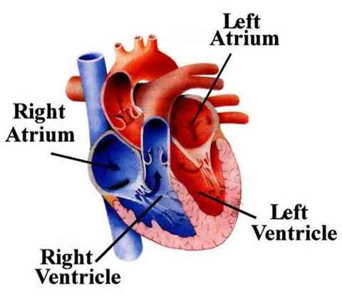
2.2.3: Describe the anatomy of the heart with reference to the heart chambers, valves, and major blood vessels - 4 chambers
Upper chambers: left and right atrium
Lower chambers: left and right ventricle
What are these 4 labels?
The 4 chambers of the heart
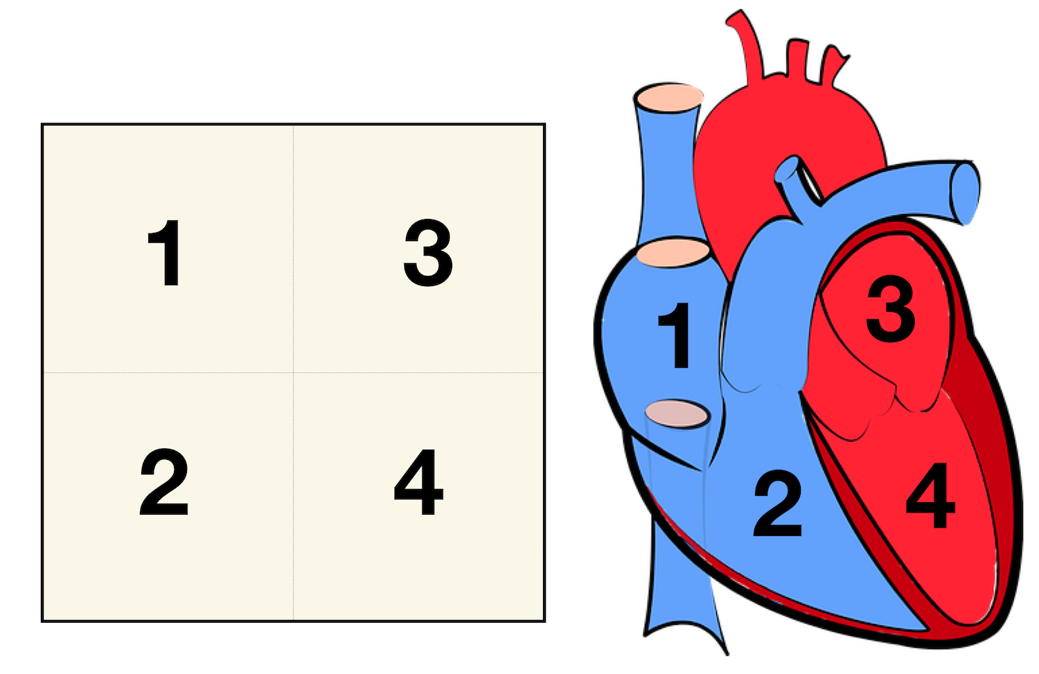
What label corresponds to number 1?
Right atrium

What label corresponds to number 2?
Right ventricle

What label corresponds to number 3?
Left atrium

What label corresponds to number 4?
Left ventricle
2.2.3: Describe the anatomy of the heart with reference to the heart chambers, valves, and major blood vessels - 4 valves
Bicuspid (mitral), tricuspid, aortic and pulmonary valve
The names of the four chambers, four valves (bicuspid, tricuspid, aortic and pulmonary valve) and the four major blood vessels (vena cava, pulmonary vein, the aorta and pulmonary artery) of the pulmonary and systemic circulation are required. The heart has its own blood supply via the coronary arteries, however the names of the coronary arteries are not required.