exactly 28 - . Functional morphology and physiological characteristics of the conduction system of the heart. Automaticity. Cardiac Rhythm. Conduction abnormalities. Physiological characteristics of the working myocardium. Excitation and contraction. Refractory periods. Extrasystoles, flutter and fibrillation. Myocardial metabolism.
1/16
There's no tags or description
Looks like no tags are added yet.
Name | Mastery | Learn | Test | Matching | Spaced |
|---|
No study sessions yet.
17 Terms
sections
physiological characteristics of heart (working myocardium)
structure of myocardium
excitation and contraction
main electro physiological properties of contractive myocardium are
extrasystoles
atrial flutter
atrial fibrillation
physiological characteristics of heart (working myocardium)
The heart contains 4 chambers
the upper atria receives the venous blood to the two lower ventricles that eject blood into the arteries.
The right ventricle pumps deoxygenated blood to the lungs, to become oxygenated and the left oxygenated blood goes rest of the body.
The right atria and ventricle are separated from the left V and A by a septum- prevents mixture of the oxygenated and deoxygenated blood.
The right atrium receives blood from the SVC and IVC
left receives from the left the pulmonary veins
structure of myocardium
Made of three layers of cells
purkinje cells
cardiomyocytes
myocardial endocrine cells
-cells of myocardium have actin and myosin filaments (involuntary skeletal muscle)
-They receive contact by sliding filament mechanism. Cells electrically linked by gap junction which allows electrical impulses to spread across the whole mass
excitation and contraction
Cardiac muscle fibers contract via excitation-contraction coupling,
using a mechanism called calcium-induced calcium release.
The stages of contraction are:
Electrical signal starts in the SA node (the heart’s pacemaker) and travels to the AV node, then spreads to heart muscle cells through gap junctions.
Action potential causes Na⁺ and Ca²⁺ to enter the heart muscle cells via T-tubules.
Calcium binds to troponin-C, which moves the troponin complex and exposes actin binding sites.
Myosin binds to actin and pulls it inward using energy from ATP, causing muscle contraction.
Calcium is removed by the sarcoplasmic reticulum, troponin blocks actin again, and the muscle relaxes.
-…………………………………………………………………………………
Metabolism- ATP needed for contraction and Ca2+
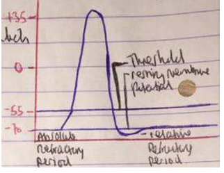
refractory periods
Refractory periods:
absolute refractory period
relative refractory period
-……………………………………………………………………………………………
plateau it what is responsible for the long refractory period
Absolute refractory period: The absolute refractory period is when no stimulus—no matter how strong—can trigger a new action potential. It lasts from the start of repolarization through rapid repolarization, covering all of systole and early diastole, during which the heart muscle can’t be re-excited.
Relative refractory period, is the interval during which a second action potential can be generated but only at a higher stimulus intensity.
…………………………………………………………………………………
Fast Na+ channels open (Na+ influx, depolarization)
Na+ channels close, K+ and slow Ca2+ channels open, the plateau phase elongating the refractory period
Slow Ca2+ channels close hyperpolarization/repolarization occurs
When resting potential is reached, K+ channels close
extrasystoles
premature contractions of the heart which temporarily disturb cardiac rhythm – has lower amp of regular one and pause for next regular systole so next systole has higher amp
atrial flutter
very rapid contraction of the atria with regular rhythm and rate 250 to 350 bpm
atrial fibrillation
irregular very rapid ineffective contractions of section of atrial muscles caused by impulses with a rate of 300 to 600 per min
section
cells of heart
natural pacemaker
5 elements of heart
conduction system
automaticity
automatic and cardiac rhythm
cause of increased heart rate
cause of decrease in heart rate
cells of heart
made of cardiomyocytes- do not require such a stimulus as they have leaking Na+ and k+ ion channels that allow for the entry and exit of Na+ and K+ ions. This allows for constant AP generation.
natural pacemaker
Sinoatrial node is the hearts natural pacemaker - pacemaker cellls
it generates its own action potential
causes the positive increase in voltage across the membrane.
5 elements of heart
SAN- in wall of the right atria—60-90 impulses
AVN-base of the right atrium- 40-60 impulses
Bundle of His- 30-40 impulses
Tawara fibres
Purkinje fibres- 20-40 impulses- spreads up to the ventricles
conduction system
conduction system:The heart has a special system that sends electrical signals to make it beat
It includes: SA node (sinus node), AV node, Bundle of His, and Purkinje fibers.
Automaticity means some heart cells (like in the SA node) can start signals on their own — without brain signals.
automaticity
Automaticity means some heart cells (like in the SA node) can start signals on their own — without brain signals.
automatic and cardiac rhythm
Sinoatrial node is the hearts natural pacemaker
generates its own action potential
causes the positive increase in voltage across the membrane. the pacemaker potential therefore controls the rate of the heart.
Normal cardiac rhythtm- 60-90bpm at rest
Tachycardia- increased HR >100 bpm
Bradycardia- decrease HR. <60bpm
cause of increased heart rate
High temperature
Sympathetic stimulation
By hormones, T3/T4, circulation of catecholamines (adrenaline)
Increased Ca2
cause of decrease in heart rate
cardiac conduction disease- disruption of electrical impulses to the heart