Animal Diversity - Practical 1 - Features, Ecology, and Body Structures
1/58
There's no tags or description
Looks like no tags are added yet.
Name | Mastery | Learn | Test | Matching | Spaced |
|---|
No study sessions yet.
59 Terms
Choanocyte:
create a current to move water through a sponge via a central flagellum
Spicule:
Sharp and hard spike-like structures that build the endoskeleton of some sponges
Spongin:
fibers that help build the endoskeleton in sponges; found in Demospongiae
Asconoid:
simplest sponge canal type
Water flow: ostium → spongocoel → osculum
chaonocyte location: spongocoel
Syconoid:
larger, “pleated” versions of asconoids
Water flow: ostium → incurrent canal → prosopyl → radial canal → apopyle → spongocoel → osculum
choanocyte location: radial canals
Leuconoid:
Largest, most complex, and most common sponge canal type
Water flow: ostia → incurrent canal → flagellated chamber(s) → excurrent canal → osculum. Water goes through several flagellated chambers before exiting.
Choanocyte location: flagellated chambers
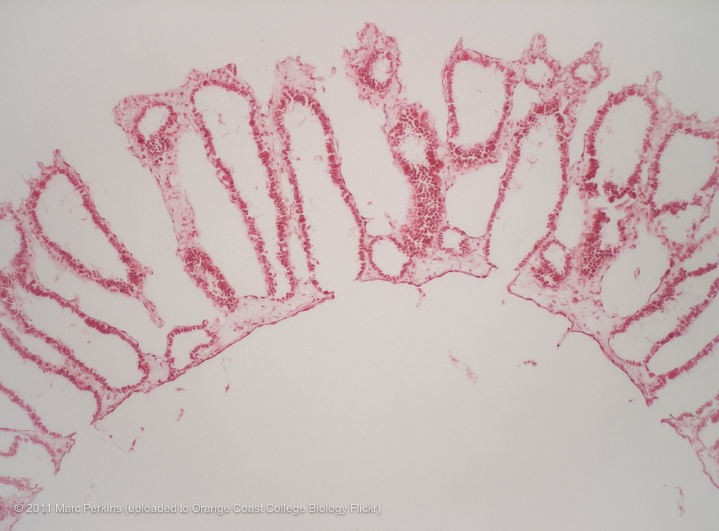
What canal system does this sponge have?
Syconoid
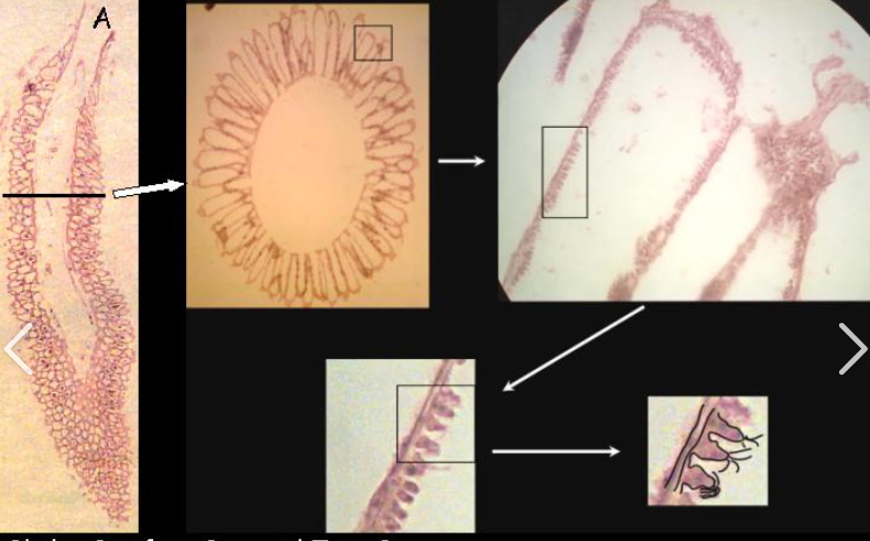
1.) what type of canal system does this sponge have?
2.) What is the structure being highlighted?
3.) What is the structure labeled “A”?
1.) Syconoid
2.) Choanocytes
3.) Osculum
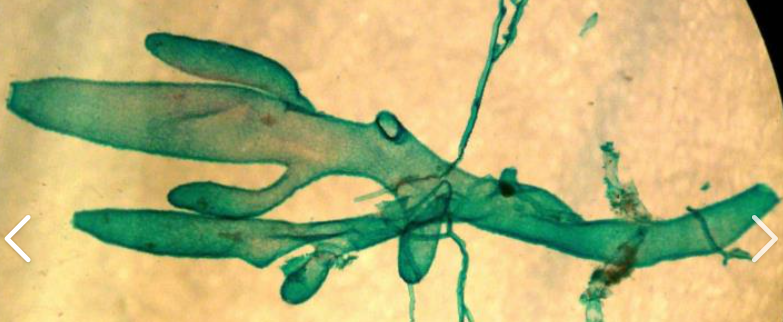
What kind of canal system does this sponge have?
Asconoid
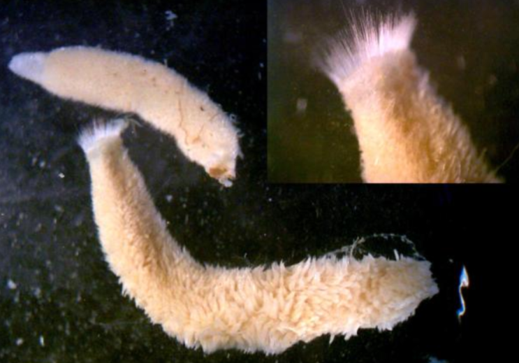
What kind of canal system does this sponge have?
Syconoid
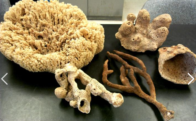
What kind of canal system do these sponges have?
Leuconoid
Spicules in Calispongiae are made of _____ and have how many points?
Calcium carbonate; mostly triradiate (3 points), some 2 or 4-pointed
Spicules in Demospongiae, if present, are made of _____? What else makes up the endoskeleton of demosponges?
Silica carbonate; spongin fibers
Spongocoel:
central cavity of a sponge
Osculum:
Exit hole of a sponge
Incurrent canal:
Canals connecting dermal pores (ostia) to internal chambers in sponges
Prosopyle:
Porocyte cell that allows water to enter the:
radial canal in syconoid sponges
flagellated chamber in leuconoid sponges
from the incurrent canal.
Apopyle:
Porocyte cell that allows water to exit the radial canal (syconoids) / flagellated chamber (leuconoids) and enter the spongocoel/excurrent canal.
Ostia/ostium:
Dermal pores on sponges that allow water to enter the body
Radial canal:
A canal found in syconoid sponges where choanocytes are located
Mesohyl:
Non-living cell matrix in sponges
Amebocyte:
Mobile cells in the mesohyl of sponges that transport food, store food, eliminate waste, and can differentiate into all other cell types (stem cells).
Pinacocyte:
Epithelial-like cells that line the outer surface of the sponge; collectively the pinacoderm
Gemmule:
An “internal bud” formed within the mesohyl in sponges; a bundle of amebocytes surrounded by spicules that become a new sponge once matured.
What cells can develop into sperm in sponges?
Choanocytes
What cells can develop into oocytes (eggs) in sponges?
Choanocytes and amebocytes (in some species)
Monoecious:
A single organism contains only one type of gametes; cannot self-fertilize.
Dioecious:
A single organism contains both male and female gametes; able to self-fertilize
What is a monaxon spicule?
A spicule that is straight with no branching
What is a triradiate spicule?
A spicule with 3 equally distanced points
What is a polyaxon spicule?
A spicule with more than 3 points
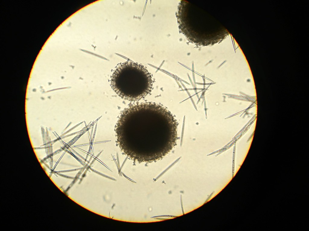
What’s this?
Gemmules found in Spongilla (Demospongiae)
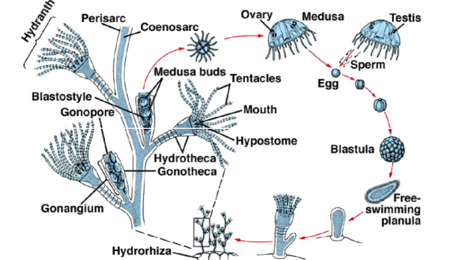
Describe the life cycle of an Obelia colony.
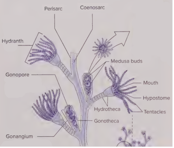
Gonangium releases medusae buds through the gonopore → develop into mature medusae → mature medusae sexually reproduce → egg becomes blastula → blastula becomes free-swimming planula → planula attaches to substrate via hydrorhiza → planula asexually reproduces (buds) to create new colony
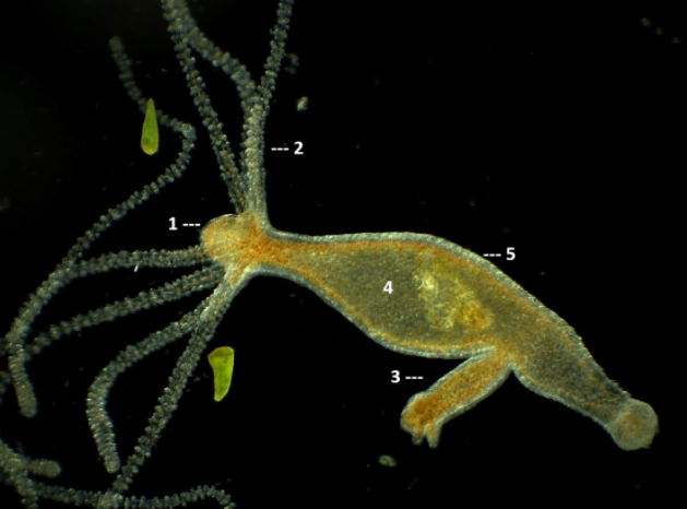
Label this diagram of a hydra.
Hypostome
Tentacle
Bud
Gastrovascular cavity
Ectoderm
Where are the cnidocytes located on hydras?
the epidermis
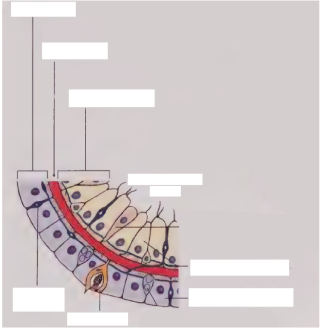
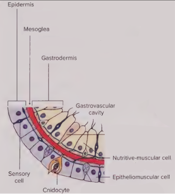
Fill in the blanks of this diagram of a Hydra.
See picture
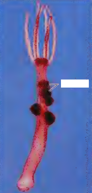
What is the structure being highlighted on this hydra?
Testes
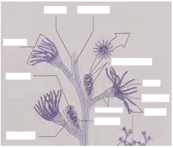
Label the structures on this Obelia diagram.
See picture.
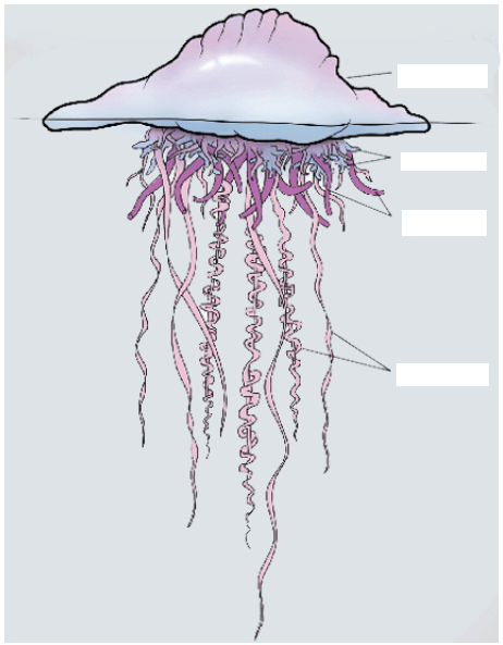
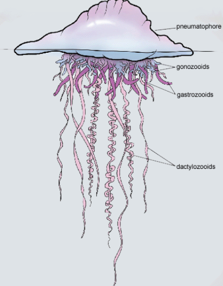
Label this diagram of a Portuguese Man o’ War.
See picture.
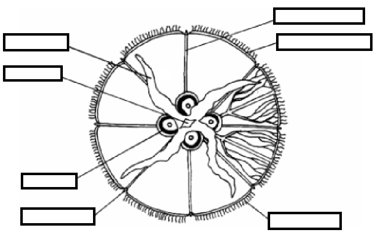
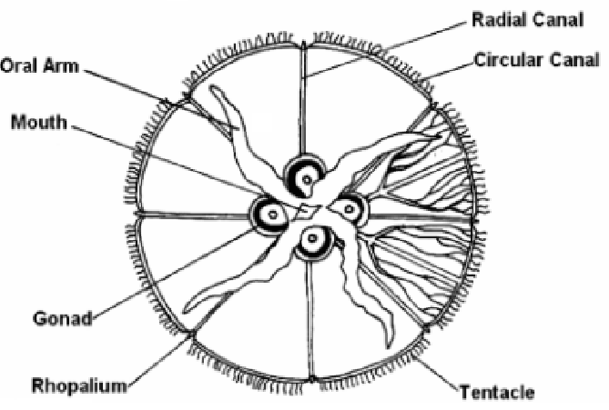
Label this diagram of a mature Aurelia medusa.
See picture.
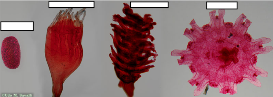
Label the various life stages of Aurelia depicted in the photo.
See picture.
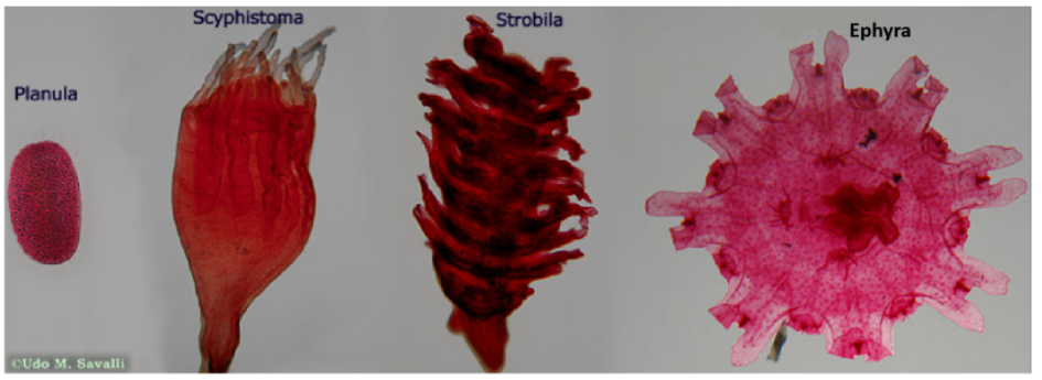
Describe the life cycle of Aurelia.
Mature medusae sexually reproduce → zygote → becomes planula larva → planula settles in substrate → becomes scyphistoma → becomes strobila → releases ephyra → ephyra matures into medusa
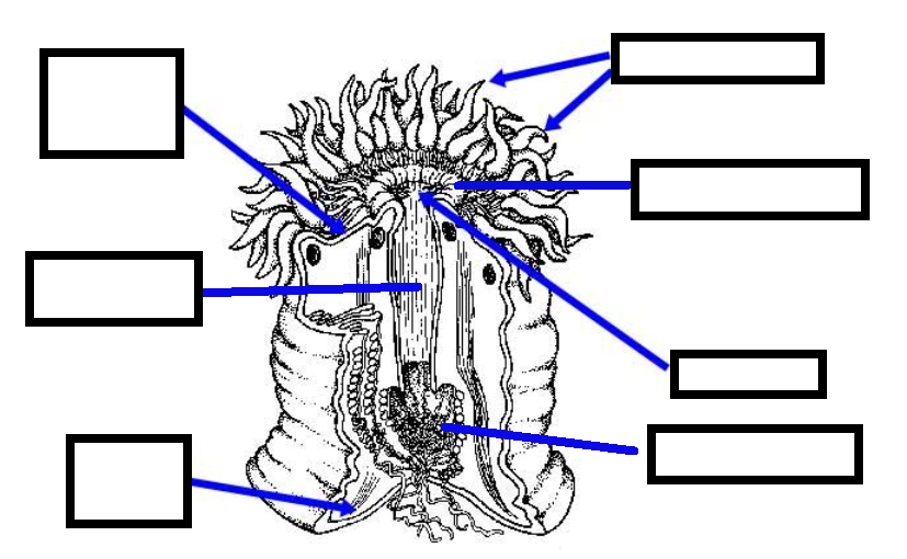
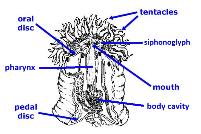
Label this diagram of Metridium.
See picture.
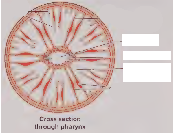
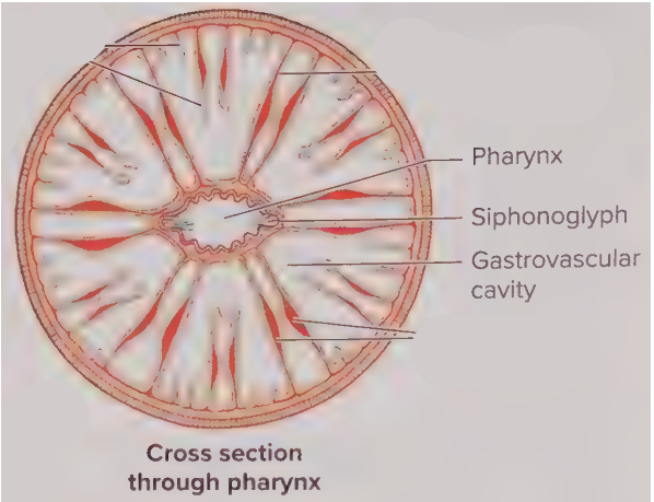
Label this cross section of a Metridium (sea anemone).
See picture.
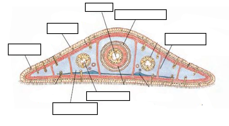
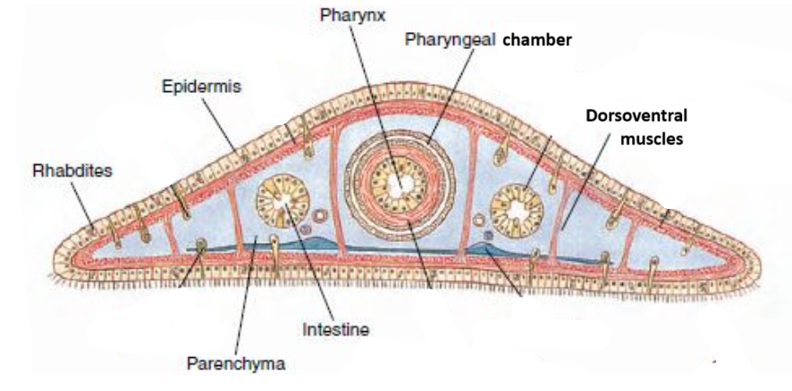
Label this cross section of a planarian (Turbellaria).
See picture.
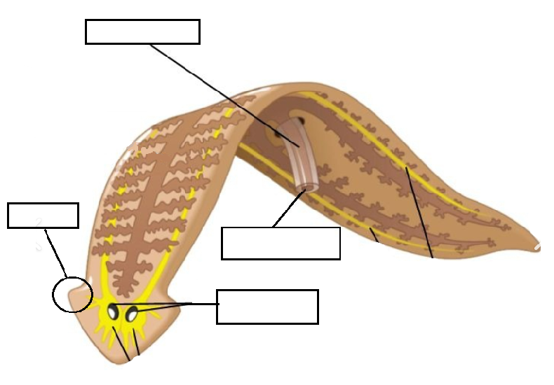
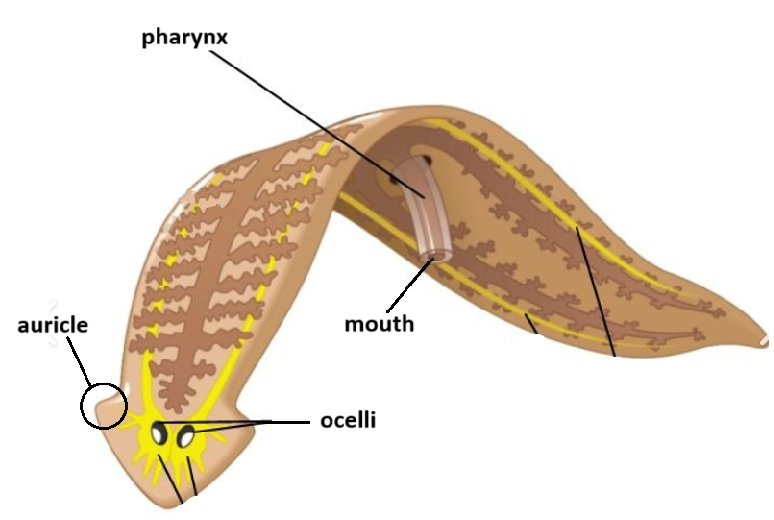
Label this diagram of a planarian.
See picture.
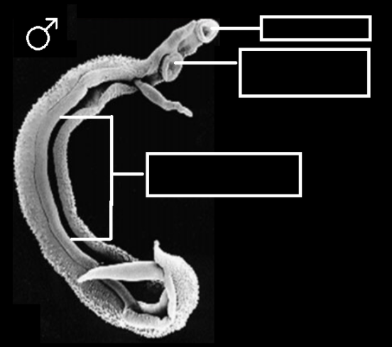
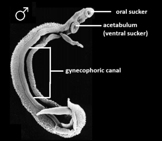
Label this diagram of a male Schistosoma mansoni.
See picture.
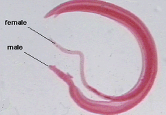
How would you describe what’s happening between these to trematodes?
They are in copula; the female is slotted within the gynecophoric canal of the male.
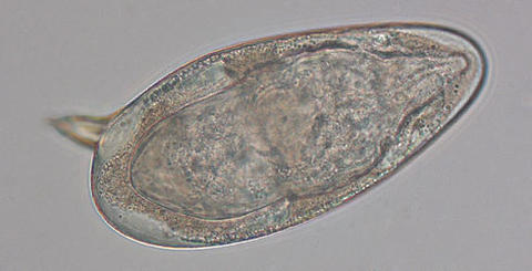
What is this?
An egg of Schystosoma mansoni
Describe the life cycle of Schystosoma mansoni.
Eggs are released in feces
miracidium hatches in water
Miracidium becomes free swimming, penetrates intermediate host (usually a snail)
Miracidium becomes sporocycst, which makes rediae (daughter sporocysts)
Sporocycts produce cercariae en masse
Cercariae leave intermediate host and become free-swimming
Free-swimming cercariae penetrate definitive host (usually human)
Cercariae develop into adult flukes
Adult flukes mate in blood vessels
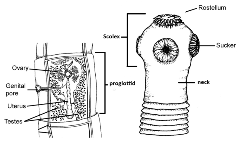
Label this diagram of Taenia pisiformis.
See picture.
Describe the life cycle of Taenia pisiformis
Adult within definitive host shed fertilized proglottids through feces
Proglottids dry out and crack open, full of eggs
Intermediate host (usually a rabbit/some other lagomorph) eats eggs
Eggs develop into larval form (Cysticercus)
Definitive host (often a dog) eats intermediate host and becomes infected
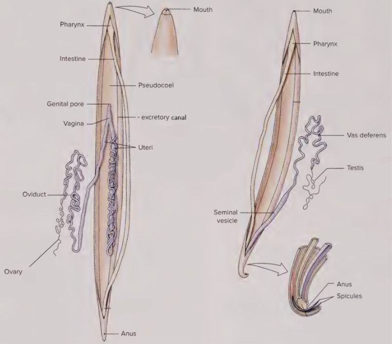
Label the diagrams of Ascaris. Which is male and which is female?
See picture. Left is female, right is male.
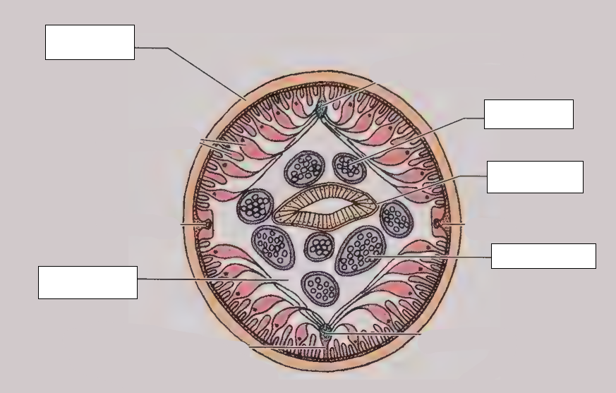
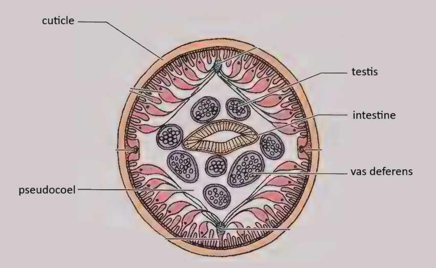
Label this cross section of a male Ascaris.
See picture.
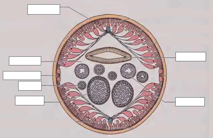
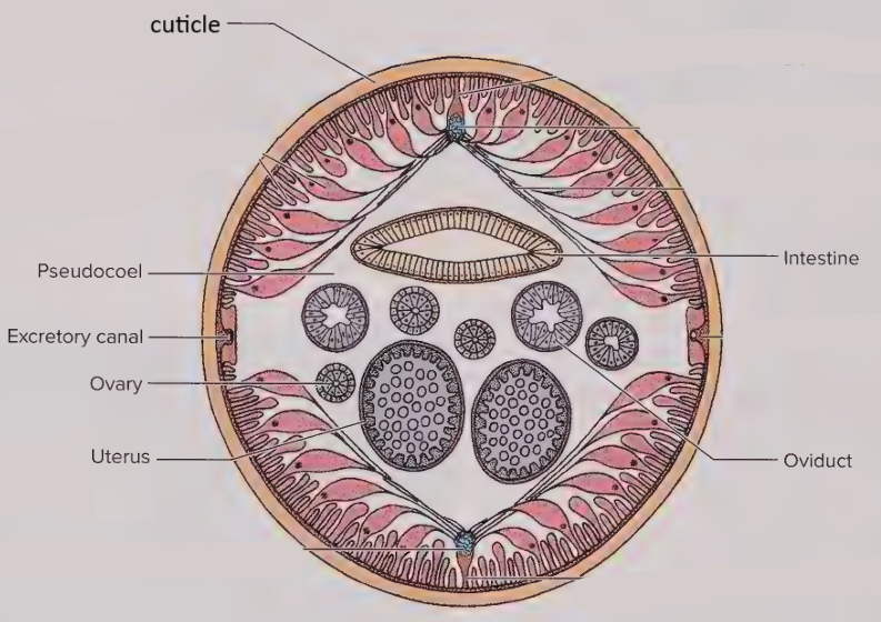
Fill in this cross section of a female Ascaris.
See picture.
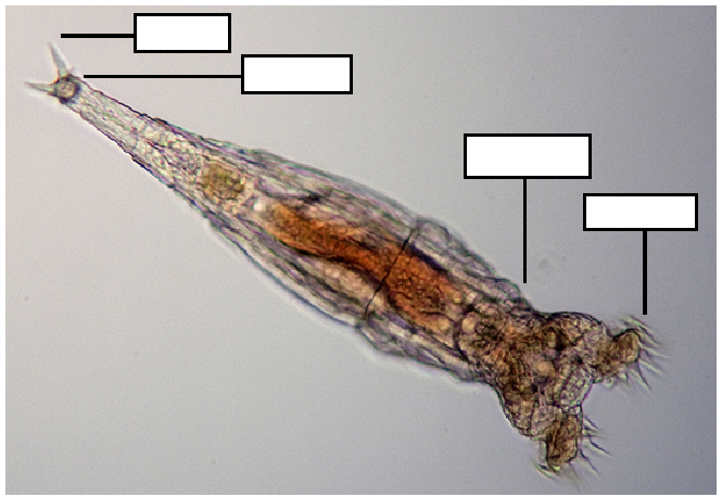
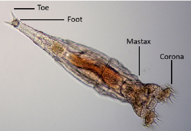
Fill out this diagram of a rotifer.
See picture.
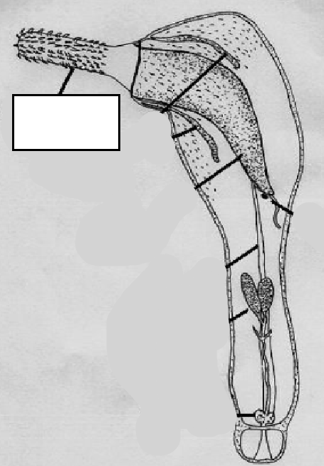
What is this structure? Does it always look like this?
Proboscis; in this diagram it is everted, but it can be inverted (pulled in) too.
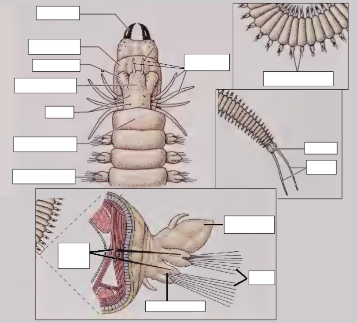
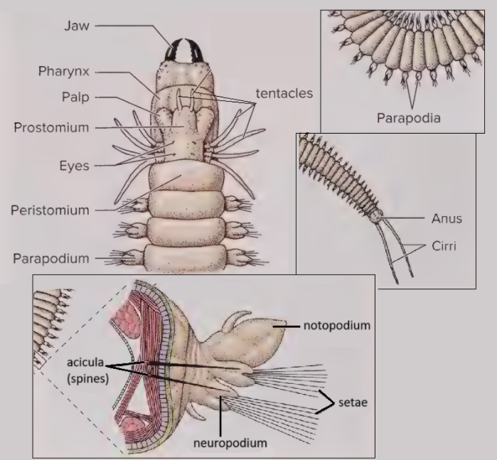
Label this diagram of Nereis.
See picture.
