Lower Motor Neurones
1/23
There's no tags or description
Looks like no tags are added yet.
Name | Mastery | Learn | Test | Matching | Spaced |
|---|
No study sessions yet.
24 Terms
Lower motor neurons mode of action
Reflexes occur at level of spinal cords regardless of upper input
Definition of LMN (= common final pathway)
Somatic motor pathway
Neuron cell bodies located in ventral column/horn of gray matter of spinal cord
[axons come out of the ventral ROOT]
LMN also within motor nuclei of CNs containing efferent component
e.g. facial nerve innervating muscles in head (orbicularis oculi)
Brainstem → connection to other nuclei
Ventrally located neurons that stimulate skeletal muscle
LMN connection/synapses
interneurones/relay → up and down SC
ascending tracts (ipsilateral or contralateral) → sensory
descending tracts (ipsilateral or contralateral)→ motor
commiss-ural tracts (contralateral)- connect left and right brain hemispheres
LMN Influences
UMN
First order neurons, initiating and modulating muscle contraction
Pyramidal tracts
both inhibitory and facilitatory
Sensors from muscle/tendons → sensory receptors, creating inputs (muscle spindles, golgi tendon organs)
sensors → spinocerebellar tract → cerebellum ← → cerebrum (primary motor cortex) → pyramidal (corticospinal or cortionuclear) tracts (UMN) → short relay → LMN
contralateral primary motor cortex → ipsilateral cerebellum → controls fine motor control of ipsilateral muscle
LMN functions
Regulate and control skeletal muscles
Regulated through voluntary centres in brain (primary motor cortex)
Lower motor neuron types
Alpha motor neurons
Fibers innervate ordinary muscle fibres (extrafusal)
Gamma motor neurons
Fibers innervate intrafusal muscle fibres (muscle spindles/sensors)
Intrafusal fibre
Specialised muscle fiber sandwiched between normal muscle fibres
Central region without myofibrils
Muscle spindles wrapped around central region of intrafusal fiber
When muscle spindle contracts → central region pulled
activates sensory input, engaging spinal cord
Rate of muscle spindle stretch increases (due to muscle contraction) → either inhibits or stimulates alpha motor neuron depending on whether muscle stretches or contracts
Coactivation and stimulation of skeletal muscles
Upper motor neuron stimulates both alpha (extrafusal) and gamma (intrafusal) motor neurons → COACTIVATION
Activation of alpha motor neurons → extrafusal fibres stimulated
Activation of gamma motor neurons → spindle contracts (stretches) → synchronises stretch between spindle and regular muscle for sensory purposes
Brain (somatomotor cortex) → UMN → LMN
Spinal cord → alpha and gamma motor neurones
Sensory feedback → smooth skeletal muscle control and movement
Muscle spindles
Golgi tendon organs
Skin receptors (pain, touch, temp)
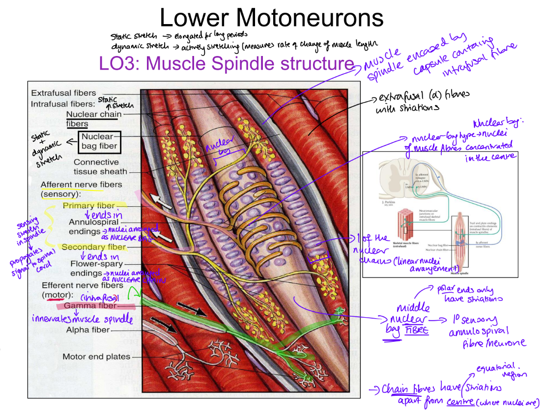
Muscle spindle structure part 1
Thin intrafusal fibres
Run parallel to thick extrafusal fibres to insert on either side of tendon
No contractile apparatus in middle of fibre BUT concentration of nuclei
Intrafusal fibre types [2]
1) nuclear bag → primary annulo-spiral neurones
wraps around central region
sensitive to dynamic change → relative length change
2) nuclear chain → flower spray neurones
sensitive to sustained stretch (static change) → persistent long length
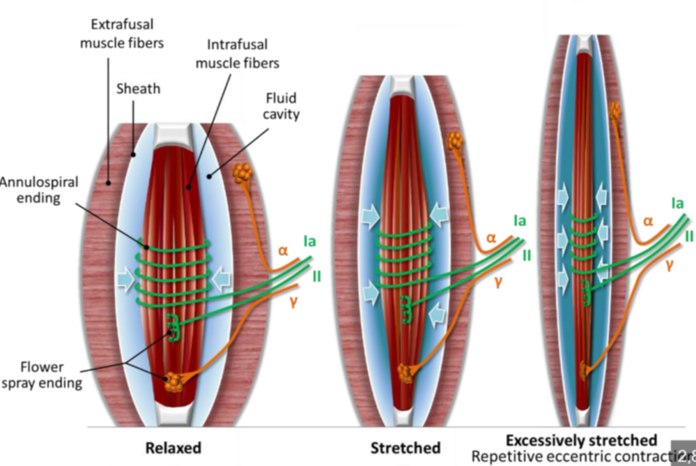
Muscle spindle structure - summary diagram
Before activation of alpha motor neurone (during passively stretch)
Muscle belly and muscle spindle under the same tension
spindle passively stretches with muscle
spindle detects changes in tension differences btw muscle and spindle
there is no change in tension→ spindle & therefore gamma neurone is not activated
Contraction → reduction in size of muscle belly
(concentric contraction)
Muscle contraction (due to alpha neurone stimulation) causes bulge in centre of belly as the extrafusal muscle shortens
intrafusal fibres in the centre go slack (bend) concomittantly/simultaneously with muscle contraction
this triggers nuclear bag and chain
signal sent to spinal cord
gamma motorneurones discharge
muscle spindle shortens as polar, striated ends of spindle contracts
new length of muscle set (slackness removed)
Golgi tendon organ functions
Monitor tendon tension caused by contraction of muscle
Overstretching → sensory signals reach spinal cord relay neuron
Relay neuron sends inhibitory signal to alpha neurons → prevents further muscle stretch
Aim - eliminate muscle tension by inhibiting contraction or shifting position
Found exclusively in tendons
Act on postural muscles
key difference btw golgi tendon organ and muscle spindle
MS - both sensory and motor neurones
GTO - sensory component only
Golgi tendon organ before sensory neuron activation
When no active tension/non-excessive tendon stretching GTO sends inhibitory signals
no contraction of muscle
communicates to spinal cord - no overstretching taking place
GTO when overstretching of muscle
GTO activates sensory neuron
Sensory neuron signals to spinal cord relay neuron
Discharge to alpha motor neuron for muscle contraction → prevents excessive overstretch of tendon and muscle
Patella reflex (mono-synaptic reflex control)
NO RELAY NEURONE (one synapse)
sensory x motor → excitatory
striking patellar ligament → stretches tendon and quadriceps femoris
stretch activates muscle spindles
sensory neurone activated
sensory neurone DIRECTLY activates alpha motorneurone
alpha motorneurone stimulates extrafusal fibres
quadriceps (femoris) contracts
UMN - can intervene → interrupts common pathway → quadriceps remain relaxed
Di-synaptic reflex control
2 synapses
sensory x relay → stimulatory
relay x motor → inhibitory
Overstretching on muscle stretches tendon
Golgi tendon organ senses tension
Sensory neuron activation
Sensory neurone stimulates relay neurone
Relay neurone inhibits ALPHA motor neurone
Tension on tendon reduced due to lack of extrafusal contraction
Multi-synaptic reflex control - reciprocal innervation
Agonist muscle contracts via monosynaptic reflex (stimulatory synapse)
sensor = muscle spindle → slackness
Antagonist muscle relaxes via disynaptic reflex (inhibitory relay x motor synapse)
sensor = golgi tendon organ → over-stretch
Patellar reflex example
quads contract via monosynaptic (stifle extensors)
hamstrings relax (inhibited) via disynpatic (stifle flexors)
Another example
biceps and triceps
any two opposing muscles
Posture control is multi-synaptic
Long spinal reflex
Different segments of spinal cord involved in execution of reflex (or postural control)
Example: reflex in response to pain or pinching is called the withdrawal reflex (also known as the flexor reflex).
Sensory recognition of pain (spinoreticular or spinothalamic tracts) → go into somatosensory cortex by travelling up spinal cord
Reflex aspect remains in spinal cord
Crossed extensor reflex → postural control
Lift one foot up → other foot goes down to compensate by weightbearing
Rising (ipsilateral) foot: (e.g. due to withdrawal reflex/pain)
Flexor contracts
Extensor relaxes
Lowering (contralateral) foot:
Extensor contracts
Flexor relaxes
Only IPSILATERAL sensory neuron activated
Relay neurons on both ipsilateral and contralateral sides (decussate) allow bilateral innervation
contralateral decussating interneurone = commissural neurone
LMN lesion potential clinical signs [5]
Muscle atrophy (if nerve cut through or injured)
Fasciculations (muscle twitching) if muscle overstimulated
Decreased reflexes
Decreased muscle tone
Flaccid paralysis (if no stimulation received)
LMN potential disorders
Polio-myelitis → selective alpha motor neuron destruction
Amyo-trophic lateral sclerosis → degeneration of LMNs at ALL levels of spinal cord → muscle atrophy, paralysis, death (diaphragm/larynx → breathing and swallowing)
Poly-myositis (cause not well known)
muscle disease that causes progressive, symmetric muscle weakness, mainly affecting the proximal muscles (like the hips, thighs, shoulders, and neck).
Myasthenia gravis → autoimmune targeting of ACh receptors (type 2 hypersensitivity → antibody blocking)