Principles of science- protein
1/9
Earn XP
Description and Tags
Name | Mastery | Learn | Test | Matching | Spaced | Call with Kai |
|---|
No analytics yet
Send a link to your students to track their progress
10 Terms
what is meant by a protein structure hierarchy
-primary, secondary, tertiary, quaternary(eg 2 beta and 2 alpha globin polypeptides with a heme group)
what type of amino acids are mammalian proteins
how many amino acids
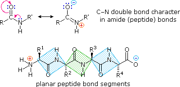
what is the peptide bond classified by
how many amino acids makes a protein
shape of peptide bond
mammalian proteins are alpha amino acids
20 amino acids
carboxyl group reacts with amino group to form a peptide bond and lose a water molecule
catalysed by peptidyl transferase (28s ribozyme-ribonucleic acid enzymes)
within a polypeptide chain, individual amino acids = residues
when >50 amino acids are in a polypeptide is a protein
peptide bond is planar (partial double bond between carbonyl O and N)
bonding determining protein structure
covalent primary
hydrogen secondary
hydrophobic+VDW tertiary
electrostatic within tertiary helps maintain
VDW/ electrostatic quaternary
alpha helix
what runs parallel to the helix axis
what winds around the axis
what does htis lead to
how many amino acid residues per complete turn
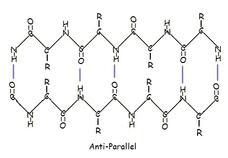
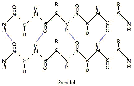
beta sheets
what orientation do H bonds run to the chain direction
alpha helix- H bonds run parallel to helix axis. alpha carbon backbone winds around an axis so that each carbonyl O atom is H bonded to each amino N of the amino acid located 4 residues closer to C terminus
The standard alpha helix has approximately 3.6 amino acid residues per complete turn
beta sheets- H bonds run perpendicular to chain direction
parallel and antiparallel
types of r groups
nonpolar, polar, electrically charged
cysteine
can form disulphide bonds as it has an SH group
examples of tertiary proteins
-catalase
-triose phosphate isomerase
-actin
tertiary domain
the smallest stable unit of a tertiary structure. a domain is defined as that region of a polypeptide chain that can fold into an autonomous stable tertiary structure
domain shuffling- when domains have been switched around between proteins through evolution
quaternary
haemoglobin- needs to deliver oxygen tissues. needs high enough affinity to pick up oxygen but low enough to release it.
t state- oxygen unbound
r state- oxygen bound. so that the 3 other units have higher affinity
small changes in oxygen concentration can dramatically affect binding to haemoglobin giving a sigmoidal curve of oxygen binding
collagen
what type of protein
what is the general structure
extracellular fibrous protein but not an alpha helix
3 helical chains that wind around a central axis
general structure is Gly-X-Y where X can be any amino acid especially proline lysine or hydroxyproline
Glycine is small so glycines from each chain fit at the centre of each helix
H bonds between the chains