CNS hlsc exam 2
1/23
There's no tags or description
Looks like no tags are added yet.
Name | Mastery | Learn | Test | Matching | Spaced |
|---|
No study sessions yet.
24 Terms
distinguish between the afferent and efferent divisions of the peripheral nervous system
afferent nervous system carries information to the CNS about changes in external and internal environment. ex) hunger, ambient temperature
efferent nervous system communicates instructions from CNS to effector organs (muscles or glands) to carry out the provided instructions
what are the two subdivisions of the efferent nervous system?
somatic nervous system consisting of nerve fibres and motor neurons that supply skeletal muscles.
autonomic nervous system consisting of nerve fibres that innervate smooth muscle, cardiac muscle, and glands.
what are the two subdivisions of the autonomic nervous system?
sympathetic nervous system initiates “fight or flight” response due to stress
parasympathetic nervous system initiates relaxation response “peace”
what are the three functional classes of neurons?
afferent neurons : a typically afferent neuron has a sensory receptor at its peripheral end that generates action potentials as a response to stimuli. The cell body of these neurons are located adjacent to the spinal cord.
efferent neurons : primarily in the PNS, with their cell bodies originating in the CNS, where many centrally located presynaptic inputs converge on them to influence output to effector organs. efferent axons (fibres) meet up with muscles or glands that they innervate.
interneurons : lie entirely within the CNS, and make up most of the neurons of the body. They lie between afferent and efferent neurons and thus integrate peripheral responses to peripheral information.

major functions are sensory perception, voluntary control of movement, language, personality traits, and sophisticated mental events (thinking, memory, decision making, creativity, self consciousness)
cerebral cortex

major functions are inhibition. of muscle tone, coordination of slow, sustained movements, and suppression of useless patterns of movement
basal nuclei

is the relay station for all synaptic input, crude awareness of sensation, some degree of consciousness, and has a role in motor control
thalamus
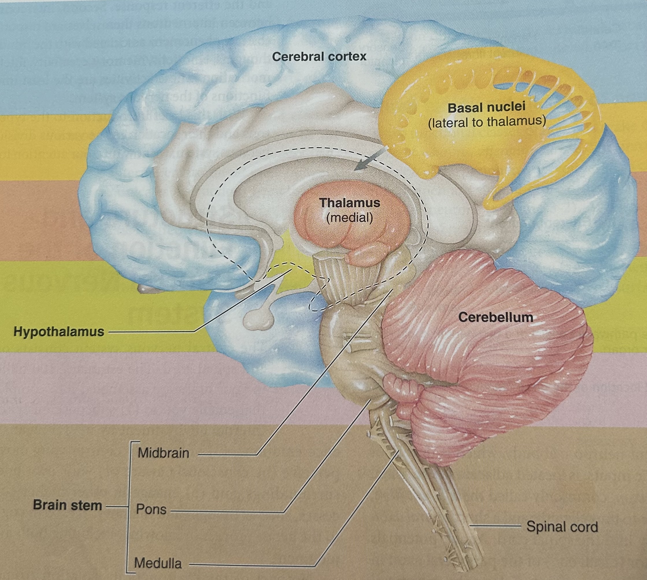
regulation of many homeostatic functions (temperature, thirst, urine, and food), is an important link between nervous and endocrine systems, involved with emotions and basic behavioural patterns, and plays a role in sleep-wake cycle.
hypothalamus

maintenance of balance, enhancement of muscle tone, and coordination and planning of skilled voluntary muscle activity (proprioception)
cerebellum

origin of majority of peripheral cranial nerves, cardiovascular and respiratory and digestive control centres, regulates muscle reflexes involved with equilibrium and posture, reception and integration of all synaptic output from spinal cord, plays a role in sleep-wake cycle (vegetative functions)
brain stem
what are glial cells (neuroglia)?
-glial cells make up ~90% of the cells within the CNS
-do not initiate or conduct nerve impulses, BUT do communicate with neurons and themselves via chemical signals.
-are the connective tissue of the CNS (support neurons physically and metabolically)
- maintain the composition of extracellular environment surrounding neurons (specialized) narrow limits for optimal functioning for them to homeostatically regulate

which type of glia: CNS: holds neurons at right distance apart, guide neurons to their proper spot during fetal brain development, induce small blood vessels of the brain to establish the blood brain barrier, take up and degrade GABA, excitatory and inhibitory neurotransmitters, take up excess K+ from brain ECF to maintain ion concentration, and form neural scar tissue?
astrocytes

which glia: CNS form myelin sheaths on neurons
oligodendrocytes
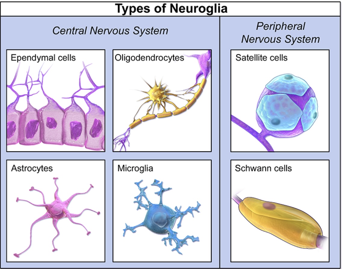
which glia: CNS acts as phagocytic scavengers for brain defence, and releases nerve growth factor to help other nerve and glial cells thrive and survive, and when activated release destructive chemicals for assault against their target?
microglia

which glia: line internal cavities of brain and spinal cord and contribute to formation of cerebrospinal spinal fluid, have cilia to aid in CSF flow throughout the four brain ventricles, and are neural stem cells either potential to form new neurons and glial cells?
ependymal cells
what are four structures that protect the central nervous system from injury?
cranium and vertebral column act as hard bony structures closing the CNS
the meninges (three membranes) that lie between bony covering and nervous tissue (dura, arachnoid, and pia mater)
CSF is a special cushioning fluid that allows the brain to float
blood brain barrier s highly selective and limits access of materials into brain tissue by
explain the three layers of the meninges in terms of anatomy and function
dura mater is MOST OUTER. it is tough and inelastic, and made up of two layers. usually layers are close together but in some areas the layers are separated to make up dural sinuses which are small blood filled cavities, or large blood filled cavities called venous sinuses. venous blood to be returned to the heart from the brain empties into these sinuses, then drains into the internal jugular vein. they also participate in the circulation of CSF. examples: superior saggital, transverse, and cavernous sinuses.
arachnoid mater is MIDDLE layer. has a cobweb like appearance and is highly vascularized. the subarachnoid space is filled with CSF. arachnoid tissue has protrusions called arachnoid villi that penetrate through gaps in dura and project into dural sinuses which is how CSF reabsorbs across villi and into the blood within the dura sinuses to circulate.
pia mater is MOST INNER. it is very fragile and highly vascular and closely adheres to surface of the brain and spinal cord. In some areas it dips deeply into brain to bring a rich blood supply into close contact with ependymal cells that line the ventricles. This aids in the formation of CSF.
where is cerebral spinal fluid formed and why is it important
CSF is formed primarily by choroid plexuses found in some ventricle cavities of the brain (in dura mater). choroid plexus consists of richly vascularized cauliflower like masses of pia mater tissue that dips into pockets formed by ependymal cells. selective membrane transport of the choroid plexuses forms CSF. CSF is not like plasma… CSF is lower in potassium, protein, and glucose, and higher in chloride and magnesium. CSF aids in the exchange of materials between neural cells and surrounding interstitial fluid. brain interstitial fluid comes into direct contact with neurons and glial cells, CSF does NOT. thus, CSF composition is closely regulated as a change in CSF causes a change in brain interstitial fluid.
how does CSF flow through ventricles
CSF flows from the lateral ventricles through the third and fourth ventricles, and into the subarachnoid space, where it is absorbed into the blood. it continuously flows and is driven by pulsation movement from ciliary beating and circulatory and postural factors (CSF pressure is approximately 10mmHg) if excess CSF accumulates from dysfunction of this cycle, hydrocephalus occurs, which increases CSF pressure and may lead to brain damage and mental retardation if untreated. treatment would include surgically shunting the excess CSF to veins elsewhere in the body.

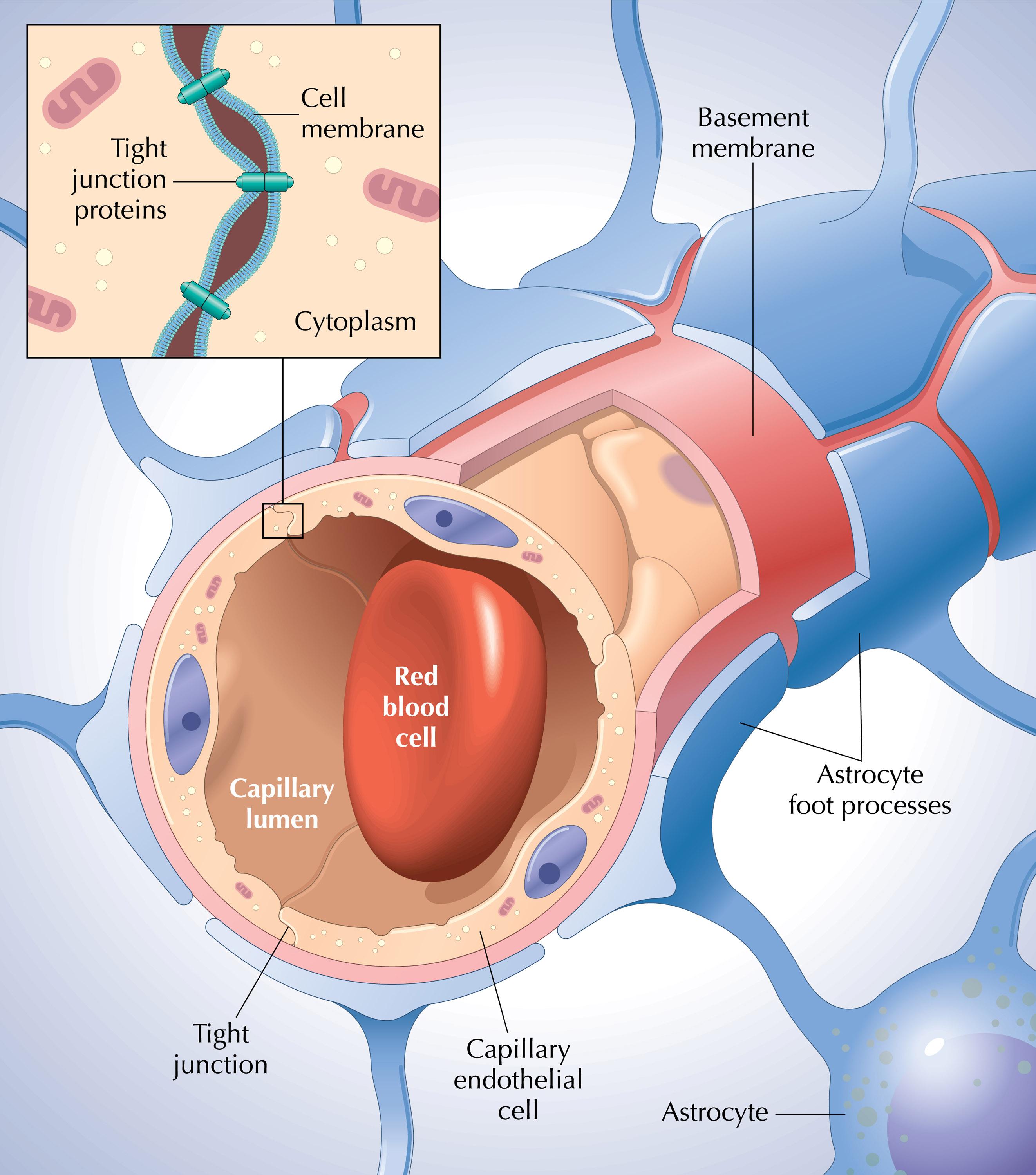
what is function of blood brain barrier and what is the structure
the BBB is highly selective and carefully shields the brain from harmful changes in the blood. it consists of endothelial cells (single layer cells that regulate blood flow and pressure, prevent blood clot formation, and control permeability). in brain capillaries, cells are joined by tight junctions, therefore exchange may only occur through the capillary cells themselves. things that can easily penetrate: lipid soluble: oxygen, carbon dioxide, alcohol, steroid hormones . small water molecules can diffuse rapidly.
negative of BBB: can limit the use of drugs for treatment of brain and spinal al cord disorders due due drug inability to penetrate barrier.
astrocytes surround brain capillaries and help to 1. signal the cells forming the brain capillaries to get tight for tight junctions and 2. astrocytes participate in the cross cellular transport of some substances such as potassium.
what are circumventricular organs
circumventricular organs ar certain areas of the brain that do not have the blood brain barrier. this allows these organs to sample the blood and adjust their output accordingly to maintain homeostasis. one output may be hormones that must enter the capillaries to be transported to their sites of action. these capillaries are not sealed by tight junctions. these organs include: the area postrema, subfornical orgn, and pineal gland (located around third and fourth ventricles)
why are oxygen and glucose so important in the brain
the brain needs a constant blood supply because brain tissue is unable to to resort to anaerobic metabolism to produce ATP. therefore, an oxygen binding protein called neuroglobin in the brain to assist the carrying of oxygen throughout brain tissues. the brain does not store glucose, thus it relies on the constant transport of glucose to do its functions.
what are white and grey matter called in the CNS and what are the functions of each?
white matter of the CNS surrounds the butterfly of grey matter of the spinal cord and is called tracts and consist of bundles of nerve fibres (axons of long interneurons). thre are ascending tracts that send info from afferent input to the brain, and there are descending tracts that relay messages from the brain to efferent neurons. ex) ventral spinocerebellar tract is an ascending pathway that runs up the ventral margin of spinal cord and eventually reaches the cerebellum. contrast ex) the ventral corticospinal tract is a descending pathway that originates in motor region of cerebral cortex travels down ventral spinal cord and terminates on cell bodies of efferent motor neurons supplying skeletal muscles.
grey matter of the CNS looks like a grey butterfly on the spinal cord and is called nuclei (made up of cell bodies and dendrites). central canal is filled with CSF is in the centre of grey matter. the grey matter is divided into a dorsal horn, a ventral horn, and a lateral horn.
dorsal horn: cell bodies of interneurons on which afferent neurons terminate
ventral horn: cell bodies of efferent motor motor neurons supplying skeletal muscles
lateral horn: cell bodies of autonomic nerve fibres supplying cardiac and smooth muscle and exocrine glands
dorsal vs ventral root of spinal nerves
dorsal root: afferent fibres carrying incoming signals from peripheral receptors enter spinal cord via dorsal root. cell bodies of afferent neurons are clustered in a dorsal root ganglion (PNS)
ventral root: cell bodies for efferent neurons originate in grey matter and send axons through ventral root and out to muscles and glands
both the dorsal and ventral roots join to form a spinal nerve that then emerges from the spinal column (spinal nerve contains both efferent and afferent fibres that traverse between spinal cord)