control of heart rate
1/26
There's no tags or description
Looks like no tags are added yet.
Name | Mastery | Learn | Test | Matching | Spaced | Call with Kai |
|---|
No analytics yet
Send a link to your students to track their progress
27 Terms
Myogenic
When a muscle (cardiac muscle) can contract and relax without receiving signals from nerves
Sinoatrial node
Located in right atrium and is known as the pacemaker
releases wave of depolarisation across the atria, causing muscles to contract
Atrioventricular node
Located near the border of the right/left ventricles within the atria
releases another wave of depolarisation after a short delay when it detects the first wave from the SAN
Bundle of His
A group of specialised muscle fibres in the heart
located in the septum between the ventricles
conducts the wave of depolarisation from the AV node down the septum
→ passes the impulse to the Purkyne fibres, causing ventricles to contract from the base upwards
Purkyne fibres
Specialised conducting fibres in the walls of the ventricles
receive the wave of depolarisation from the Bundle of His
spread the impulse through the ventricular muscle, causing it to contract from the base upwards
→ ensures efficient pumping of blood out of the heart
Role of non-conductive tissue
Found between the atria and ventricles
Prevents the wave of depolarisation from passing directly from atria to ventricles
Forces the impulse to travel through the AV node and Bundle of His
→ Creates a short delay, allowing the atria to fully contract and fill the ventricles before they contract
Importance of short delay between SAN and AVN waves of depolarisation
Ensures enough time for atria to pump all blood into ventricles before the ventricles contract
Role of the medulla oblongata
Controls heart rate via the autonomic nervous system
uses sympathetic and parasympathetic nervous system to control SAN rhythm
Chemoreceptors
Located in carotid artery and aorta
responds to pH / CO2 conc. changes
Baroreceptors
Located in carotid artery and aorta
responds to pressure changes
Response to high blood pressure
Baroreceptors in the aorta and carotid arteries detect high pressure
Send more impulses to the medulla oblongata
Medulla increases impulses along parasympathetic neurones to the SAN
Acetylcholine is released → slows SAN activity
→ Heart rate decreases, reducing blood pressure
if blood pressure is too high this can cause damage to the walls of the arteries
Response to low blood pressure
Baroreceptors in the aorta and carotid arteries detect low pressure
Send more impulses to the medulla oblongata
Medulla increases impulses along sympathetic neurones to the SAN
Noradrenaline is released → increases SAN activity
→ Heart rate increases, raising blood pressure
if blood pressure is too low there may be insufficient supply of oxygenated blood to respiring cells and removal of waste
Response to low blood pH
Chemoreceptors in the aorta and carotid arteries detect high CO2 levels (low pH)
Send more impulses to the medulla oblongata
Medulla increases impulses along sympathetic neurones to the SAN
Noradrenaline is released → increases SAN activity
→ Heart rate increases, removing more CO2 via the lungs and returning pH to normal
During high respiratory rate or exercise, CO₂/lactic acid increases → pH decreases
excess acid must be removed from blood rapidly to prevent enzymes denaturing
so increase in heart rate so carbon dioxide can diffuse into the alveoli more rapidly
Response to high blood pH
Chemoreceptors in the aorta and carotid arteries detect low CO2 levels (high pH)
Send more impulses to the medulla oblongata
Medulla increases impulses along parasympathetic neurones to the SAN
Acetylcholine is released → slows SAN activity
→ Heart rate decreases, allowing CO2 to accumulate and pH to return to normal
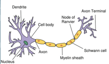
Structure of myelinated motor neurone
Resting potential
The difference between the electrical charge inside and outside the axon when a neurone is not conducting an impulse
more positive ions (Na+ /K+ ) outside axon compared to inside
inside the axon -70mV
How is resting potential established
Sodium potassium pump actively transports 3 Na+ out of the axon, 2 K+ into the axon
membrane more permeable to K+ (more channels and always open)
K+ diffuses out down conc. gradient - facilitated diffusion
membrane less permeable to Na+ (closed Na channels)
higher conc. Na+ outside
Action potential
Stimulus causes membrane to depolarise past the threshold (–55 mV)
Voltage-gated Na+ channels open → Na+ diffuses in
→ Inside becomes more positive (depolarisation)
At +40 mV, Na+ channels close and voltage- gated K+ channels open
K+ diffuses out, making the inside negative again (repolarisation)
Hyperpolarisation may occur as K+ channels are slow to close
Action potential: stimulus
Stimulus causes the membrane to
depolarise past the threshold (–55 mV)
Voltage-gated Na+ channels open → Na+ diffuses in
→ Inside becomes more positive (depolarisation)
Action potential: depolarisation
When a threshold potential is reached, an action potential is generated
more voltage-gated Na+ channels open
Na+ move by facilitated diffusion down conc. gradient into the axon
potential inside becomes more positive
Action potential: repolarisation
Na+ channels close, membrane becomes less permeable it Na+
K+ voltage-gated channels open, membrane more permeable to K+
K+ diffuses out neuron down conc. gradient
voltage rapidly decreases
Action potential: hyperpolarisation
K+ channels slow to close -> overshoot in voltage
too many K+ diffuse out of neurone
potential difference decreases to -80mV
sodium-potassium pump returns neurone to its resting potential
Action potential graph
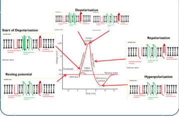
All or nothing principle
If depolarisation does not exceed -55 mV threshold, action potential is not produced
any stimulus that does trigger depolarisation to -55mVthreshold will always peak at the same maximum voltage
Importance of all or nothing principle
Ensures that only stimuli above a threshold trigger an action potential
Prevents response to small, insignificant stimuli — avoids overload
All action potentials are the same size, so information is transmitted clearly
→ Stimulus intensity is shown by frequency, not size, of impulses
Refractory period
After an action potential has been generated, the membrane enters a period where it cannot be stimulated
because Na channels are recovering and cannot be opened
Importance of refractory period
Ensures discrete impulses produced - action potentials separate and cannot be generated immediately
unidirectional - cannot generate action potential in refractory region
limits number of impulse transmissions - prevent overwhelming