Biopsychology - The Nervous System, neurons and synaptic transmission
1/40
There's no tags or description
Looks like no tags are added yet.
Name | Mastery | Learn | Test | Matching | Spaced | Call with Kai |
|---|
No analytics yet
Send a link to your students to track their progress
41 Terms
The Nervous System
A specialised network of neurones that send messages around the body by transmitting electrical impulses (action potentials) in the body in order to fulfil its two main functions: Collecting, processing + responding to information in the environment AND co-ordinating he working of different organs and cells within the body.
What are the two sub-systems of the Nervous system?
Central Nervous System (CNS) and Peripheral Nervous System (PNS)
Central Nervous System
Consists of the Brain and the Spinal Cord
Brain
Divided into 4 main areas: Cerebrum, Cerebellum, Diencephalon and Brain Stem
What does the Brain look like?
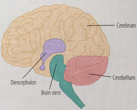
Cerebrum (brain)
Largest area of the brain that is split down the middle into two cerebral hemispheres connected by the corpus callosum. Each hemisphere has 4 lobes.
Frontal lobe (cerebrum)
Learning, thought, language
Parietal lobe - Cerebrum
Touch and taste
Temporal lobe (cerebrum)
Hearing and Memory
Occipital lobe (Cerebrum)
Vision
Cerebellum (brain)
Located beneath the cerebrum, controls motor skills and balance and coordinates muscles for precise movements.
Diencephalon (brain)
Bellow the Cerebrum but above the brain stem, consists of two key parts: Thalamus and Hypothalamus
The Thalamus
Located in Diencephalon, a relay station for information from the sensory receptors. Information is relayed from the sensory receptors to the appropriate area of the brain. (eg. sensory receptors in eyes —> occipital lobe
The Hypothalamus
Located in the Diencephalon, regulates many key bodily functions. (e.g. body temp, hunger, thirst) and is the link between the nervous system and endocrine system. The pituary gland (located here) controls hormone release.
Brain Stem (brain)
Responsible for regulation of involuntary actions (eg. breathing, digestion, heart beat) and connects to the spinal cord.
Spinal Cord
Main function is to transmit information between the brain and the peripheral nervous system. Is a part of the CNS.
The Peripheral Nervous System (PNS)
Transmits information between the brain and the CNS. Consists of two sub-systems: Autonomic Nervous System and Somatic Nervous System
The Somatic Nervous System
Part of the Peripheral Nervous System. Transmits information from the Sensory Receptors to the CNS, then from the CNS to the skeletal muscles. This allows the CNS to coordinate bodily movement.
The Autonomic Nervous System
Part of the Peripheral Nervous System. Transmits information to and from internal bodily organs. Consists of two sub-systems: Sympathetic Nervous System and Parasympathetic Nervous System
The Sympathetic Nervous System
Part of the Autonomic Nervous system. Works alongside the endocrine system in order to bring about the fight-or-flight response when an individual is faced with a threat.
The Parasympathetic Nervous System
Part of the Autonomic Nervous System. Works alongside the endocrine system to bring an individual back to their resting state (after a fight or flight response) once a threat has passed.
Neurons
Specialised Cells that transmit action potentials/electrical impulses to enable communication within the Nervous System.
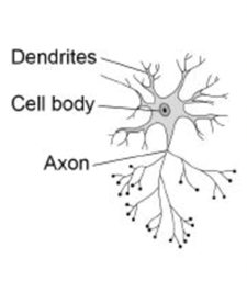
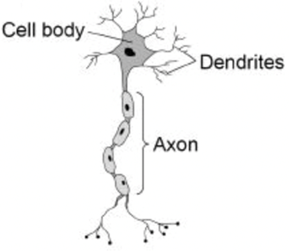
Structure of a neuron
Every neuron has 3 main parts: Dendrites, a cell body and an axon
Dendrites (Neurone Structure)
Receives impulses from the other neurones or sensory receptor cells and carries impulses towards the cell body.
Cell body (Neuron structure)
Contains a nucleus which consists of genetic material (DNA)
Axon (cell structure)
Carries impulses away from the cell body. Often covered in a fatty layer of myelin sheath (sensory+motor) which speeds up the transmission of an impulse. Axon terminals are at the end of an axon. They contain Neurotransmitters and form synaptic connections with other neurons or effector cells.
Types of neuron
The three types of neuron I must know are: Sensory neurons, Relay neurons and Motor neurons
Sensory Neurons - function
These neurons carry impulses/information from sensory receptors to the CNS - (They only allow impulses to travel one way). These impulses carry the information of environmental changes that have been detected by sensory receptors that produce the impulses.
What do sensory Neurons look like?
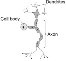
Sensory Neurons - structure
They are located in eyes, nose, ears, tongue and skin. They have short dendrites and a long axon. The axon is covered in a fatty layer of myelin sheath.
Relay Neurons - function + structure
Carry messages from one part of the CNS to another. Located in the CNS. They have short dendrites + a short axon.
What do relay Neurons look like?
Motor Neurons - function
Connects the CNS to the effectors so the effector (eg. muscle or gland) can produce a response to a detected change in the environment.
Motor Neurons - structure
They have short Dendrites and a long axon. The axon is covered in a fatty layer of Myelin Sheath.
What does a motor neuron look like?
Synapse
The small gap between the axon terminal of presynaptic neuron and the dendrite of the postsynaptic neuron. Electrical impulses are transferred across these by neurotransmitters (chemicals) diffusing across the gap
What does a synapse/synaptic transmission look like?
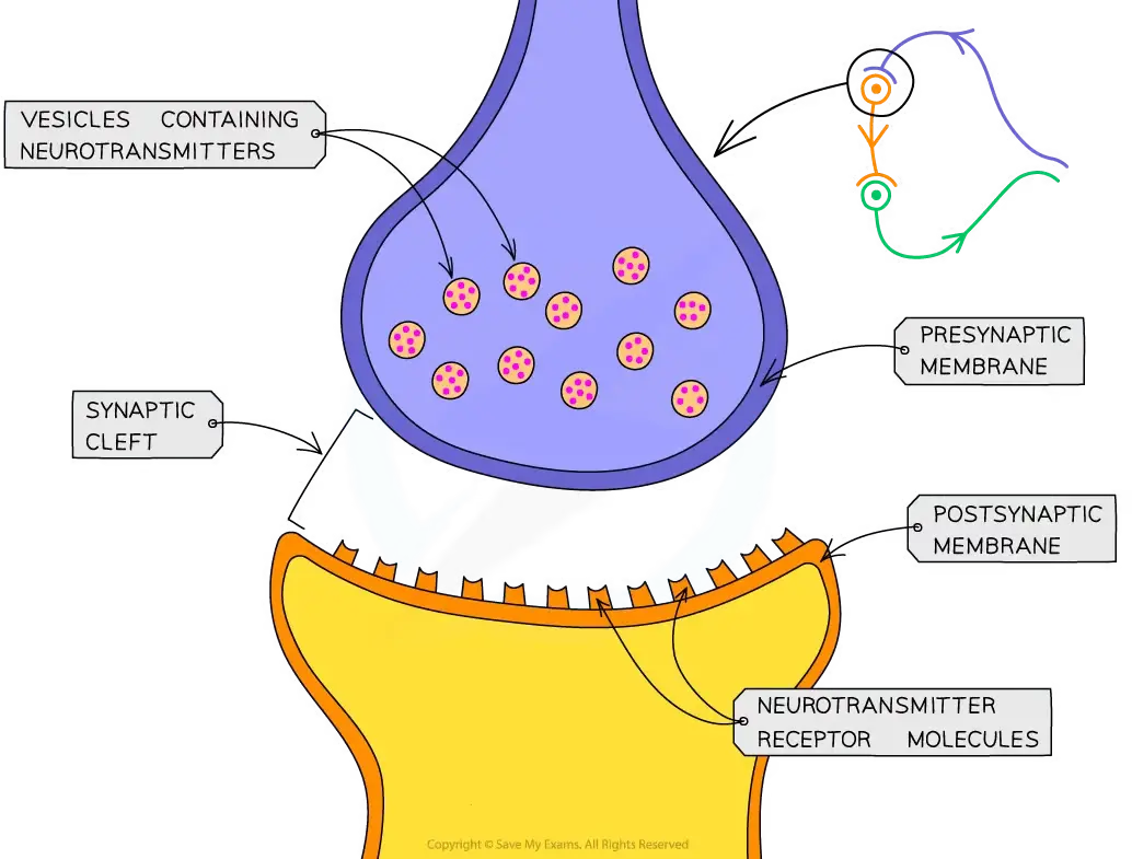
Basic explanation of synaptic transmission
An electrical impulse reaches the end of the axon terminal at the presynaptic neuron
Causes synaptic vesicles to release neurotransmitters into the synaptic cleft
Neurotransmitters diffuse across the synapse (one direction) and bind to receptor sites on dendrite of postsynaptic neuron
Chemical message is converted back into an electrical impulse
Excitatory neurotransmitters
Neurotransmitters that bind to receptor sites, open channels into the post-synaptic neuron that positively charges sodium ions (Na+) flood into. This creates an excitatory post-synaptic potential (EPSP) that means the post-synaptic neuron is more likely to fire. Firing neurons causes electrical impulses to be carried through the body + bodily processes to occur (e.g. adrenalin released).
Inhibitory Neurotransmitters
Bind to receptor sites, open channels into post-synaptic neuron that negatively charged chloride ions (Cl-) flood into. This creates an inhibitory post-synaptic potential (IPSP) that means the neuron in less likely to fire. Stopping a neuron from firing can stop unnecessary signals in the body (eg. pain) and prevent over-excitation + stress.
Summation
Both ESPS and ISPS are received at the same time by a neuron. The likelihood of a neuron firing is determined by adding up the excitatory + inhibitory synaptic input. The net result of this calculation (known as summation) determines whether the cell fires). There is a threshold for firing.