Central Nervous System III
1/80
Earn XP
Description and Tags
Biology 315: Gross and Microanatomy Laboratory
Name | Mastery | Learn | Test | Matching | Spaced | Call with Kai | Chat |
|---|
No analytics yet
Send a link to your students to track their progress
81 Terms
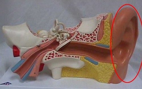
Auricle
collective structure
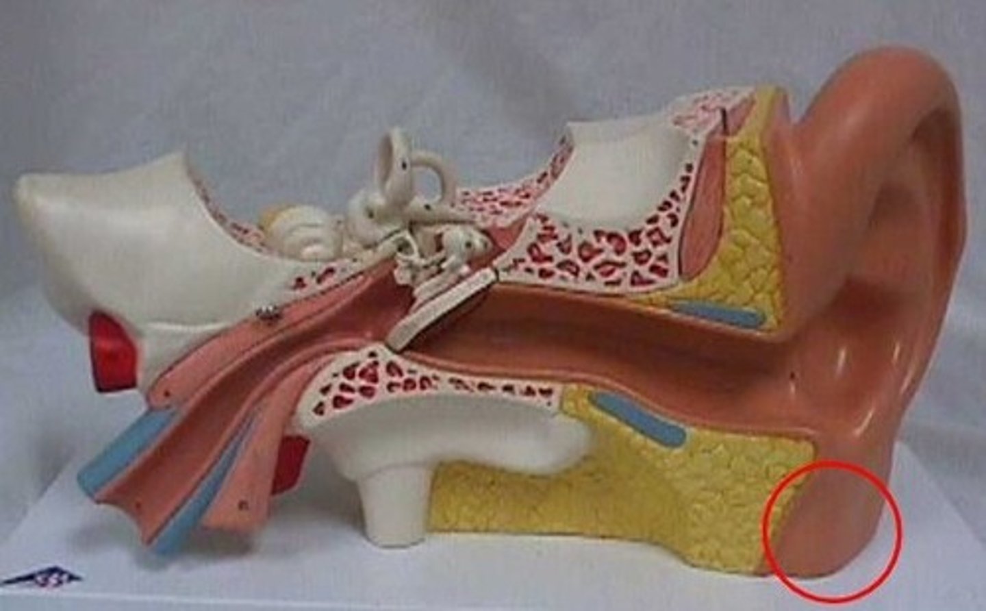
Lobule of the auricle
structure
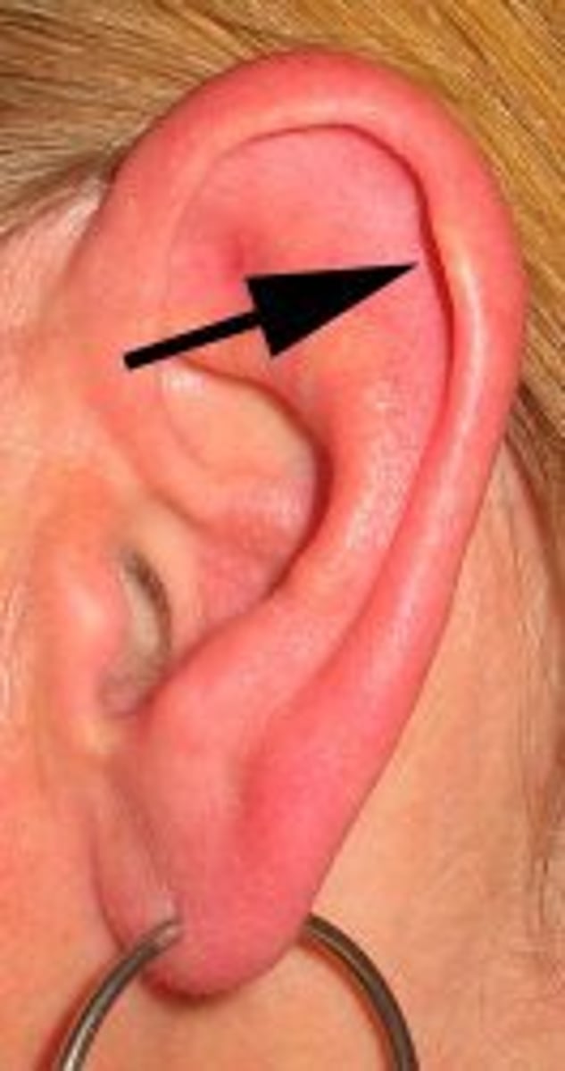
Helix of the auricle
(Structure)
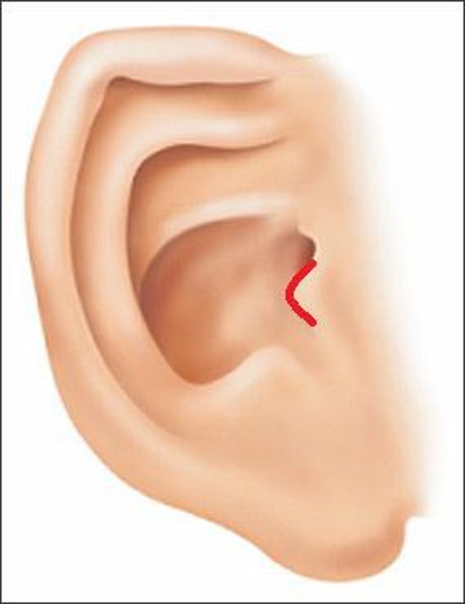
Tragus of the auricle
(Structure)
-On whole outer ear model
-Middle flippy thing
Concha of the auricle
(Depression)
-On whole outer ear model
-Most inner depression
-My piercing on the left ear
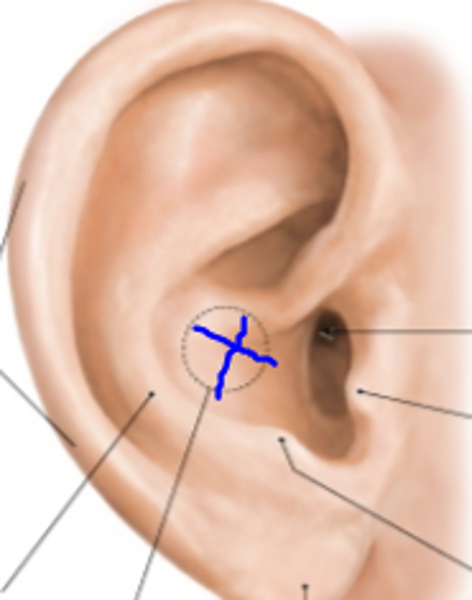
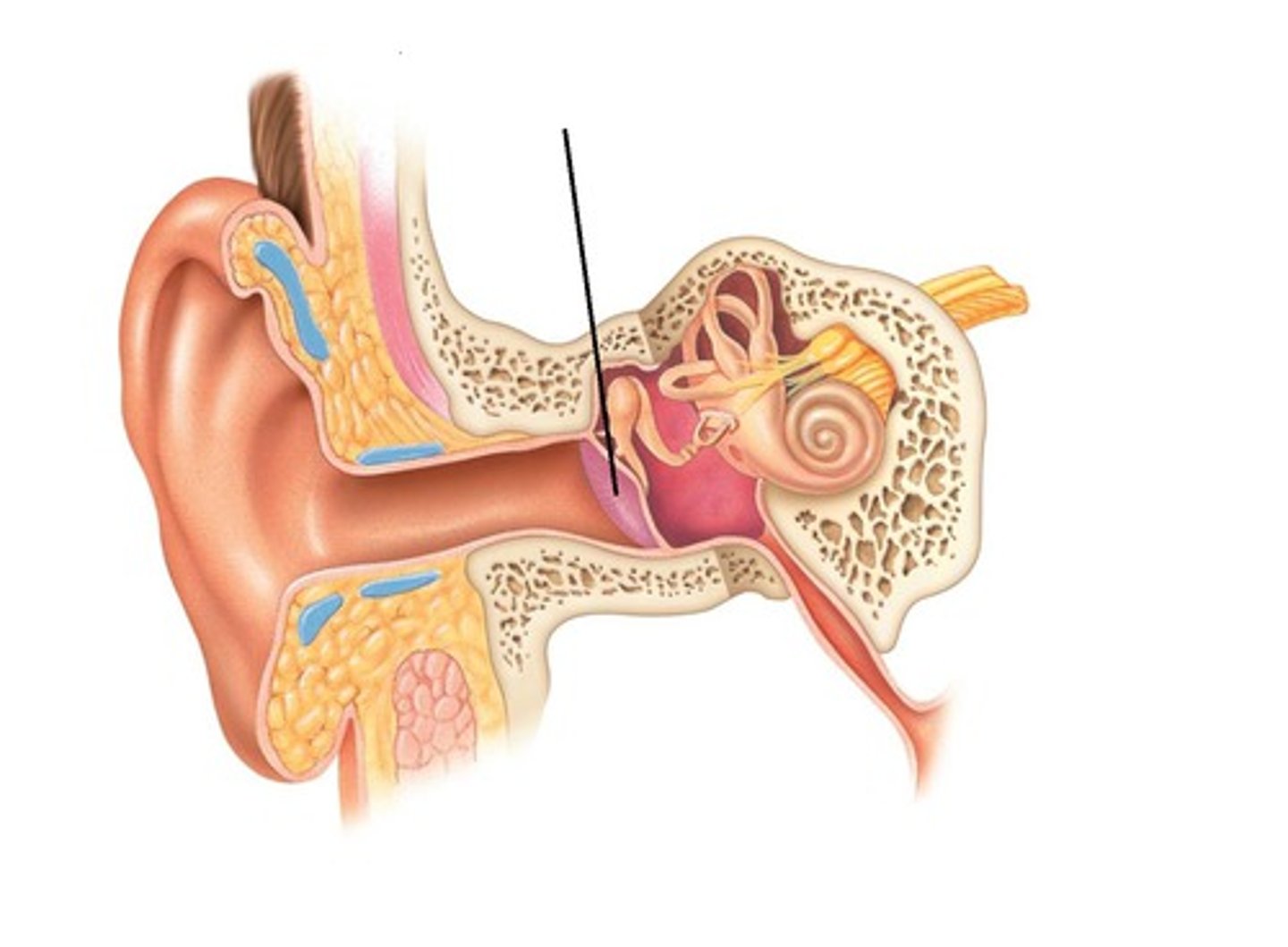
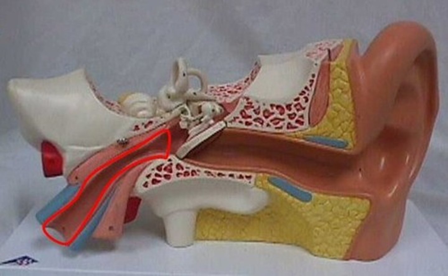
External acoustic meatus
(Passage)
-On whole outer ear model
-Probe will be going into the ear hole
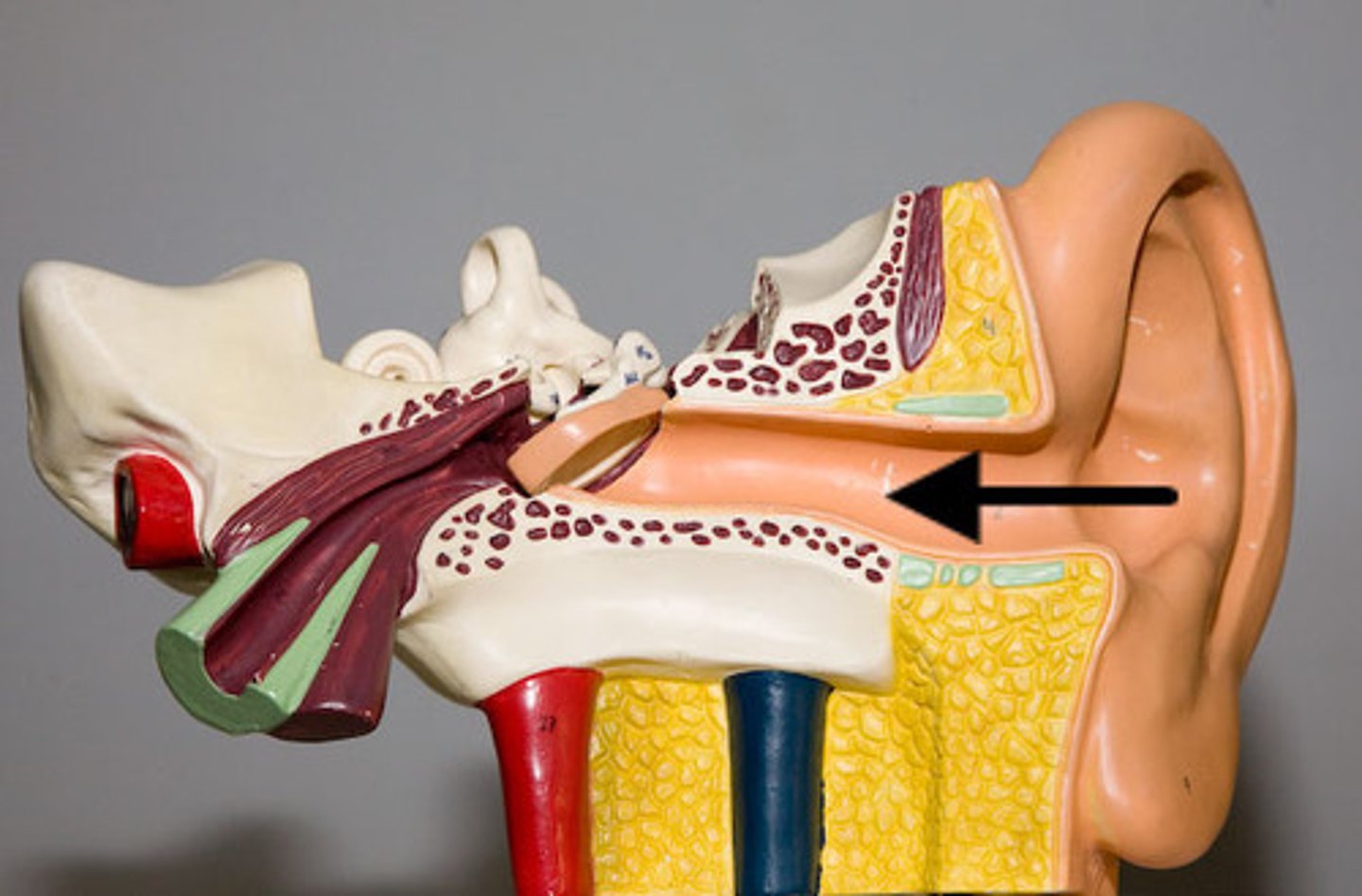
Tympanic membrane
(Structure)
-On white base model
- First thing the probe will bump into when going into the ear
-Ear drum will be taken out
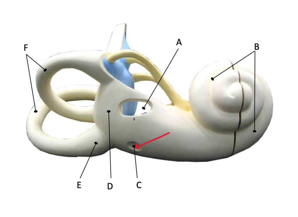
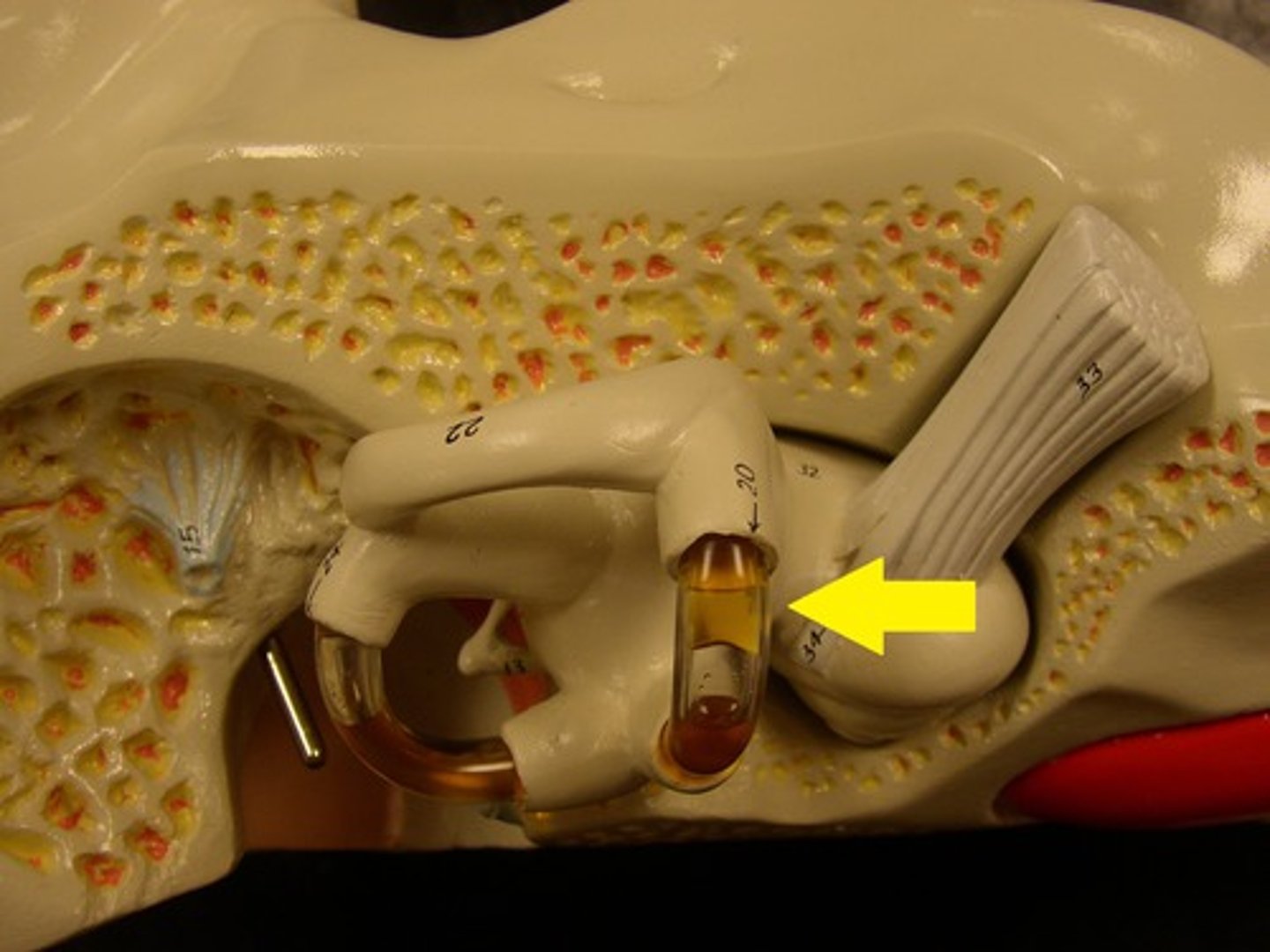
Oval window
(Opening)
-On wood base model
- Oval hole
Round window
(Opening)
-On wood base model
-Lower small circle
Mastoid cells
(Spaces)
-On wood base model
-Golf ball pink texture
Anterior wall
(Wall)
-Put piece together on white base model
Pharyngotympanic tube
(Passageway)
-On white base model
-Large tube going down
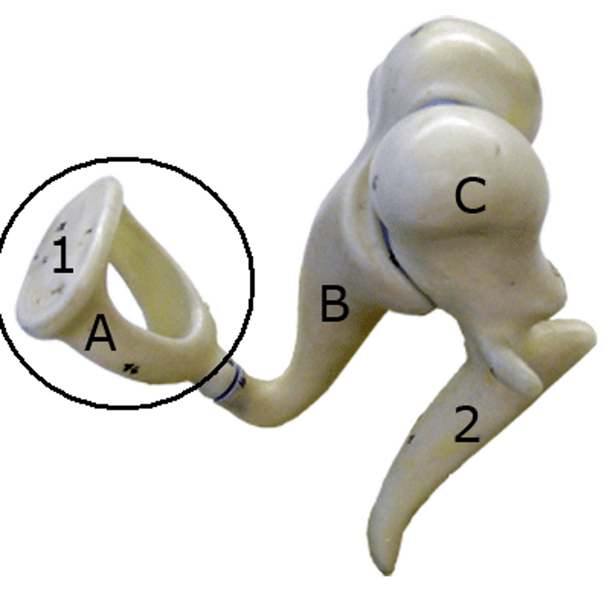
Malleus B.
(Structure)
-Attached to eardrum model
-Looks like a golf club, music note

Head of the malleus B.
(Feature)
-Attached to eardrum model
-Head of the music note

Handle of the malleus B.
(Feature)
-Attached to eardrum model
-Handle of music note
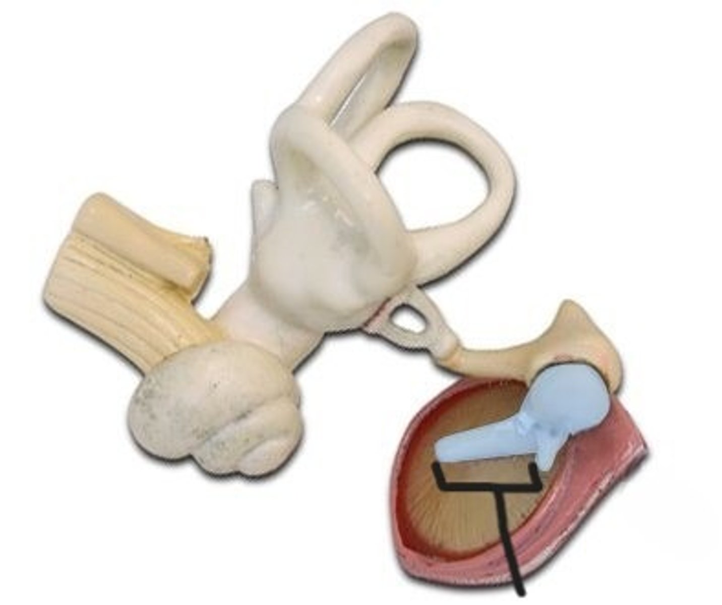
Incus B.
(Structure)
-Floating on malleus
-larger music note
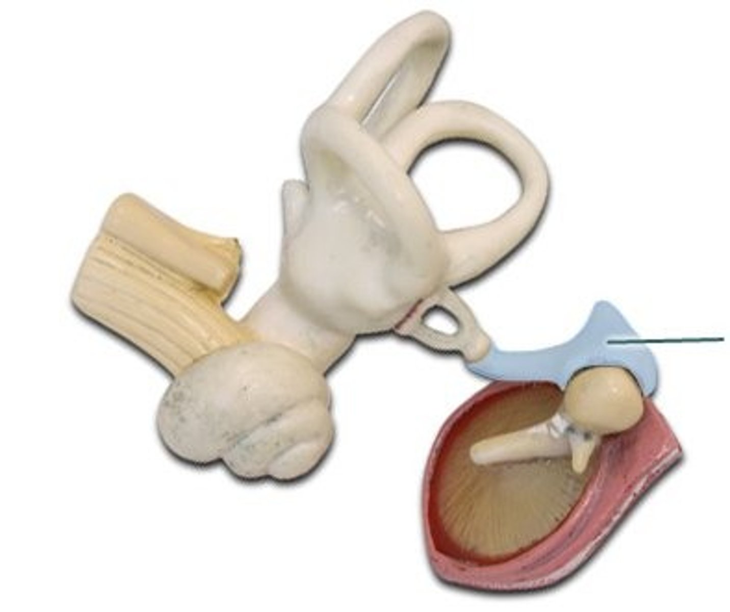
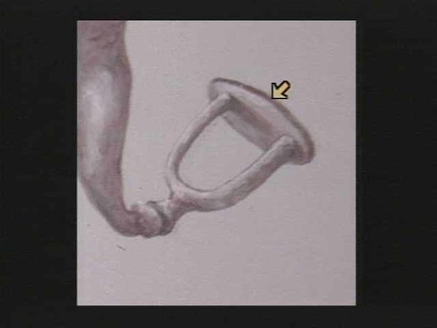
Stapes B.
(Structure)
-Attached to eardrum model
-Stirrup
-Top of all white loopy structure
Head of the stapes B.
(Feature)
-Attached to eardrum model
-Little bump on top of stirrup
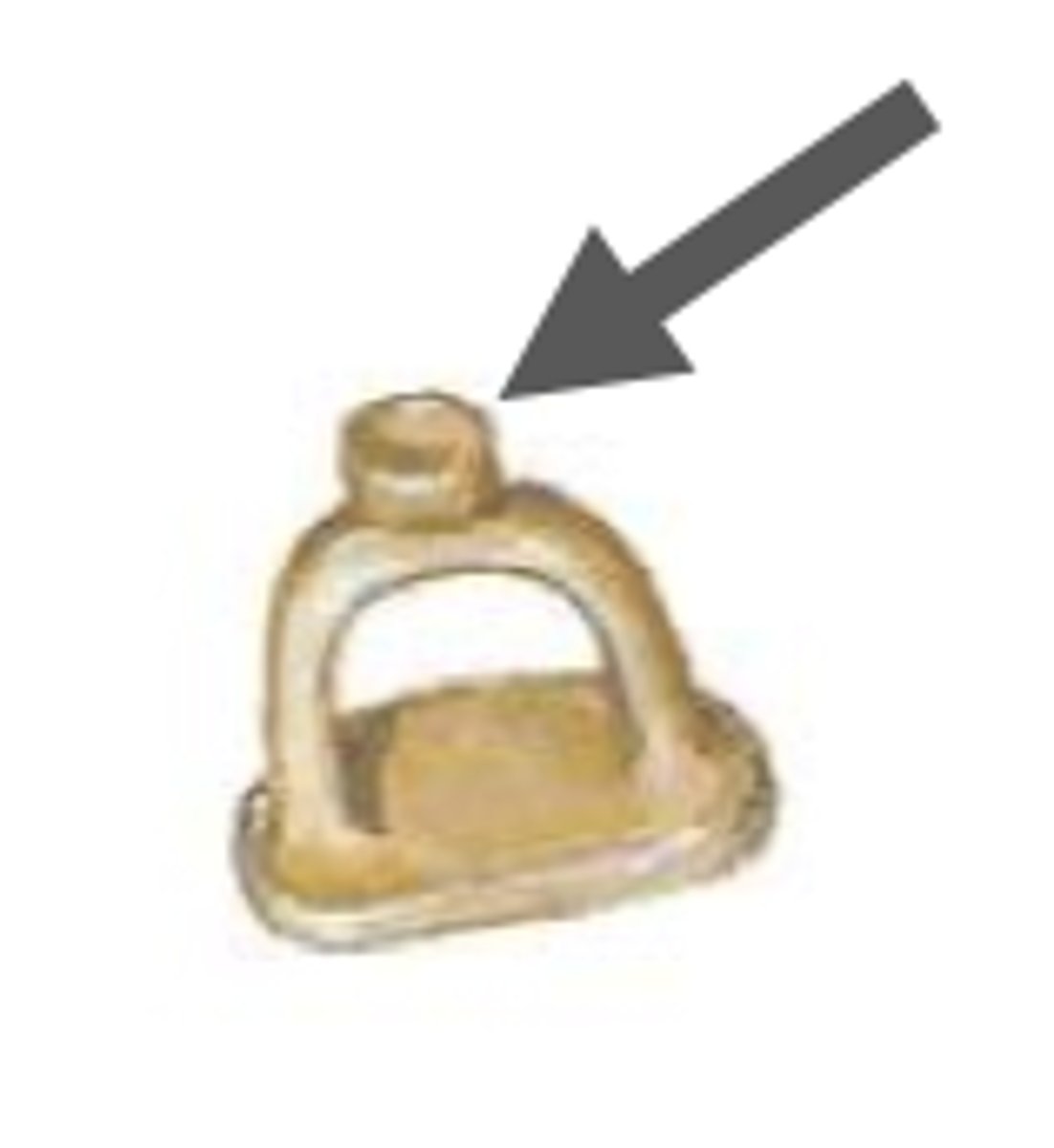
Footplate of the stapes B.
(Feature)
-Attached to eardrum model
-Where foot would be in stirrup
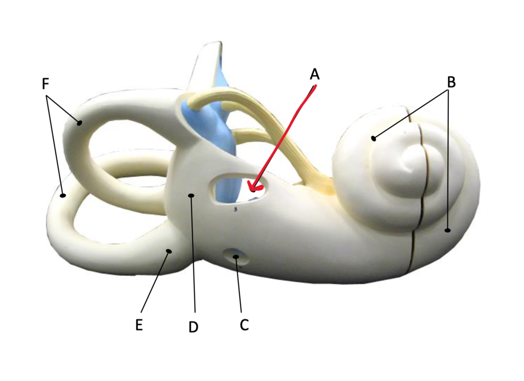
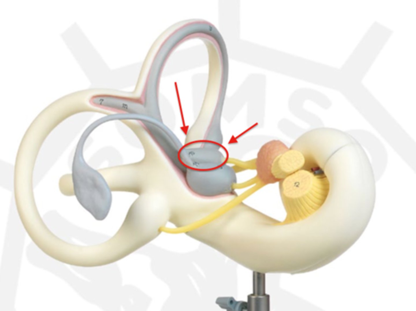
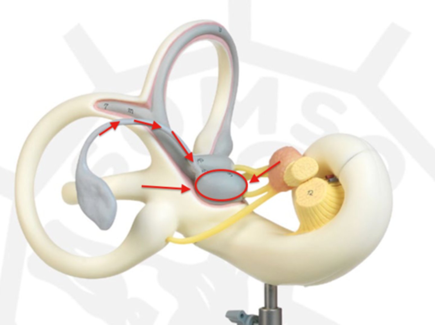
Semicircular canals
(Collective structure)
-On white loopy model
-All 3 circles
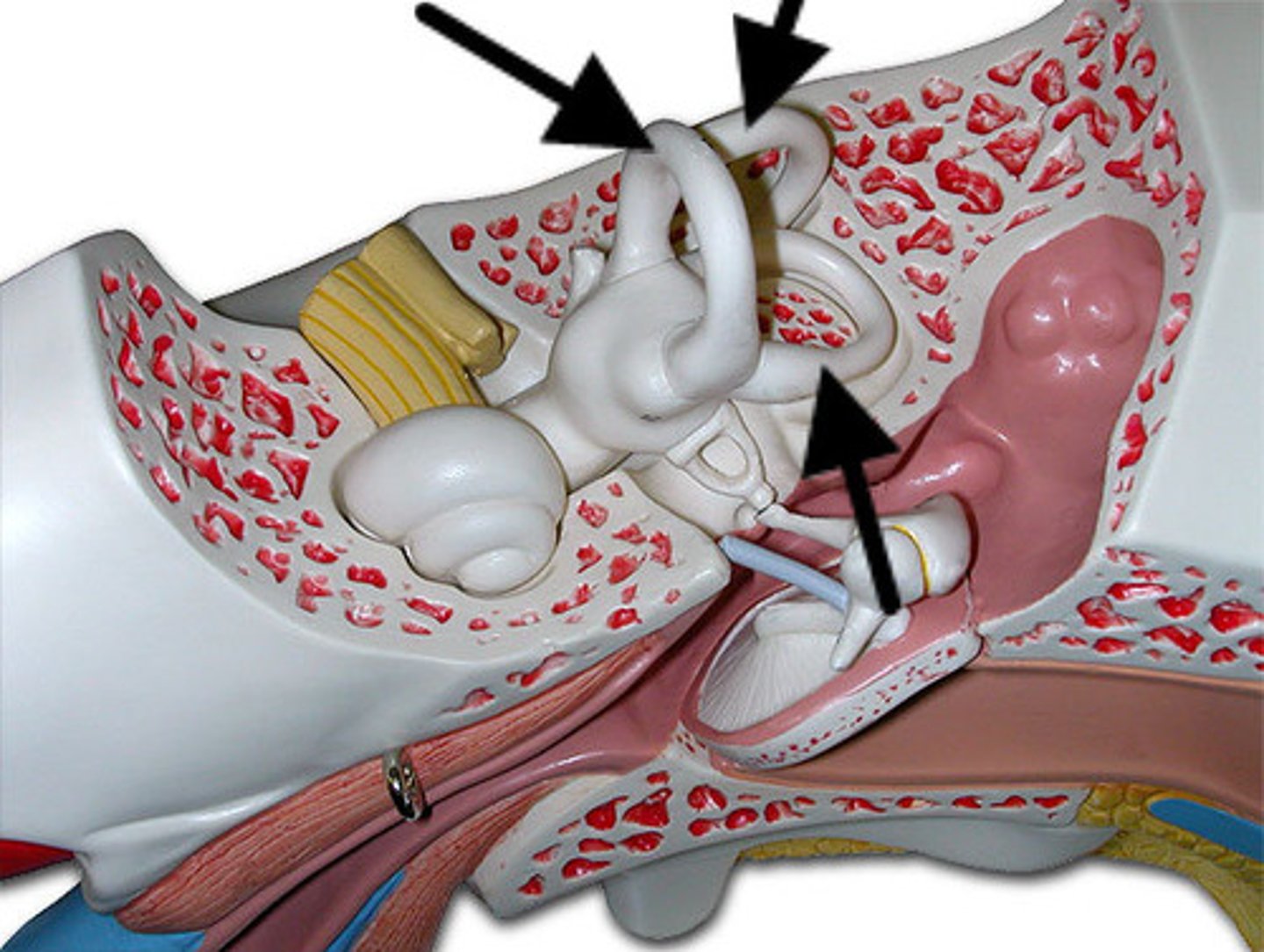
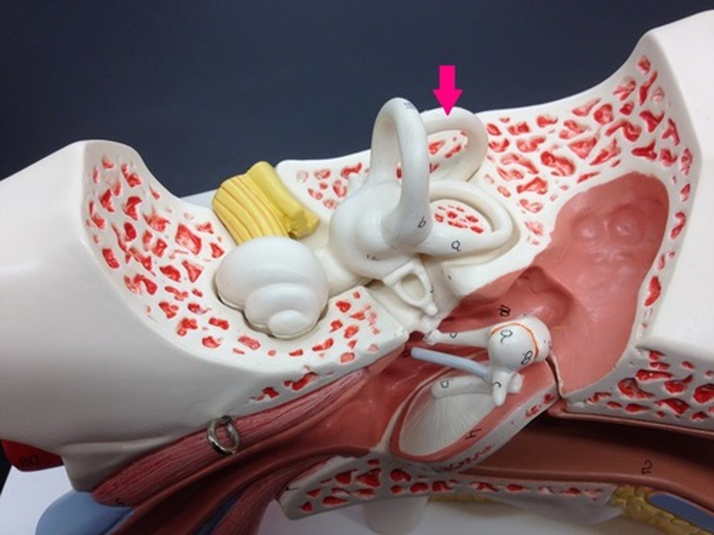
Anterior semicircular canals
(Structure)
-On white loopy model
-Back rest on chair
-#16 on model
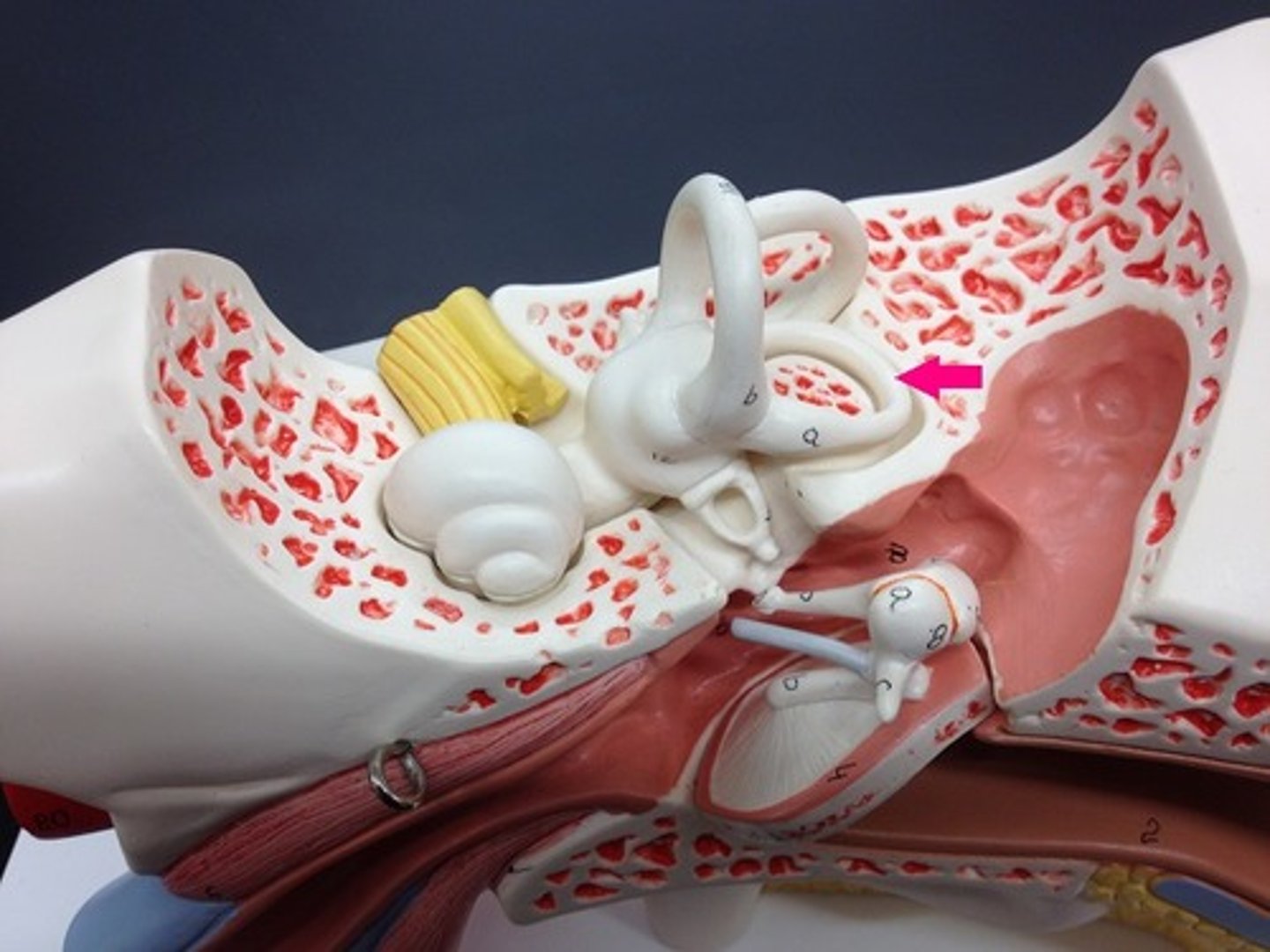
Posterior semicircular canal
(Structure)
-On white loopy model
-Armrest on chair
Lateral semicircular canal
(Structure)
-On white loopy model
-Butt on chair
-#15
Vestibule of the semicircular canals
(Junction)
-On white loopy model
-Will pinch where all 3 rings connect
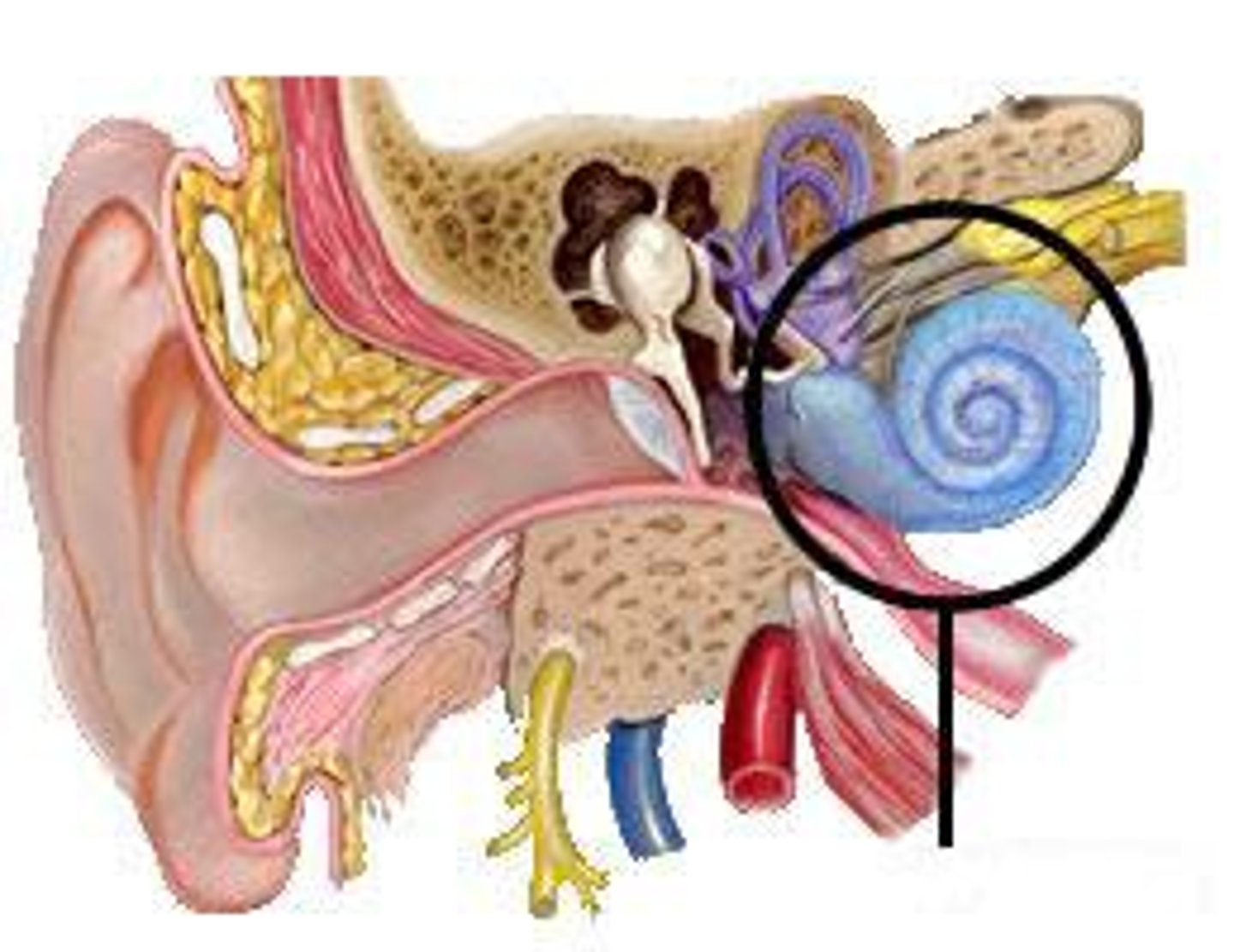
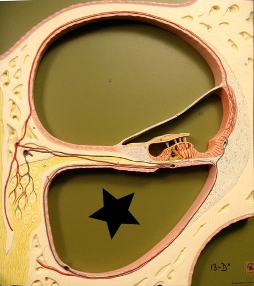
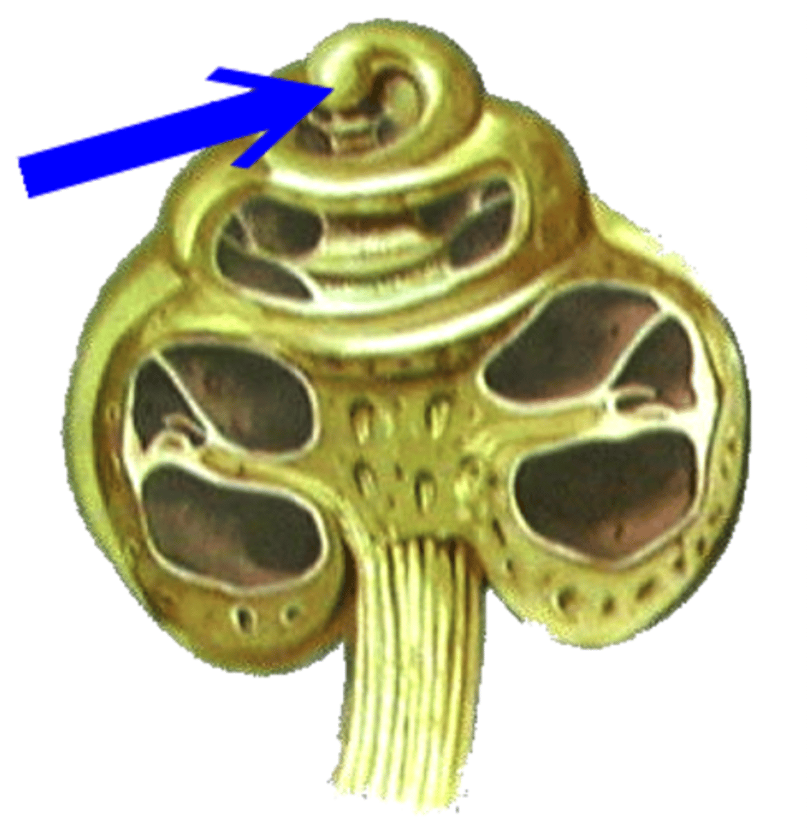
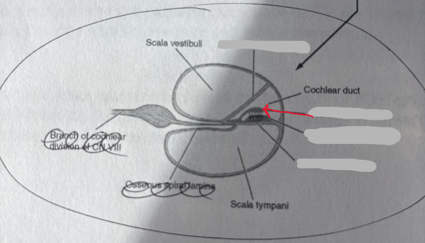
Cochlea
(Collective structure)
-Snail shell
-All lines on the shell
Modiolus of the cochlea
(Structure)
-Will open snail shell
white lines
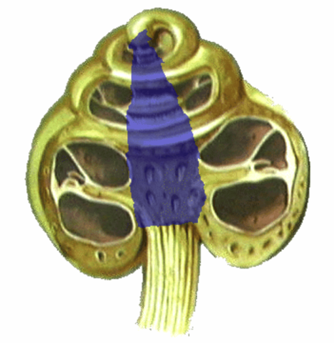
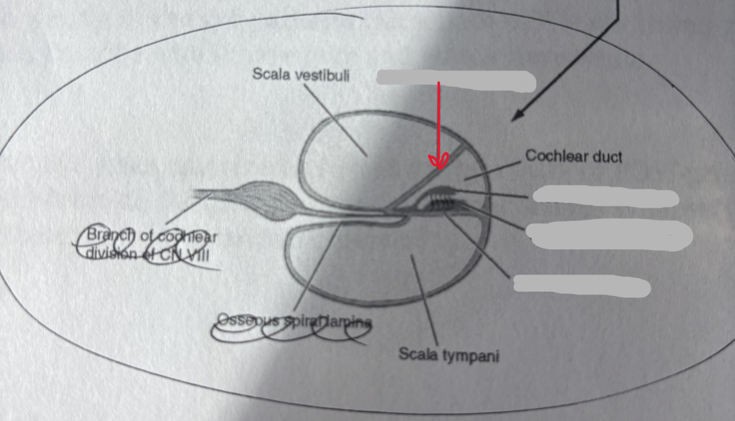
Osseous spiral lamina of the cochlea
(Covering)
-Will open snail shell
-Flower petals
beige color
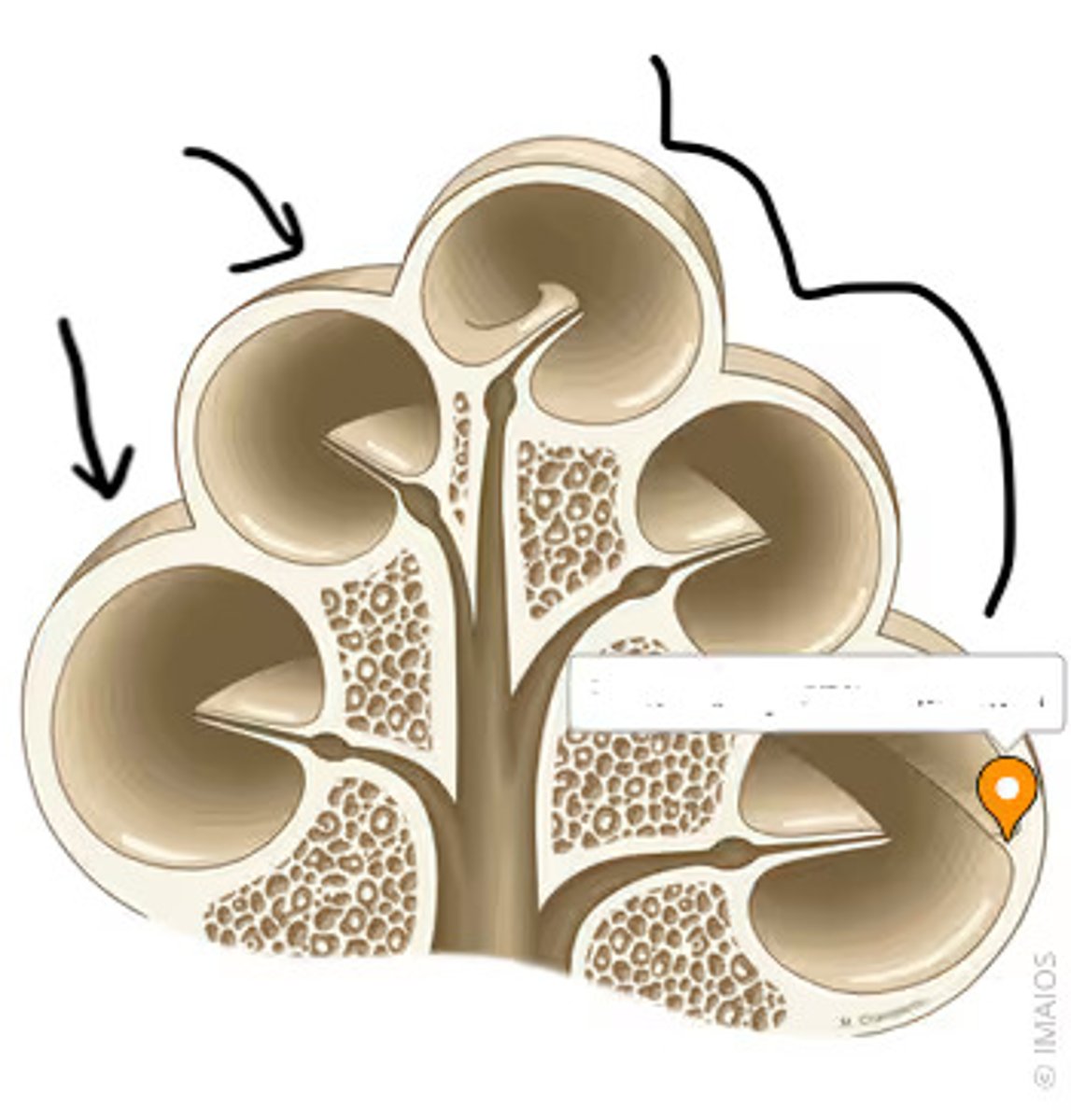
Scala vestibuli of the cochlea
structure
-Nasal look
-Right/ top space
Scala tympani of the cochlea
structure
-Nasal look
-Left/ bottom space
Helicotrema of the cochlea
structure
-Very tip of snail shell
-Looks like a nipple
Semicircular ducts
(Collective structure)
-On wood base model
-On loop pullout
-Clear tubes
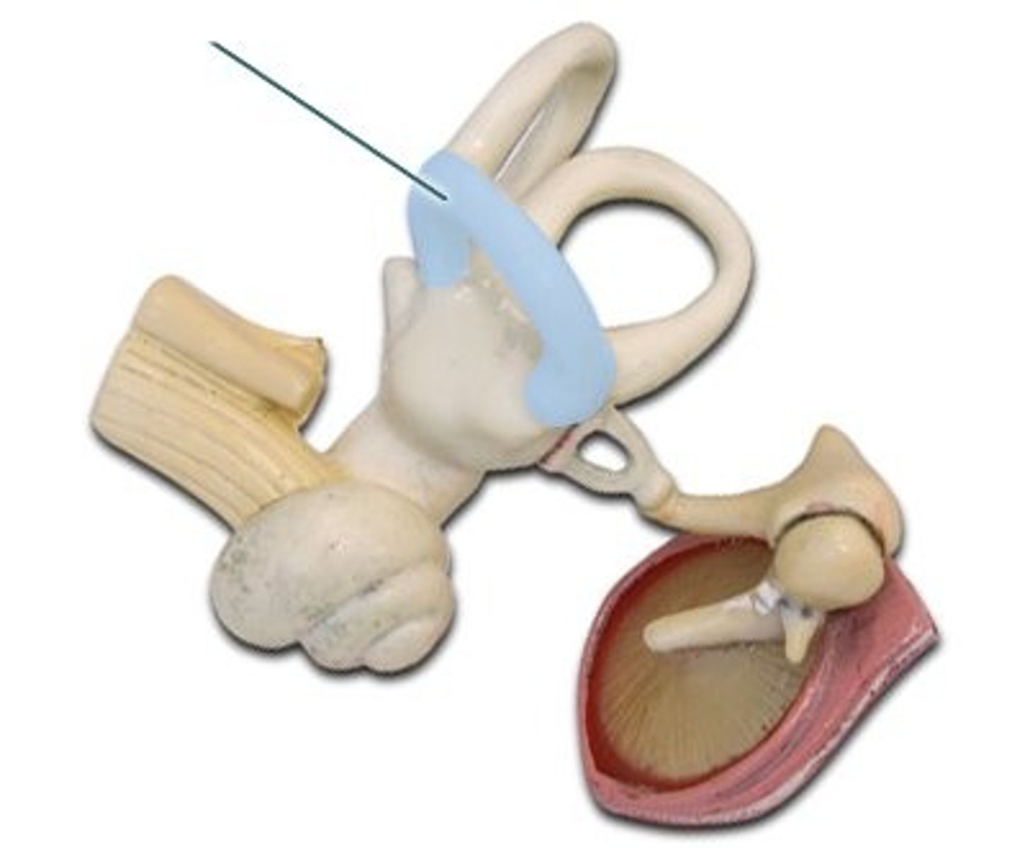
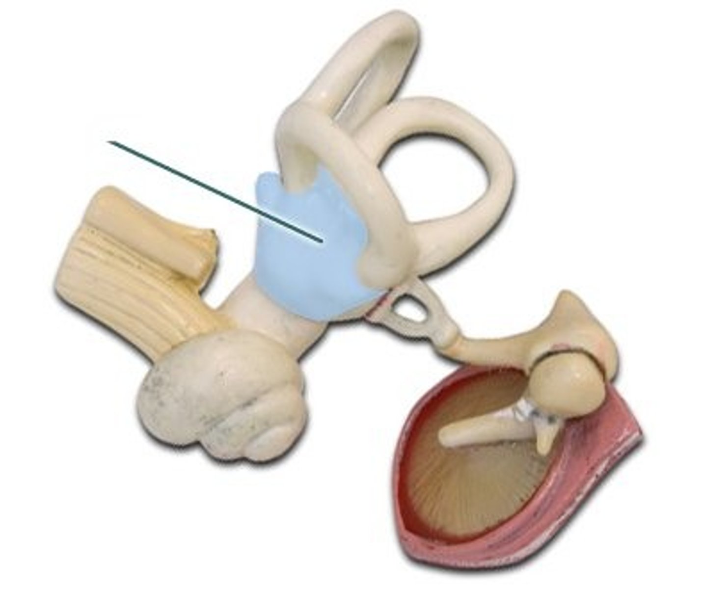
Utricle
(Structure)
-On green base model
-Baby blue color
Saccule
(Structure)
-On green base model
-Light grey under the utricle
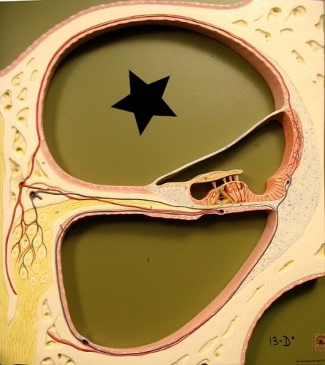
Cochlear duct
structure
blue dot
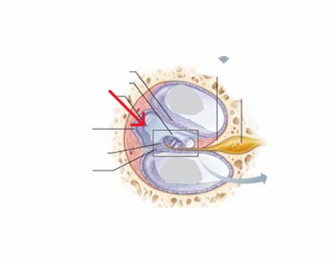
Vestibular membrane
Basilar membrane
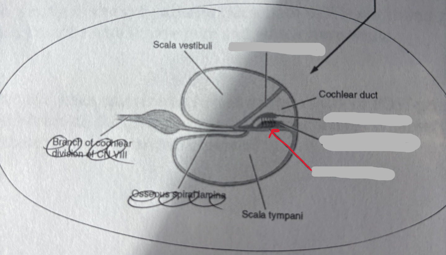
Tectorial membrane
Spiral organ of Corti

Fibrous layer of the eye
collective region
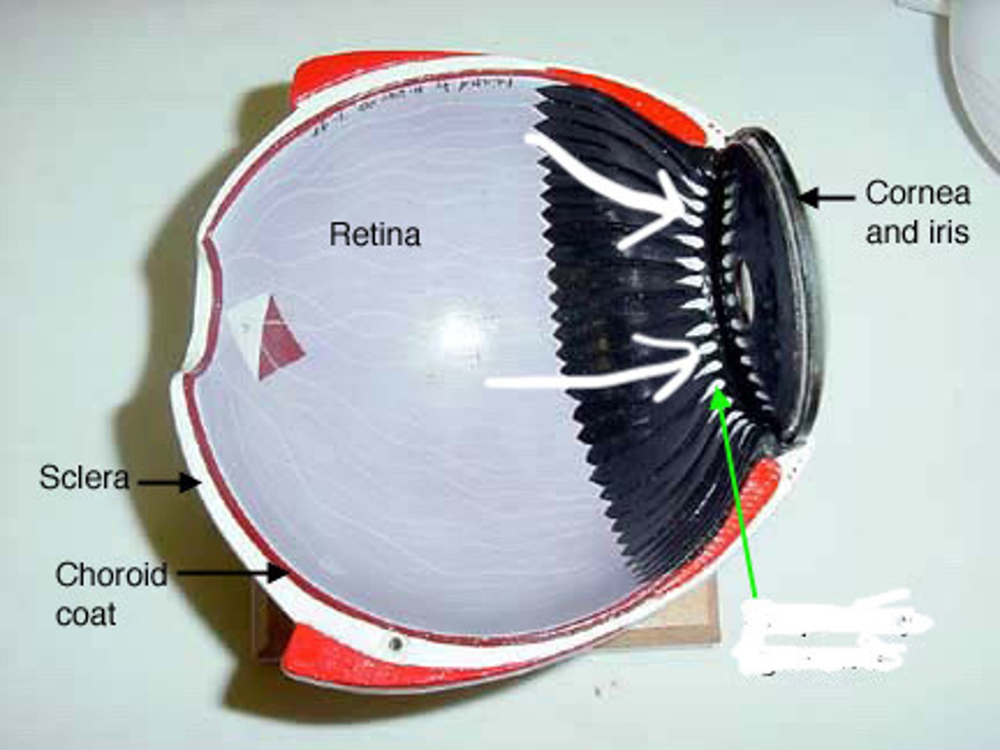
Sclera
structure
-All of white around the eye
-On green base model
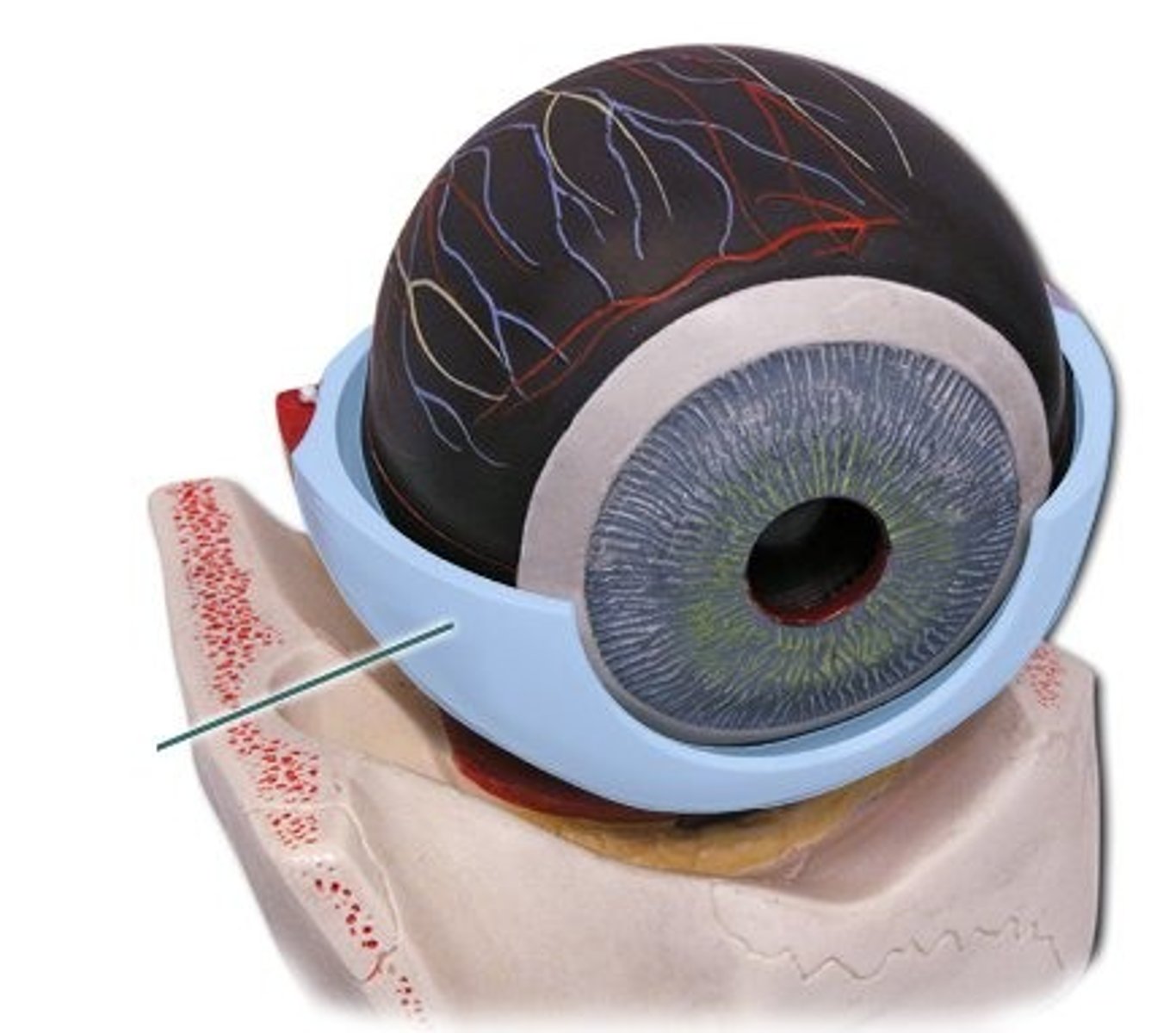
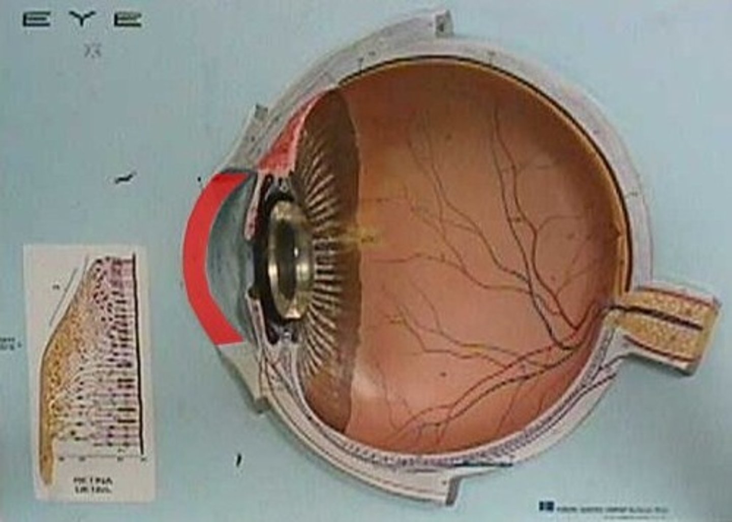
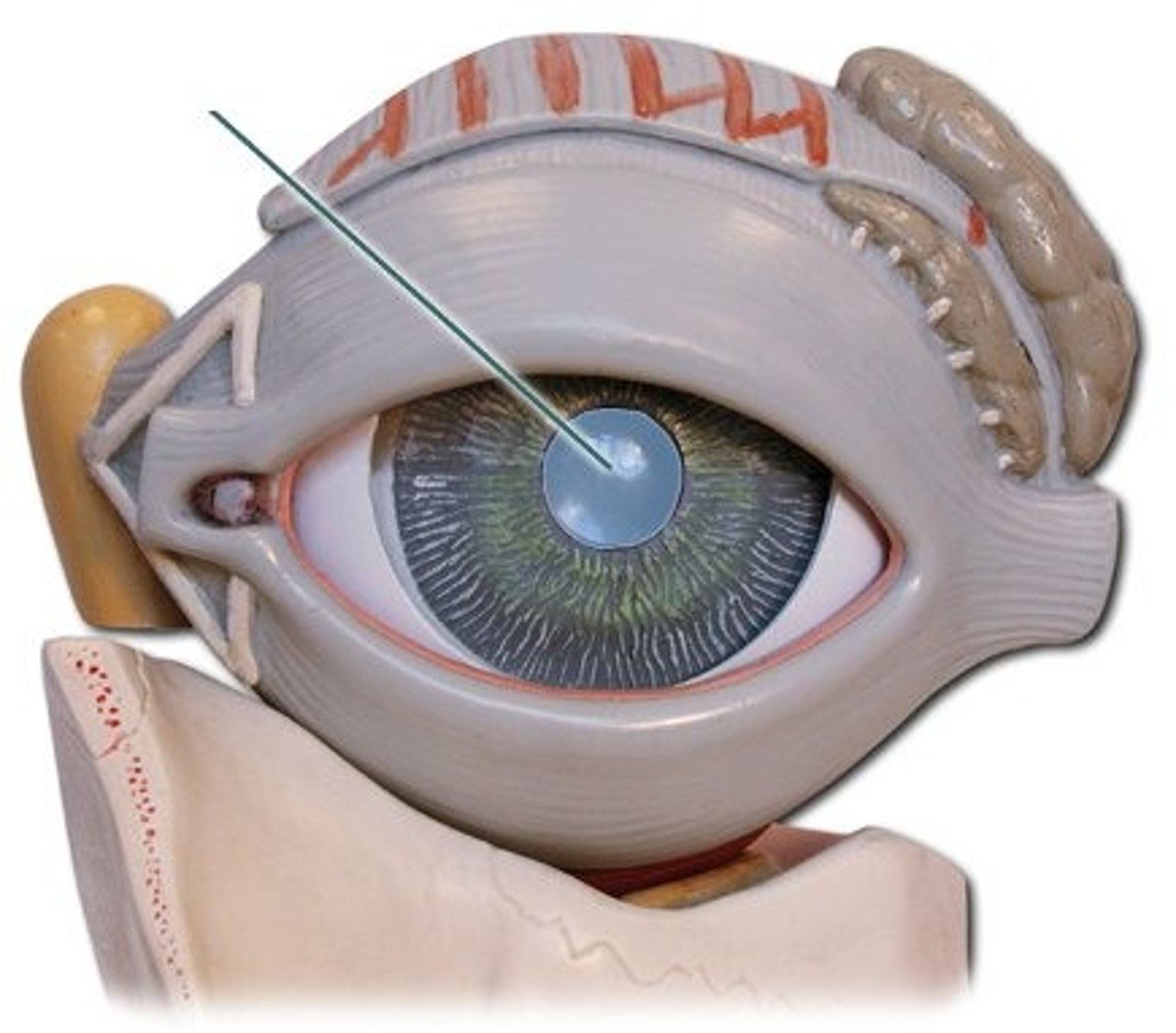
Cornea
structure
-On green base model
-Window in the front of the eye
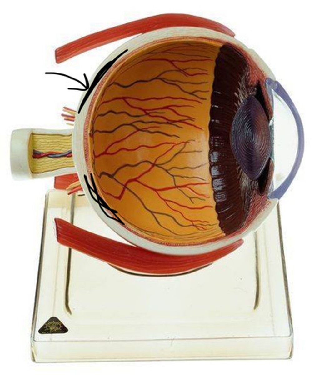
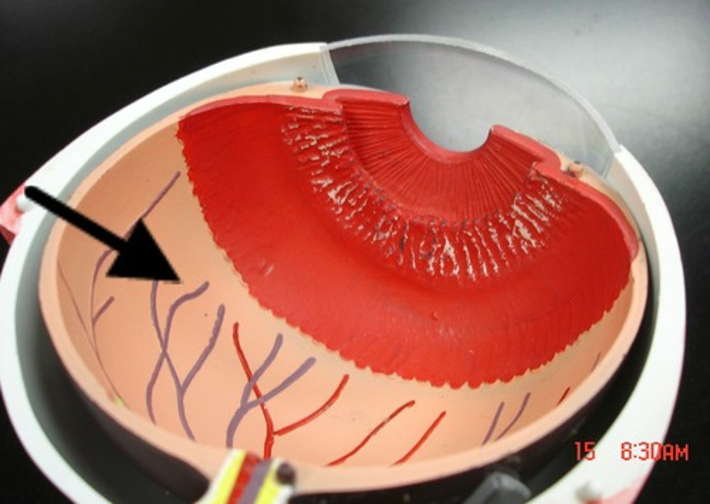
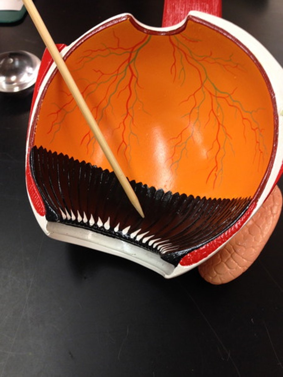
Vascular layer of the eye
(Region)
-On large eye model
-On the inside wall
-Red layer in between white/orange
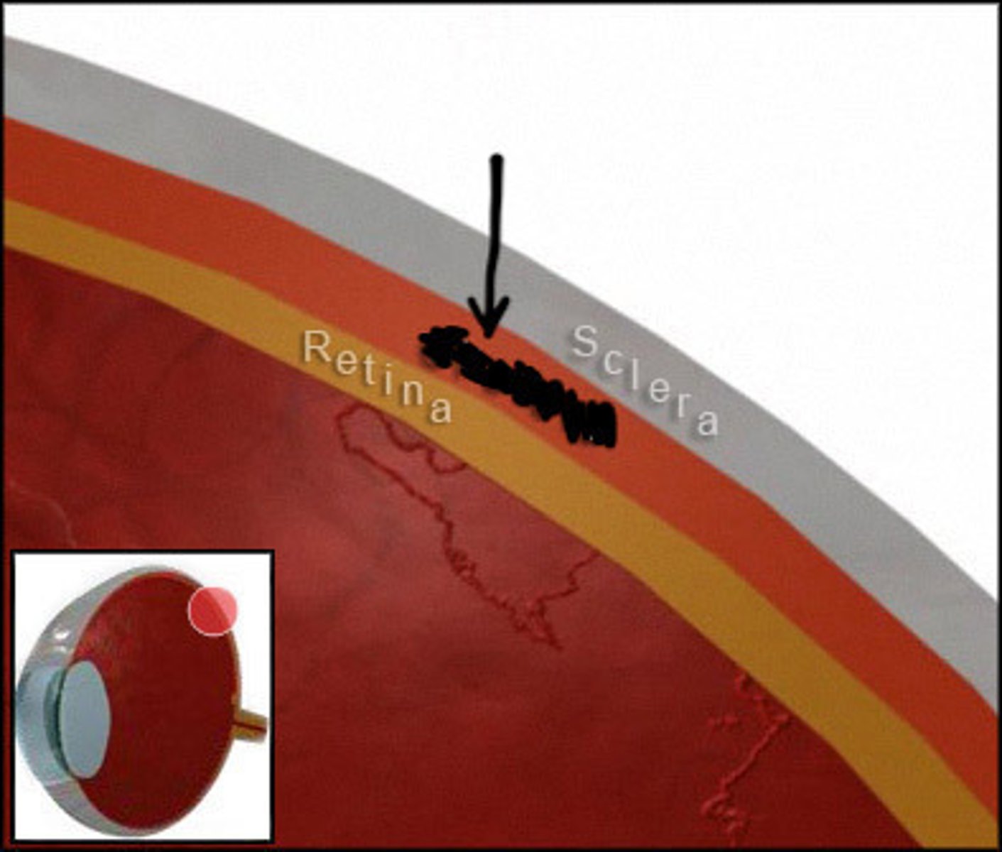
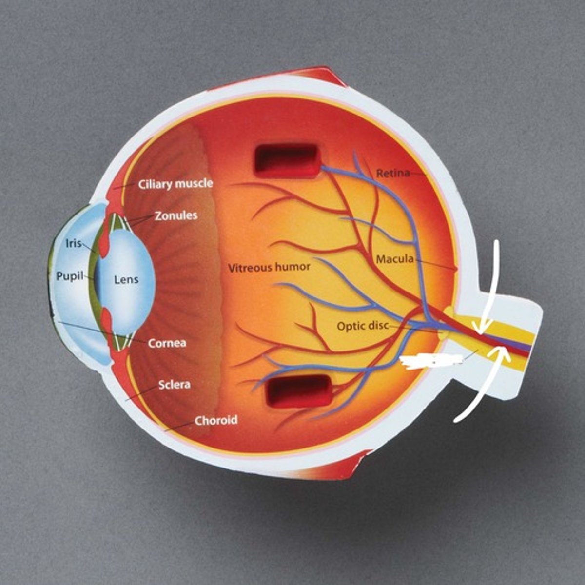
Choroid of the eye
(Structure)
-On large eye model
-Toward posterior part of the eye
-Thin part of the wall
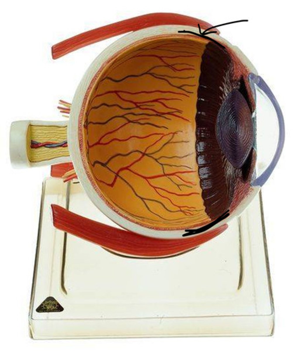
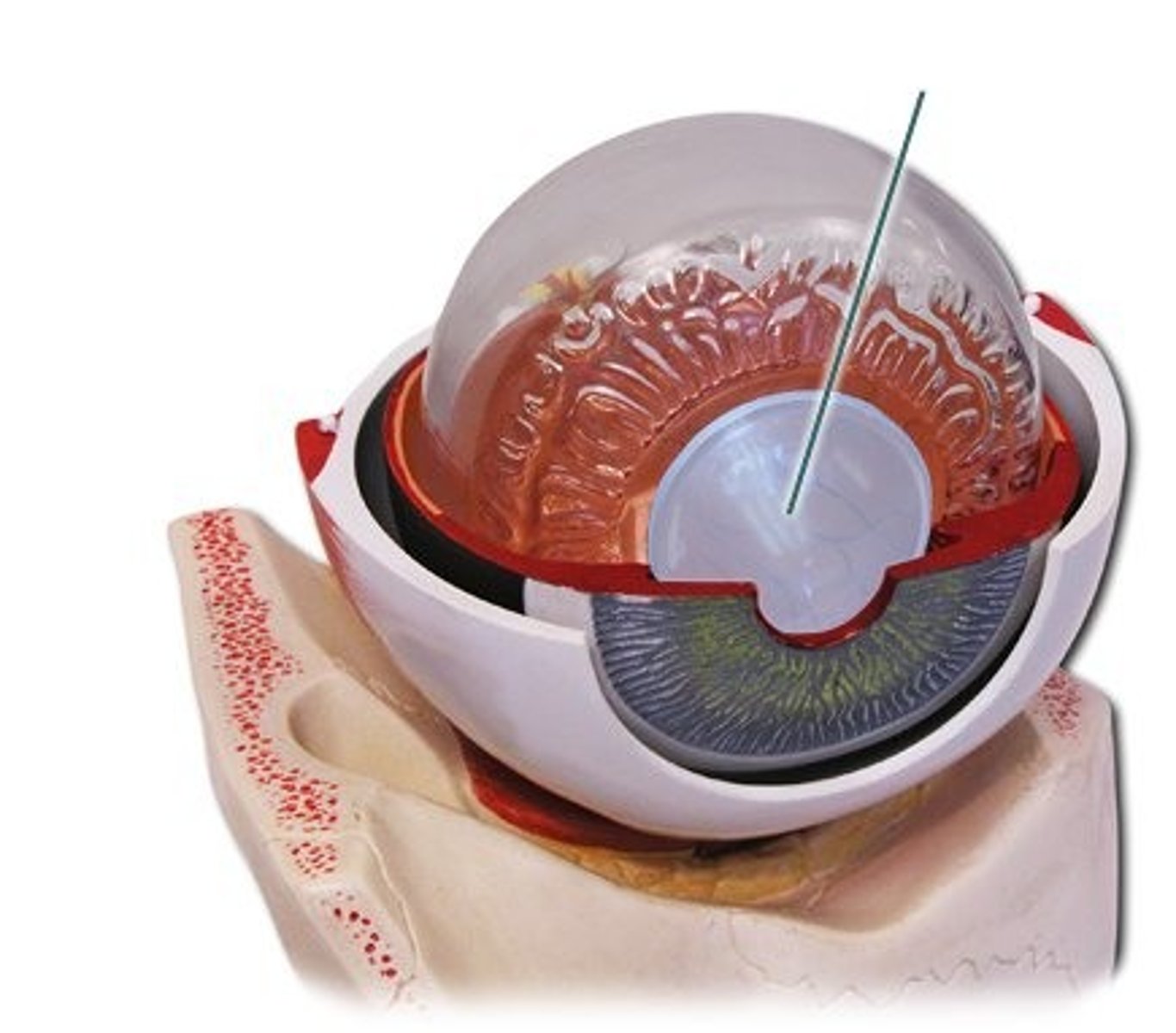
Ciliary body
(Collective structure)
-On large eye model
-Thick part of vascular layer toward the front
Iris
(Collective structure)
-On large eye model
-Colored part of eyeball
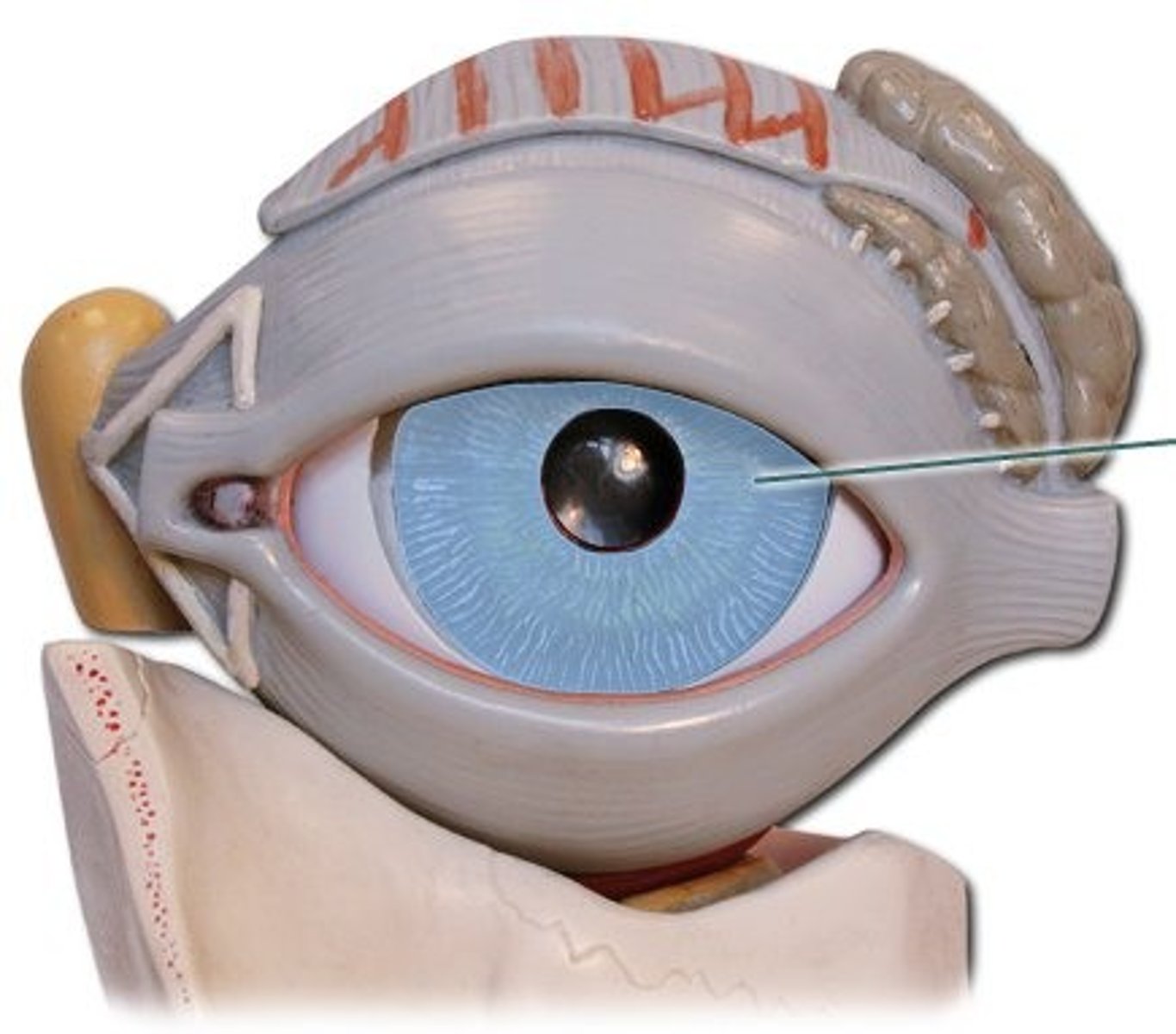
Pupil
(Space)
-On large eye model
-Middle of the iris
-Hole
Constrictor pupillae muscle
(Structure)
-On large eye model
-Lines pupil
-Probe will trace around pupil hole
-Shown almost the same as the pupil
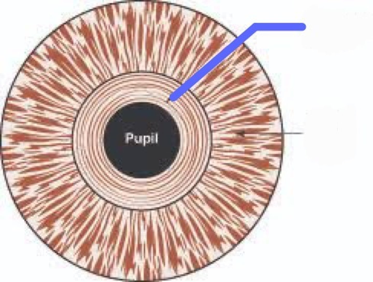
Dilator pupillae muscle
(Structure)
-On large eye model, will open up
-Lines going out of the pupil
-On closest ring to the pupil
Inner layer
(Region)
-On large eye model
-Everything on the inside

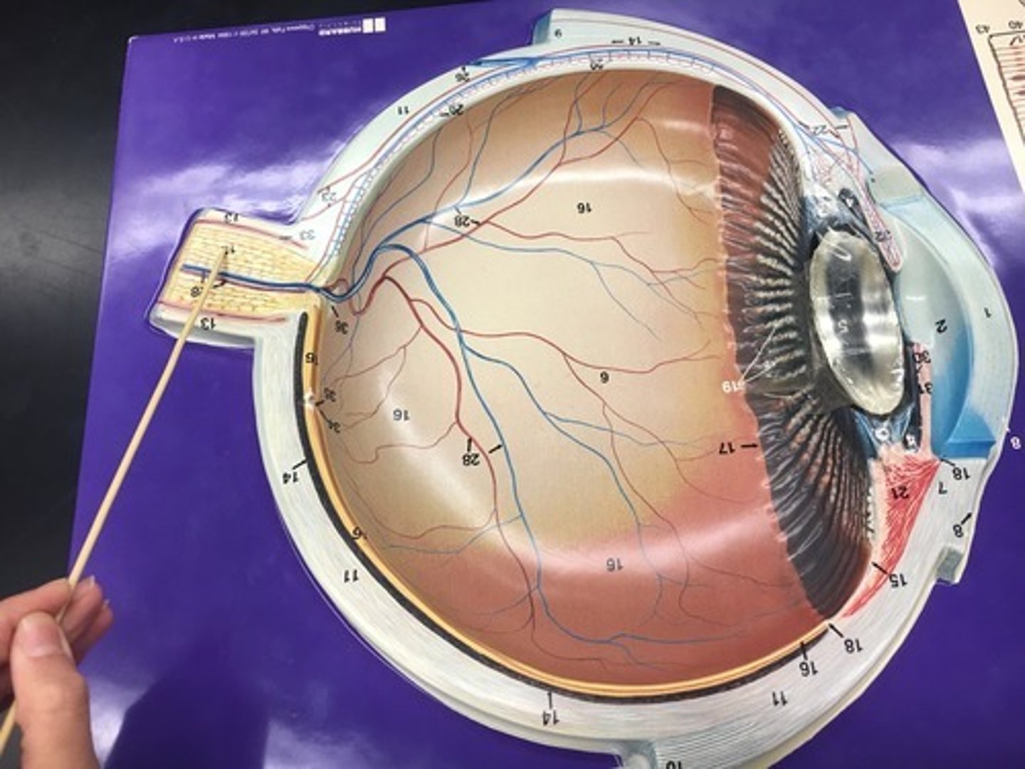
Retina
(Internal structure)
-On large eye model
-Only orange in model

Optic part of the retina
(Portion)
-On large eye model
-Orange part, same as the retina but a portion
Nonvisual part of the retina
(Portion)
-On large eye model
-All of the purple in the front layer
Macula
(Structure)
dracula
-On large eye model
-#8 on model
-Yellow circle to the left of the middle
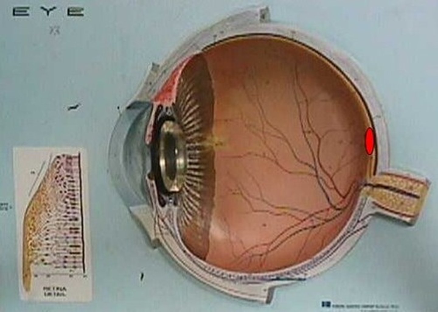
Fovea centralis
(Feature)
-On large eye model
-#9 on model
-Slightly depressed, right next to #8
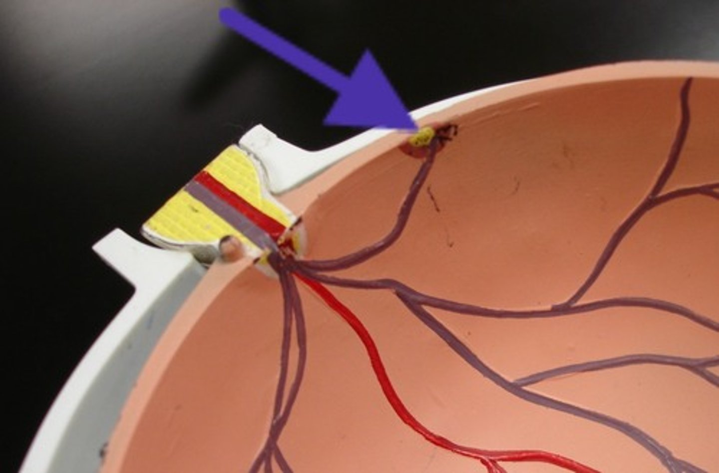
Optic disc
(Structure)
-On large eye model
-Yellow circle on back of the eye in the middle
-#7 on model
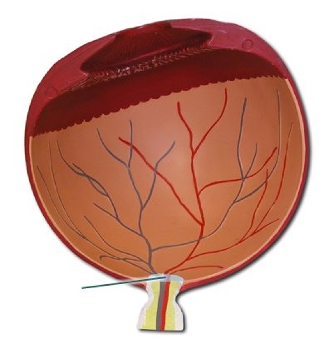
Optic nerve
(Structure)
-On large eye model
-Looks like corn
Central artery of the retina
(Structure)
-On large eye model
-Red line in middle of corn
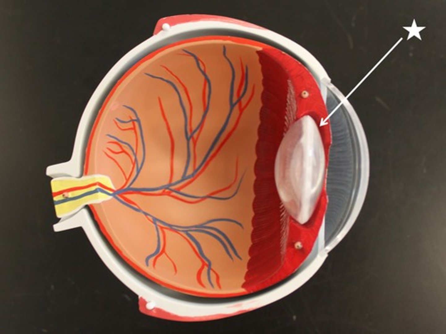
Lens
(Structure)
-Shown on stand alone model
-Glass held in hand
-Pulled out of model
Anterior chamber of the eye
(Space)
-On big eye model
-Space in between outer lens/ iris
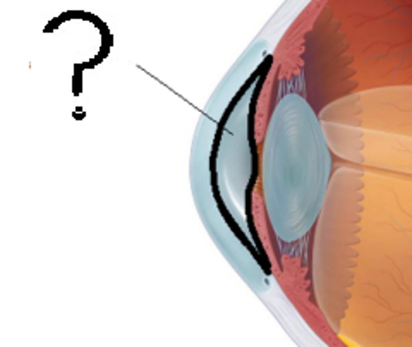
Posterior chamber of the eye
(Space)
-On big eye model
-Between inner lens and iris
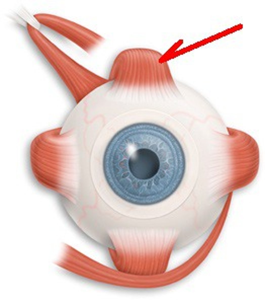
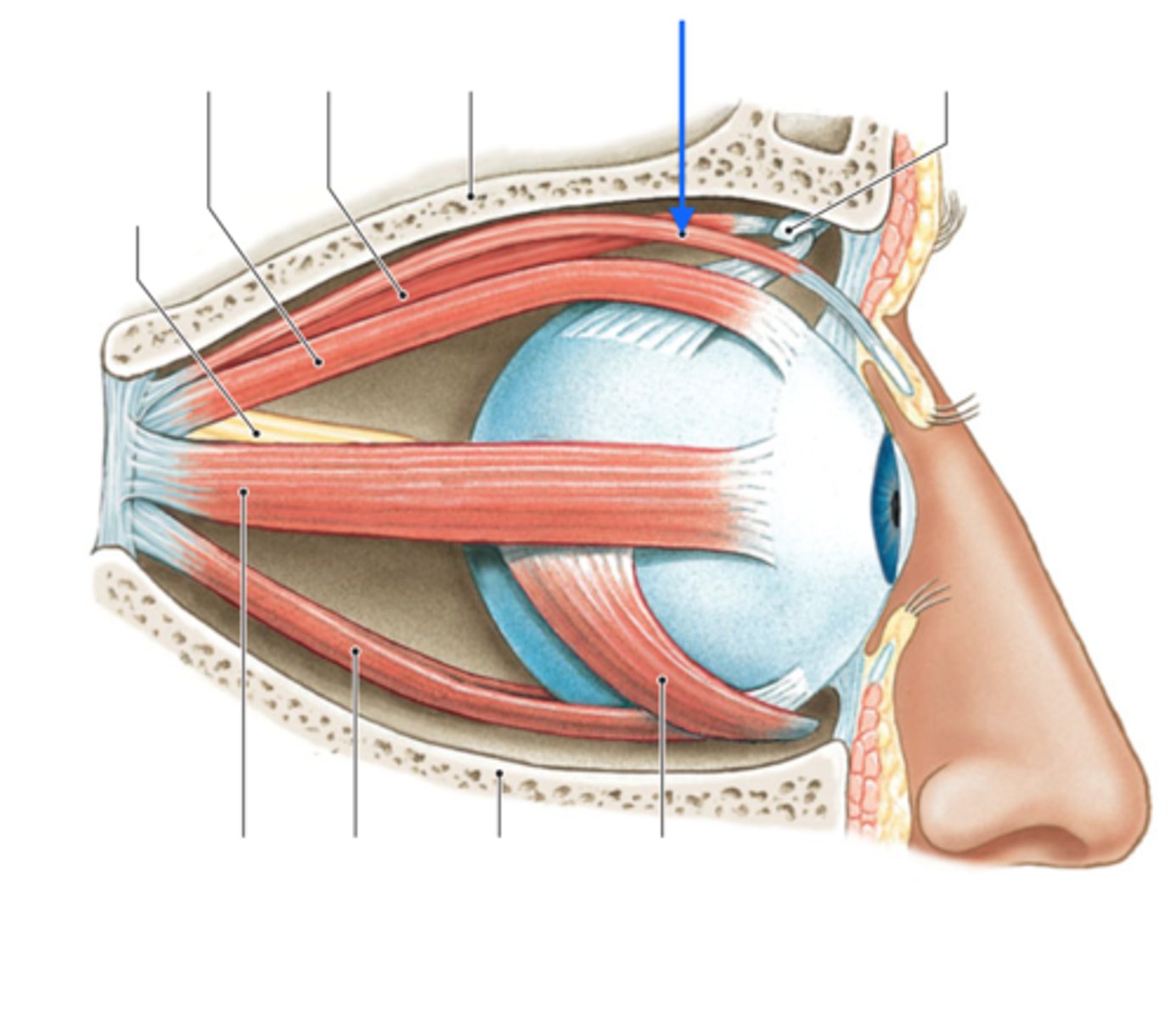
Superior rectus M.
(Structure)
-On big eye model
-Superior muscle
-On top of eye
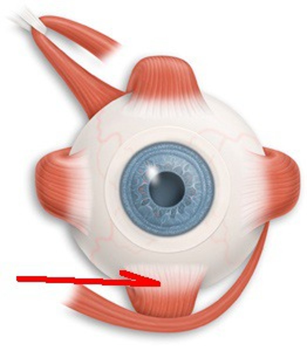
Inferior rectus M.
(Structure)
-On big eye model
-Bottom of eye
Medial rectus M.
(Structure)
-On big eye model
-Closest to trochlea
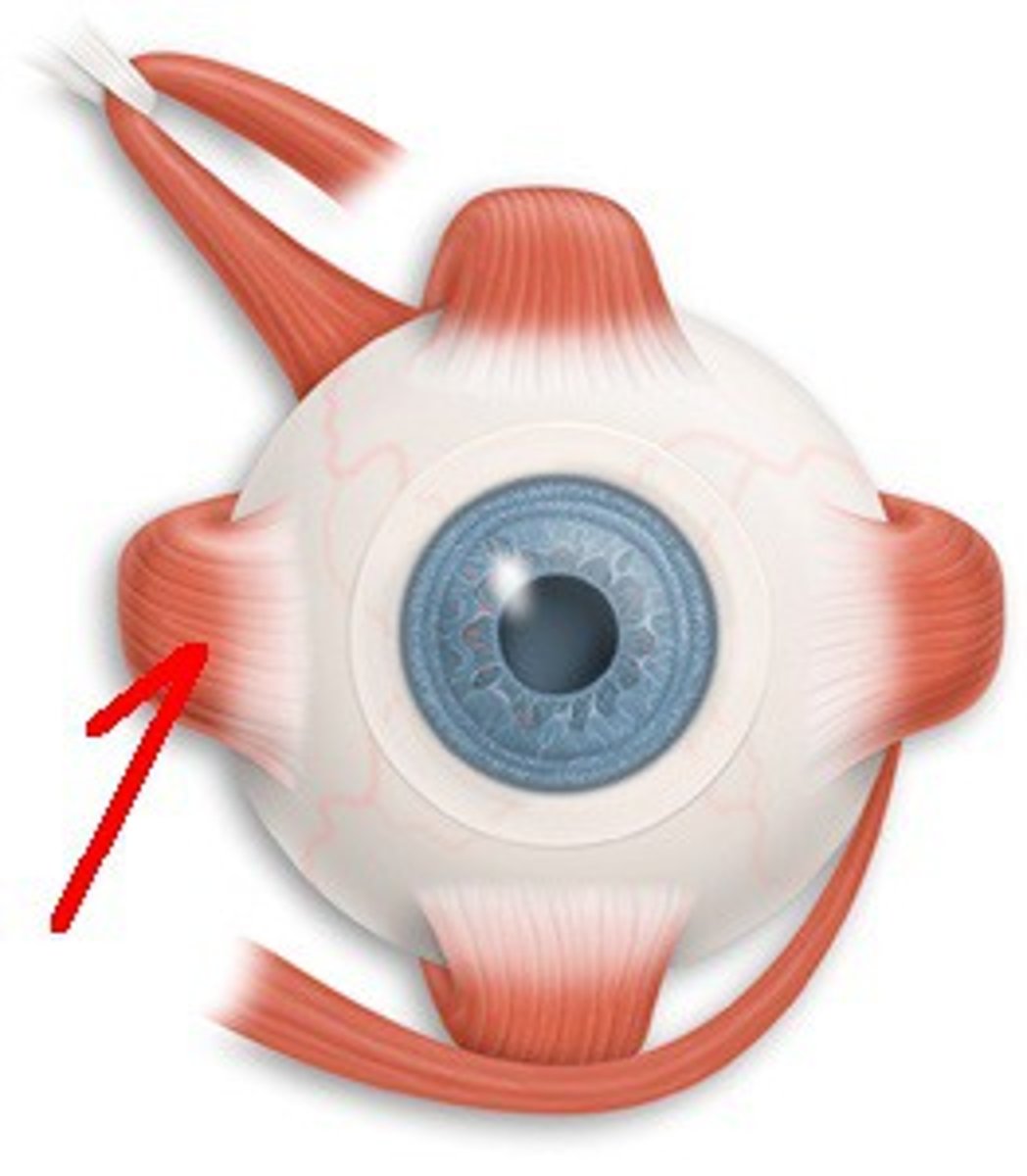
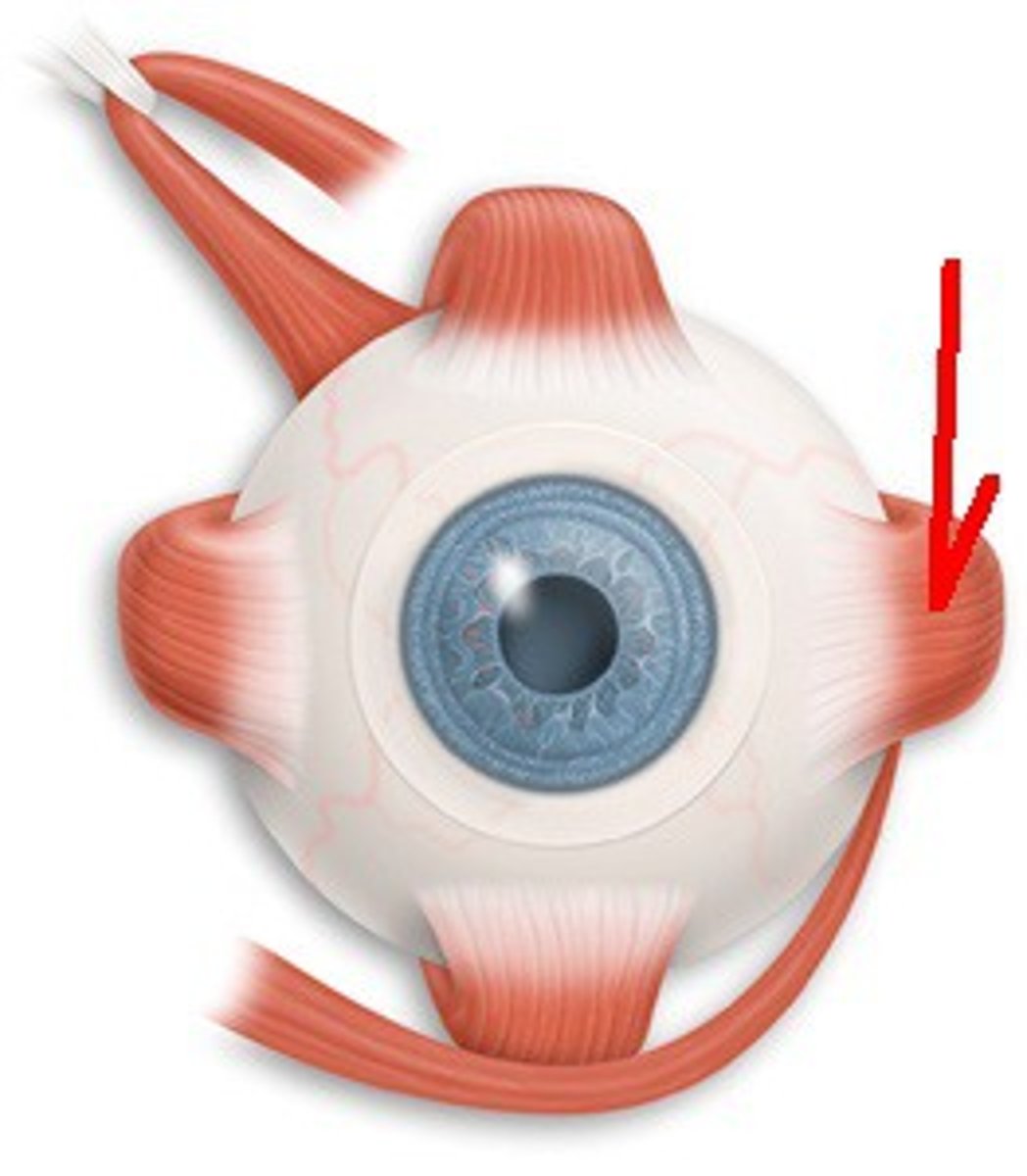
Lateral rectus M.
(Structure)
-On big eye model
-Lateral side
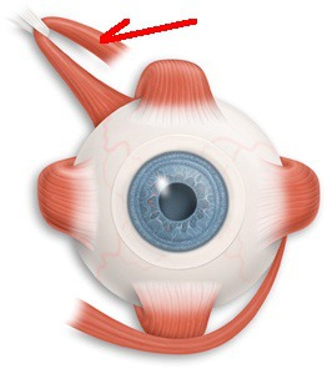
Superior oblique M.
(Structure)
-On big eye model
-Goes to trochlea
-Off center/ almost superior
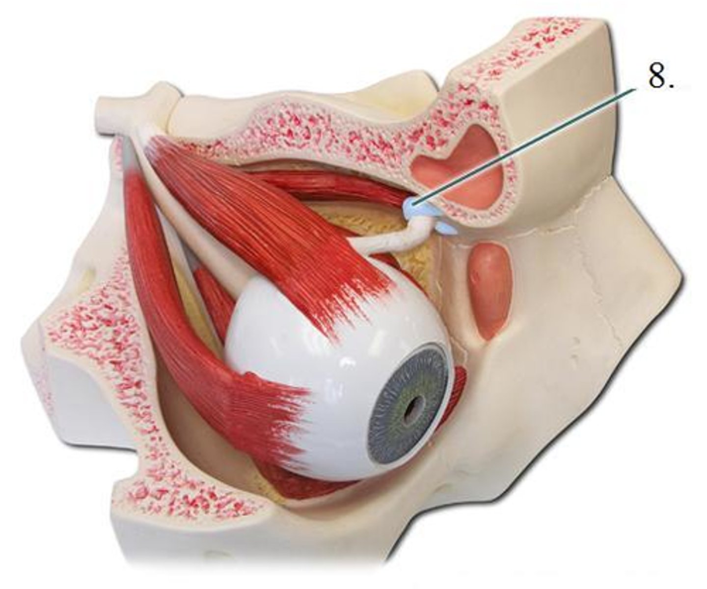
Trochlea of the eye
(Structure)
-On big eye model
-Blue on top/ right of eye
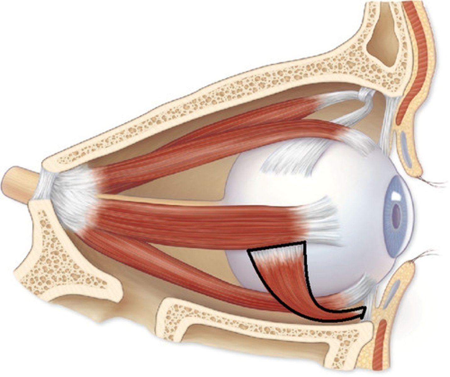
Inferior oblique M.
(Structure)
-On big eye model
-Superior/ off center
-Cut off, short muscle
Levator palpebrae superioris M.
(Structure)
-On big eye model
-Attached to part on top
-Attached to brainy part
Superior and inferior eyelids
(Structures)
-On body 23-45
-Superior = Upper eyelids
-Inferior = Lower eyelids
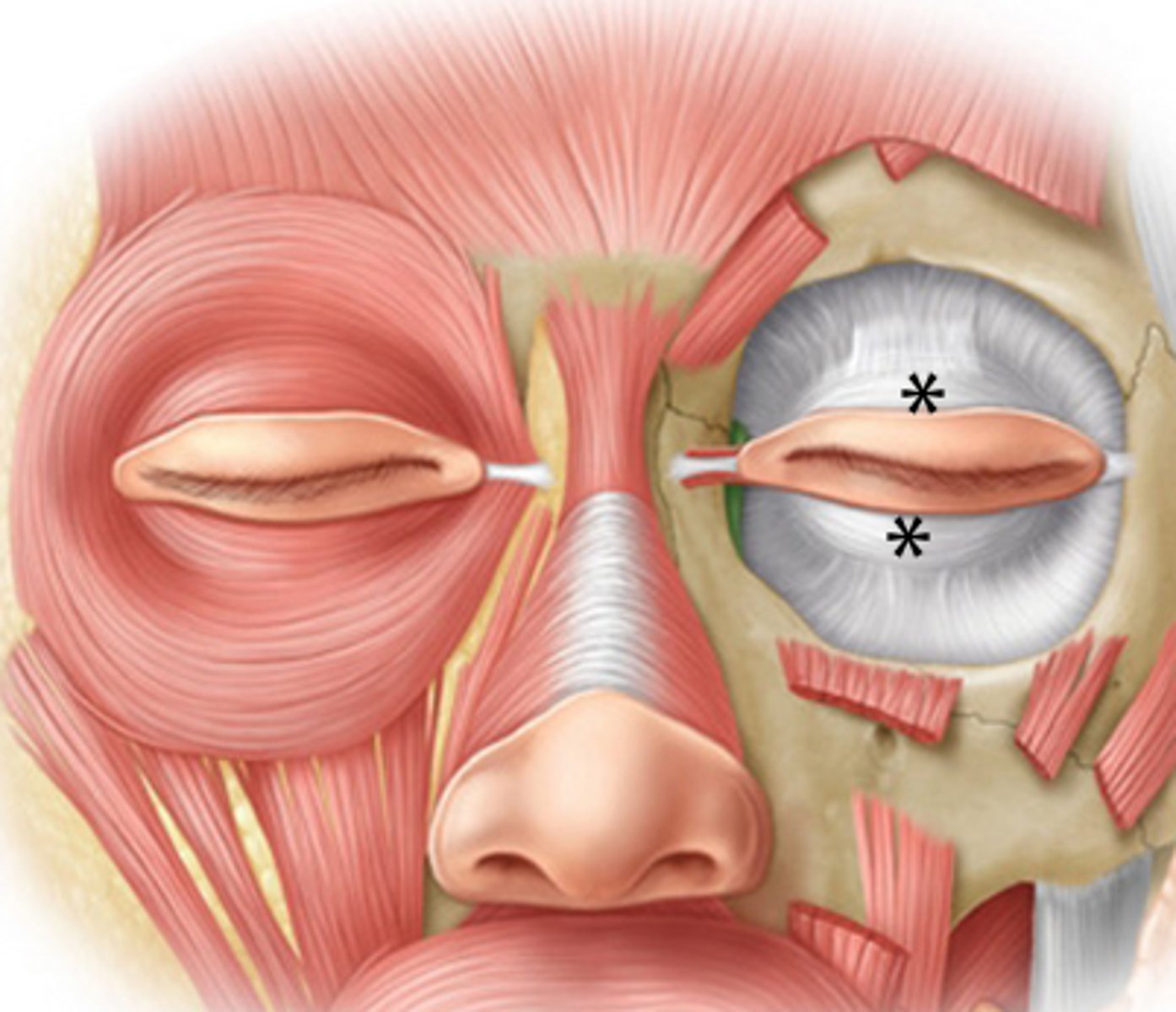
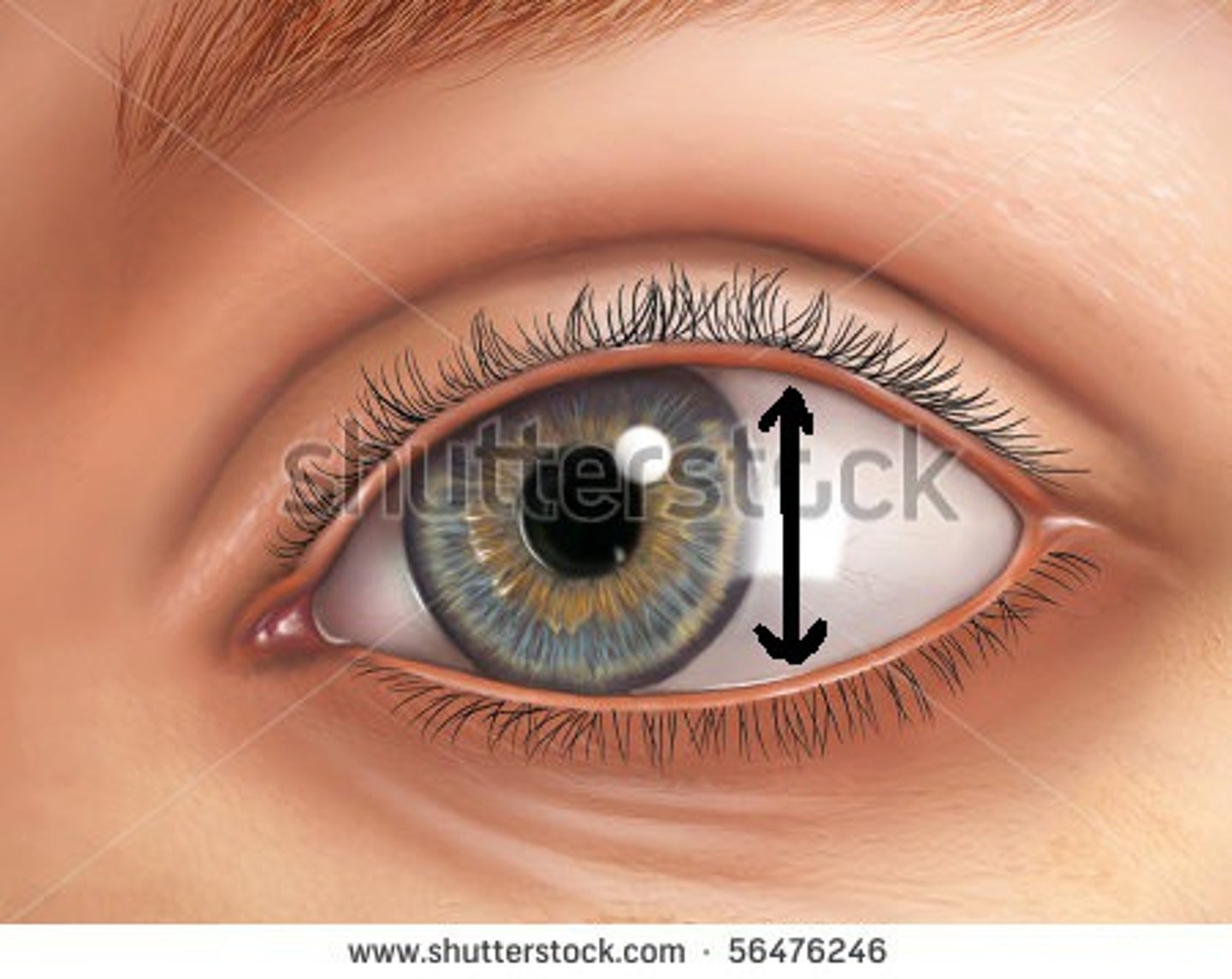
Palpebral fissure
(Space)
-On body 23-45
-In between eyelids
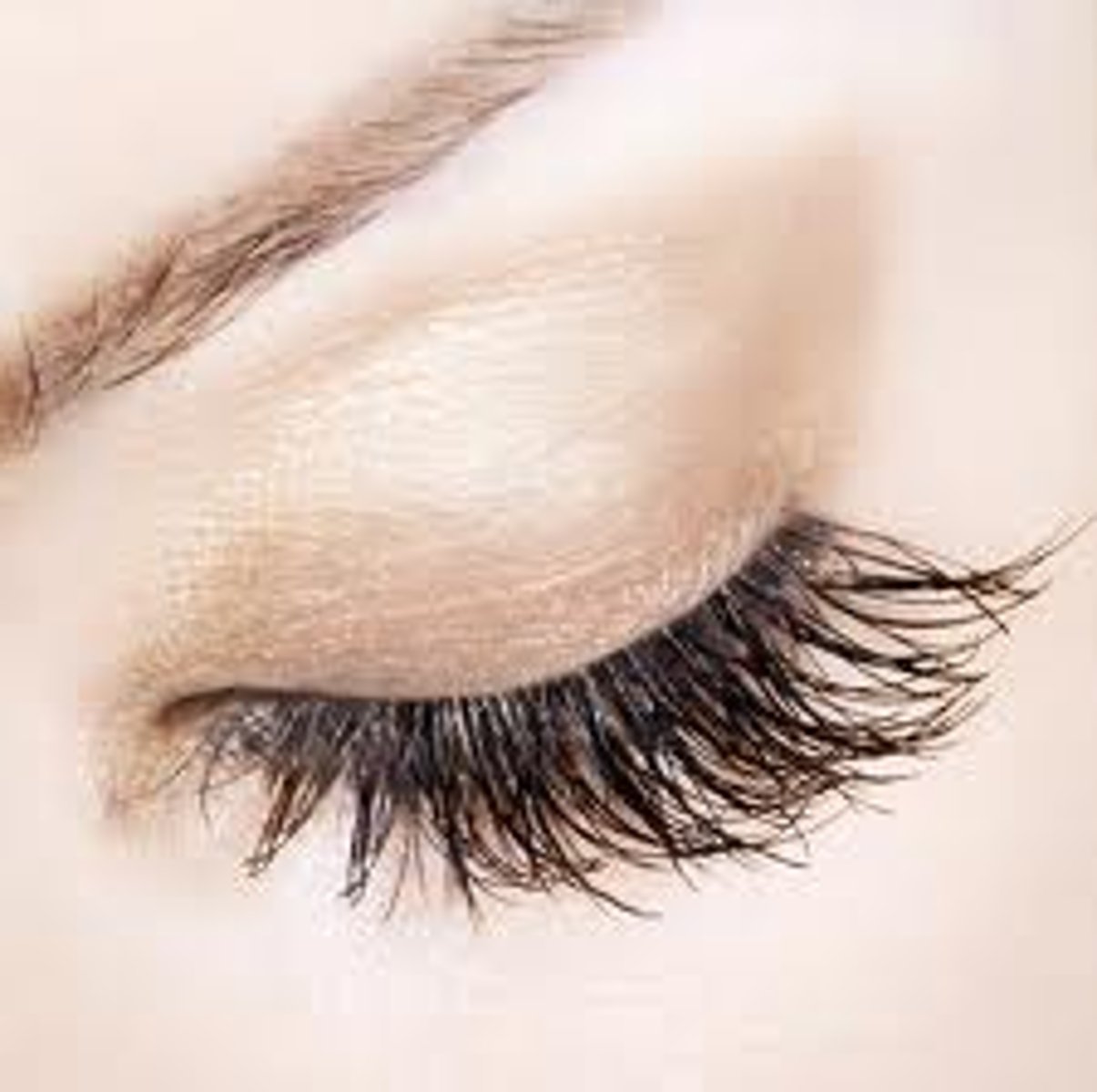
Eyelashes
(Structure)
-On body 23-45
-Will poke hairs on own body or on cadaver
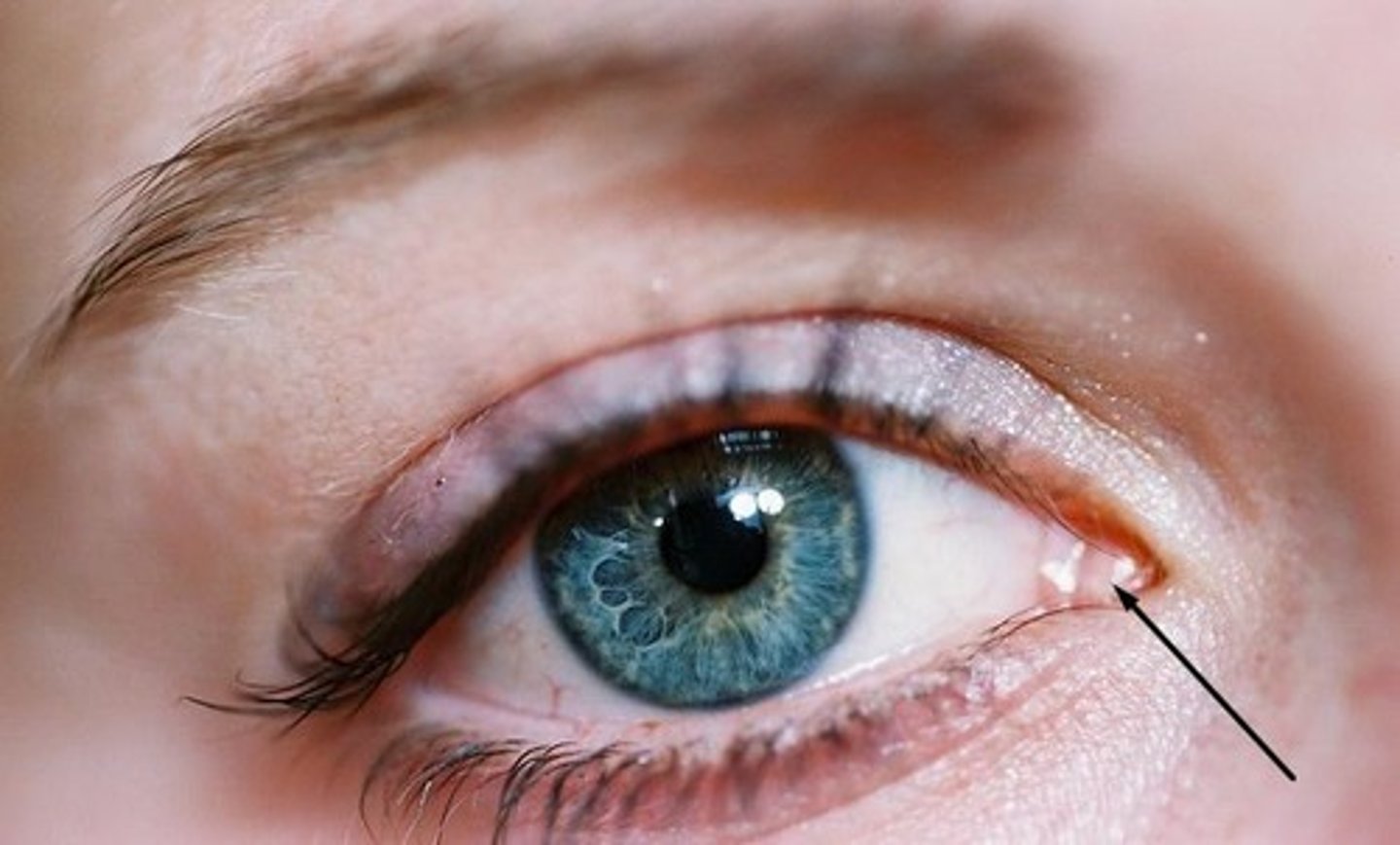
Lacrimal caruncle
(Structure)
-On body 23-45
-Medial part of the eye
-Where you get eye boogers
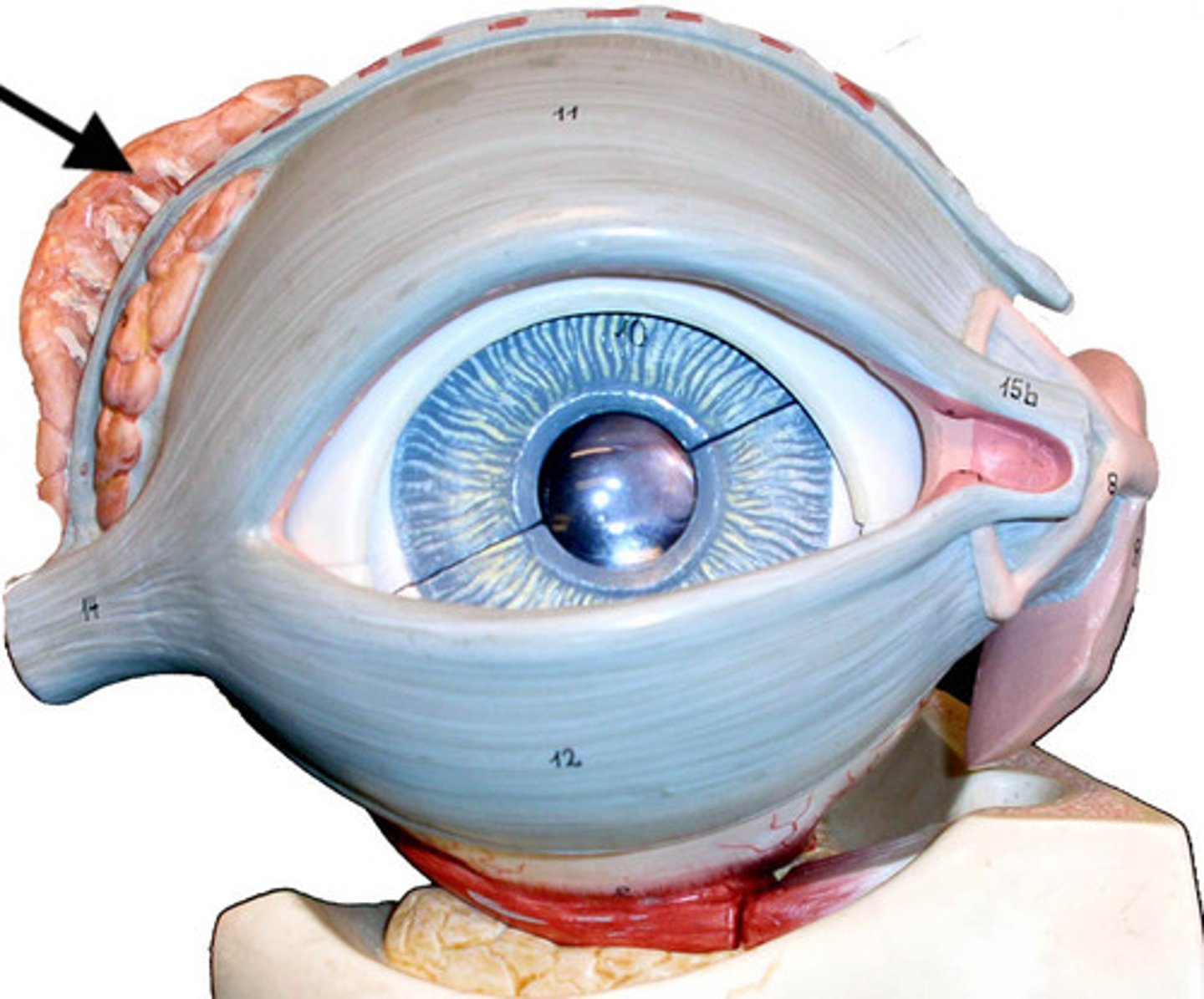
Lacrimal gland
(Structure)
-On body 23-45
-Brainy like structure
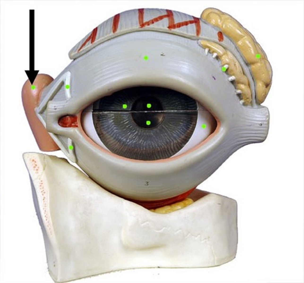
Lacrimal sac
(Structure)
-On long green base model
-Pink behind eye
-Medial side
CN III -> ciliary ganglion -> smooth muscle in eye -> constrict pupil
CN III flow
CN VII -> Pterygopalatine ganglion -> lacrimal gland -> increase tear production / CN VII -> sublingual ganglion -> submandibular gland -> increase salivation
CN VII flow (2 routes)
CN IX -> otic ganglion -> parotid gland -> increase salivation
CN IX flow
CN X -> Intramural ganglion -> cardiac muscle -> decrease heart rate / CN X -> intramural ganglion -> smooth muscle in digestive tract -> decrease blood flow
CN X flow (2 routes)
Sacral spinal cord -> intramural ganglion -> smooth muscle in pelvic organs -> increase urine excretion
sacral spinal cord flow
Upper thoracic spinal cord -> superior cervical ganglion -> smooth muscle in eye -> dilate pupil / Upper thoracic spinal cord -> upper thoracic chain ganglion -> cardiac muscle -> increase heart rate
upper thoracic spinal cord flow (2 routes)
Lower thoracic spinal cord —> celiac ganglion —> smooth muscle in stomach or intestines —> decrease blood flow
lower thoracic spinal cord flow