Cell structure A2.2
1/61
Earn XP
Description and Tags
Name | Mastery | Learn | Test | Matching | Spaced | Call with Kai |
|---|
No analytics yet
Send a link to your students to track their progress
62 Terms
cell definition
the basic structural unit of all living organisms.
principles of cell theory
all living things are made out of cells.
unicellular cells are made from one cell
Multicellular organisms have a number of specialised cells
cells are the smallest units of life
cell components cannot survive alone
organelles carryout metabolic functions in cell
cells arise from pre-existing cells
cells multiply by division (mitosis + meiosis)
all cells descended from simpler common ancestors
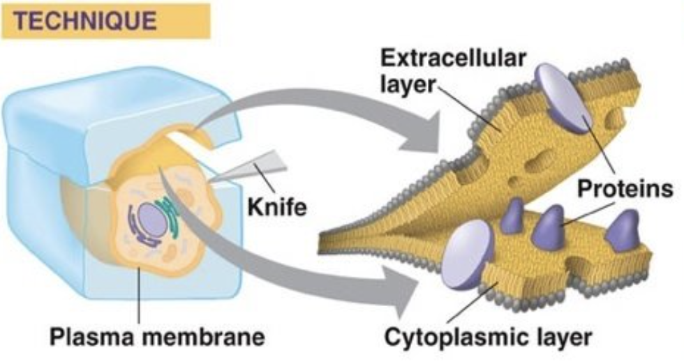
Cell (Plasma) membrane significance + function
outer boundary of cell + encloses all its contents
controls entry + exit of substances. (can pump substances in even when concentration outside is low) (keeps unwanted substances outside)
allows cell to maintain concentrations of substances that are different from those outside of the cell
permeability of plasma relies on a structure based on lipids
what is lysis?
when the plasma membrane of a cell bursts. caused by excess pressure, viruses or the cell carries it out itself (autolysis).
lysis leads to the death of the cell
Genetic material significance + function
contains information needed for a cell to carry out its functions
Many genes hold the instructions to making a protein
DNA can be copy and pasted on to daughter cells → information stored is inheritable
DNA is stored in nucleus
Bacteria don’t have a nucleus - DNA is stored in Cytoplasm
Cytoplasm Significance + function
main component → water
substances are dissolved in the water
water allows enzymes to catalyse reactions → metabolism of cell
cytoplasm must continuously break down and replace proteins due to proteins being easily damaged.
importance of metabolism in cytoplasm
provides cell with energy and produces proteins and other substances that make up the structure of the cell
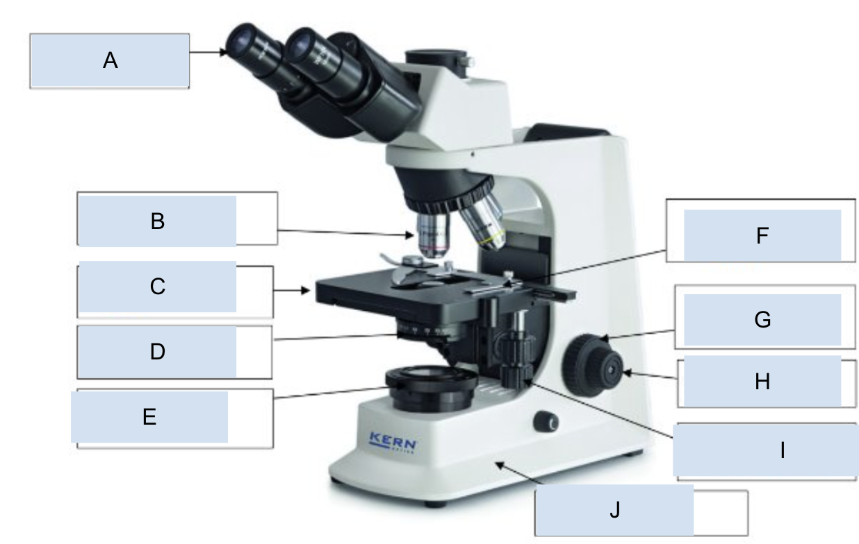
label
A - eyepiece lens
B - objective lens
C - stage
D - aperture
E - light source
F - stage clips (where slide goes)
G - coarse focus
H - fine focus
I - stage controls
J - base
eyepiece graticule vs stage micrometer
eyepiece graticule - scale in the microscope eyepiece. By itself, it has no fixed units (the "divisions" are arbitrary).
stage micrometer - a slide with a precise scale, usually 0.01 mm or 10 µm divisions
how to determine the size of a division in a eye piece
Place the stage micrometer on the microscope stage.
Focus on the scale.
Line up the eyepiece graticule scale with the stage micrometer scale.
See how many eyepiece divisions match up with a known length on the stage micrometer.
1 eye piece division = 100/number of eyepiece divisions
(⚠ Important: If you change objective lenses (magnification), you must recalibrate, since the apparent size changes.)
how to determine the size of a cell using an eye piece graticule
Replace the stage micrometer with your specimen slide after finding the length of one division.
Count how many eyepiece graticule divisions span across the cell (e.g., diameter of a nucleus, length of a cell).
Convert using your calibration.
(e.g : Cell = 12 eyepiece divisions, calibration = 2.5 µm/division → 12 × 2.5 µm = 30 µm)
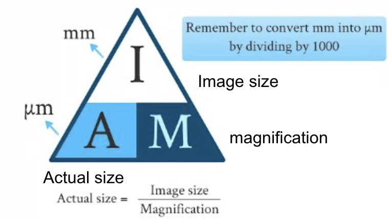
magnification triangle formula
Magnification = image size(mm)/actual size(micrometers) = measured length/scale bar length
what is resolution
the ability to distinguish between two objects very close together.
The higher the resolution of an image, the greater the detail that can be seen.
resolution is limited by the wavelength of the radiation used to view the sample
Light microscopes
magnify images only up to x1000 due to long wavelength of light
low resolution - 0.5µm (micrometers)
cells viewed under microscope are alive
images are coloured
easy to use
Electron microscopes
types - transmission + scanning
use beams of electrons → have shorter wavelength than light
high resolution - 2nm (nanometers)
high magnification - x 1,000,000
expensive
not easy to use
black + white pictures
transmission electron microscopes
2D images
samples stained with heavy metals → samples are dead
good imagery of structures in cells
electrons are scattered as they pass through a thin section of the specimen, and then detected and projected onto a fluorescent screen.
scanning electron microscopes
3D images
electrons are reflected off the surface of the specimen.
sample is stained using harsh chemicals → dead
staining with methylenblue
Molecules are colourless under electron microscopes and so stains such as methylenblue binds to DNA or RNA in order to be able to visualize the nucleus or cytoplasm.
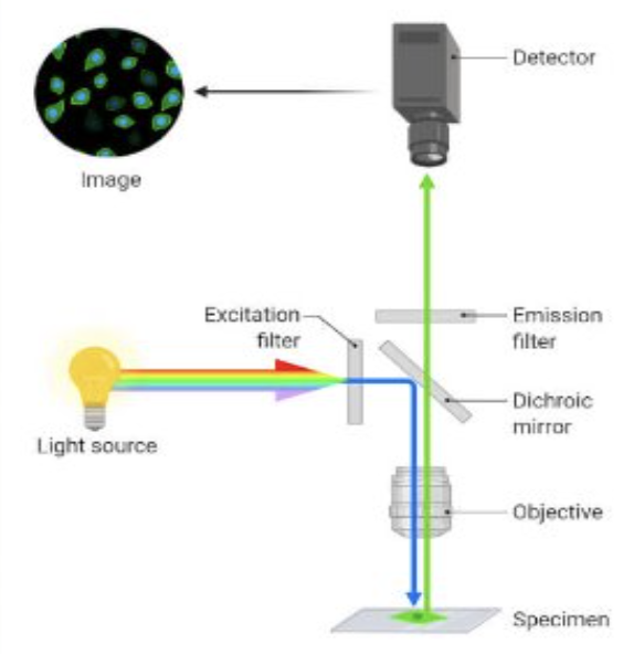
fluorescent staining
uses a higher intensity light to illuminate the sample
sample is stained with fluorescence dye which causes the light to emit back at a longer wavelength
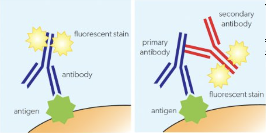
Immunofluorescent staining
uses antibodies which are equipped with a fluorescent marker.
Upon binding with an antigen a fluorescent image can be produced
cryogenic microscopy
used for researching the structures of proteins
protein is frozen and then placed in an electron microscope and patterns of many different proteins are produced
using computer algorithms a 3D image is then produced
freeze-fracture electron microscopy
used to produce images of surfaces within cells
rapid freezing of cells + fracturing allows the cell to be broken along lines of weakness (center of membrane)
any structures which appear globular are transmembrane proteins
cell wall features + functions in Prokaryote
features :
semi rigid structure
made from peptidoglycan
function:
maintains shape of the cell
protects the cell
prevents the cell from bursting
Cell membrane features + functions in Prokaryote
features:
thin
partially permeable layer of phospholipids
function:
controls the entry + exit of substances
pumps substances in and out by active transport
cytoplasm features + function in Prokaryote
features:
fluid (mostly water) that fills space inside the plasma membrane
contains many enzymes + ribosomes
does not contain any membrane bound organelles
functions:
carries out chemical reactions of metabolism using enzymes and biochemical molecules
Ribosomes features + functions in Prokaryote
features:
70S (smaller than eukaryotic ribosomes)
granular appearance in electron microscope
functions:
Synthesize (make or manufacture) proteins through transcription & translation
Nucleoid features + functions Prokaryote
features:
Central region of the cytoplasm containing naked (not wrapped around a protein) single chromosomal DNA
DNA in prokaryotes is circular
Not surrounded by a membrane
functions:
essential for controlling the activity of the cell and reproduction.
where transcription and replication of DNA take place
Eukaryotic cells
animal, plant, fungi
more complex + bigger in size
Nucleus with genetic material surrounded by a membrane
Membrane bound organelles
Unicellular or multicellular
Cell structure is compartmentalised.
plant cells and structure : plastids, cell wall, vacuole, centrioles, Undulipodia
Plastids : plastids of varied types such as chloroplasts (for photosynthesis) and amyloplasts (to store starch)
Cell wall: have walls composed of cellulose
Vacuole: have a large permanent vacuole used for storage of substances and pressurising the cell
Centrioles: Absent
Undulipodia: Absent
Animal cells and structure - plastids, cell wall, vcuole, centrioles, undulipodia
Plastids : none
Cell wall: none
Vacuole: small + temporary, used to expel water and digest food or pathogen
Centrioles: used to construct spindle that moves chromosomes in mitosis
Undulipodia: cilia + flagella present in many animal cells
Fungal cells and structure — plastids, cell wall, vacuole, centrioles, undulipodia,
Plastids : none
Cell wall: present + composed of chitin
Vacuole: large permanent vacuole used for storage of substances + pressuring cell
Centrioles: Absent
Undulipodia: Absent
eukaryotic
Nucleus + Nucleolus features
spherical with double membrane
have holes in membrane
chromatin - uncoiled chromosomes
Nucleolus consists of RNA and proteins, makes up 25% of nucleus
Nucleus + Nucleolus function
Stores genetic information in form of chromosomes (DNA and associated histones)
Nucleolus produces rRNA (ribosomal) which combine with proteins for use outside the cell to form ribosomes.
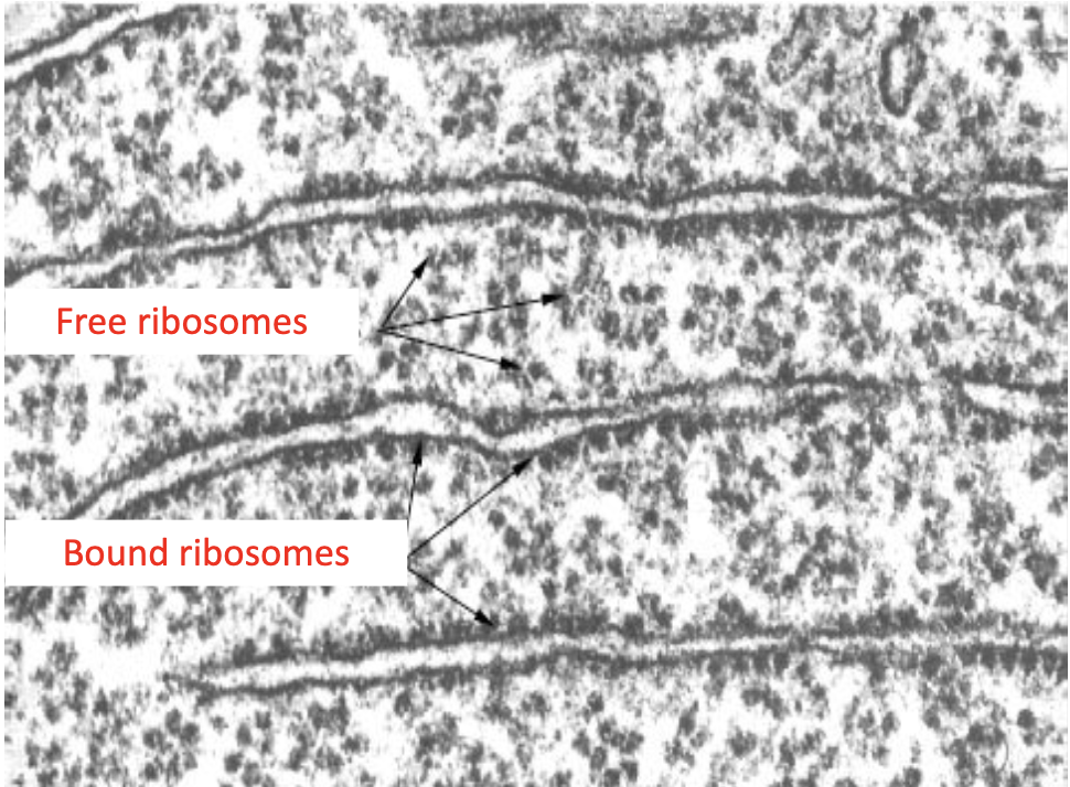
Free ribosomes features + functions
features
80S (larger than in prokaryotes), ca.20nm
No exterior membrane
Free in the cytoplasm or bound to ER
Composed of ribosomal RNA and protein
produced in the nucleolus of the nucleus
Appear as dark granules
functions
Produces proteins to function in the cytoplasm for use within the cell (enzymes)
Mitochondrion features
Has a double membrane
Outer membrane is smooth, inner membrane is folded
The folds are called “cristae”
Variable in shape and number (spherical or ovoid)
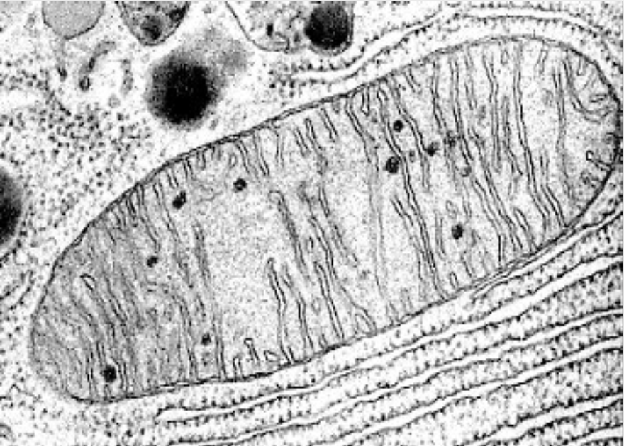
Mitochondrion function
Site of ATP production by (aerobic) cell respiration.
Fat digestion if it is used as an energy source in the cell
rough endoplasmic reticulum (rER) features
Made of flattened membrane sacs called cisternae, attached to the outside of the cisternae are ribosomes (rER)
Extensive network of tubules or channels that extends almost everywhere in the cell from the nucleus to the plasma
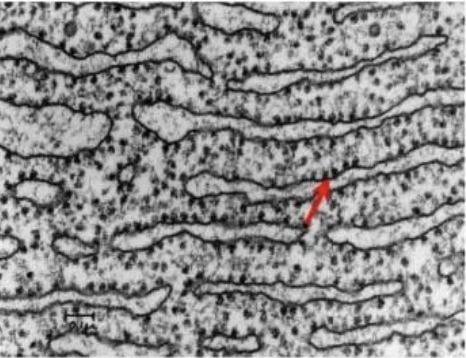
rough endoplasmic reticulum (rER) function
responsible for the production of proteins which are then transported by vesicles to the Golgi apparatus for modification.
Golgi apparatus features
Consists of flattened sacs called cisternae, which are stacked on top of one another
Has a two sides: cis-side (receives products at that site), and a trans-side (discharges products)
Transport vesicles bud off
Most of these are packaged into vesicles for secretion through the plasma membrane
Difference to rER:
No attached ribosomes
Often sited close to the plasma membrane
The cisternae are shorter and more curved than those of rER
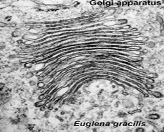
Golgi apparatus function
Processes proteins that arrive from the rER.
functions in collection, packaging, modification and distribution and transportation of materials synthesised in the cell.
chloroplasts features + functions
features
Double membrane surrounding the chloroplast
Stacks of thylakoids inside
Each thylakoid is a disc composed of a flattened membrane.
Variable shape (spherical or ovoid)
functions
Production of glucose and other organic compounds by photosynthesis
lysosomes features
features
Formed from Golgi vesicles which bud off
spherical with single membrane
High concentration of enzymes (proteins)
cause this organelle to stain heavily and hence appears dark
Only in animal cells (plants use vacuoles)
lysosomes function
Used for the breakdown of food or unwanted, damaged substances + organelles using enzymes.
vacuoles + vesicles features
Single membrane with fluid inside
Plant cells: vacuoles are large and permanent, often occupying the majority or the cell
Animal cells: Small and temporary – typically referred to as vesicles.
vacuoles + vesicles function
Vacuoles: In plant cells: Used for maintenance of water balance and internal pressure.
Vesicles: Used for transport of substances within the cell – often from rER to Golgi apparatus.
flagellum + cilia features
Whip-like structures projecting from the cell surface
Contain a ring of 9 double microtubules + 2 central ones
Flagella are larger, and only one is present, cilia are smaller and many are present
Have microtubules inside
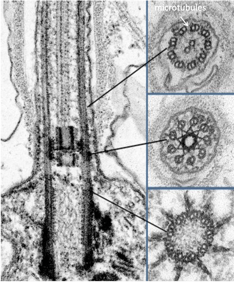
flagellum + cilia functions
Cilia move liquid over surfaces (e.g. particle-laden mucus towards throat)
For movement (sperm cells)
microtubules + centrioles features
features
Microtubules:
Small cylindrical fibres
Form core inside flagella or cilia
Composed of the polymer tubulin
centrioles:
Consist of 2 groups of 9 triple microtubules
Only in animal cells
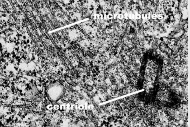
microtubules + centrioles functions
Microtubules move chromosomes to opposite sides of a cell during cell division and help to construct cell walls.
In animal cells, centrioles move towards the poles of a cell and serve as anchor points for microtubules during cell division.
cytoskeleton features + functions
features
Constructed from protein fibers like tubulin + actin, which are used to make microtubules + microfilaments.
functions
Cytoskeleton microfilaments help animal cells to maintain shape.
cell wall features + functions
features
extracellular component, not an organelle
All plant cells have a cell wall, but also fungi and some protists
Consists of the polysaccharide cellulose
functions
Permeable – does not affect transport in and out of the cell
Strong – gives support to the cell and prevents plasma membrane bursting when under pressure
prokaryotes vs eukaryotes Genetic material
Prokaryotes - DNA often circular (plasmids) and nucleoid without proteins (histones)
Eukaryotes - DNA is linear and associated with proteins (histones) to form chromatin
prokaryotes vs eukaryotes size
prokaryotes - small (0.2-0.3 micrometers)
eukaryotes - large (10-100 micrometers)
prokaryotes vs eukaryotes membrane enclosed organelles
prokaryotes - no nucleus or any membrane bound organelles Have nucleoid instead of nucleus.
eukaryotes - Always have membrane-surrounded nucleus and other membrane-bound organelles
prokaryotes vs eukaryotes uni/multicellular
prokaryotes - always unicellular
eukaryotes - often multicellular
prokaryotes vs eukaryotes examples
prokaryotes - bacterial cells
eukaryotes - plant cells, animal cells, fungal cells
prokaryotes vs eukaryotes ribosomes
prokaryotes - small 70S
eukaryotes - large 80S
characteristics + functions of life
Metabolism - sum of all biochemical reactions that occur in a living organism
Reproduction - production of offspring, sexually/asexually
Homeostasis - maintenance of a constant internal environment in an organism
Growth - an increase in size or number of cells
Response - perception of stimuli and carrying out appropriate reactions in response
Excretion - removal of waste products of metabolism from an organism
Nutrition - supplying the nutrients required for energy, growth and repair in an organism
why are Striated muscle cells atypical
have multiple nuclei (multinucleated)
why are aseptate fungal hyphae atypical
have multiple nuclei (multinucleated)
why are red blood cells atypical
dont have a nucleus
why are phloem sieve cells atypical
dont have a nucleus
have holes in cell walls
hardly have any cytoplasm