Terms list 7 and 8
1/157
There's no tags or description
Looks like no tags are added yet.
Name | Mastery | Learn | Test | Matching | Spaced | Call with Kai |
|---|
No analytics yet
Send a link to your students to track their progress
158 Terms
Cell body (soma)
2

mitochondria of axon
Produces energy for the cell
Nissl bodies
Clusters of rough endoplasmic reticulum in neurons
Dendrites
1
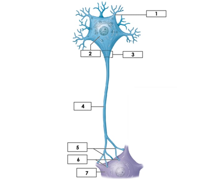
Axon: Unmyelinated
4

Axon Hillock
3

Oligodendrocytes (CNS)
Produce myelin in the central nervous system
Myelin sheath
Insulating layer around axons
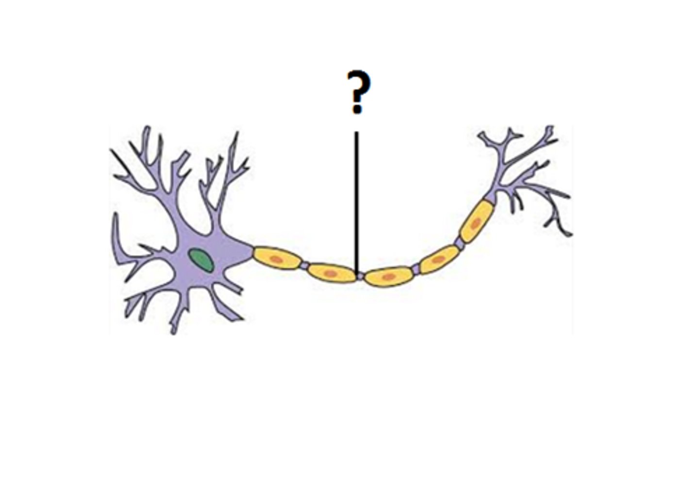
Nodes of Ranvier
3

Schwann cells (PNS) = internode
5
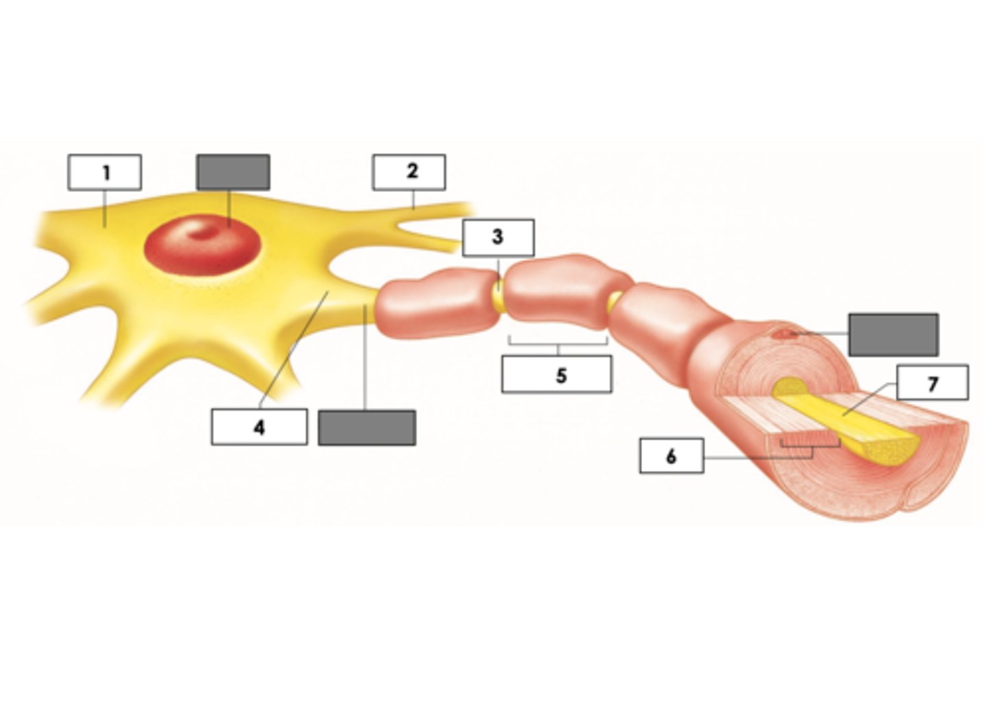
Telodendria
5

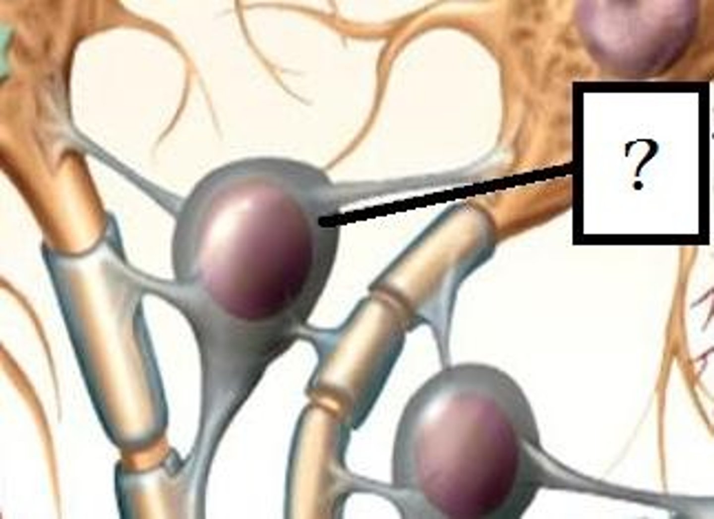
Synaptic knobs (ends of telodendria)
Enlarged structures that release neurotransmitters
Synapse
Junction between two neurons
Synaptic vesicles
3
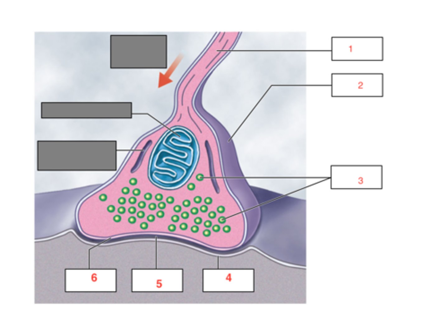
Neurotransmitter
Chemical messenger that transmits signals between neurons
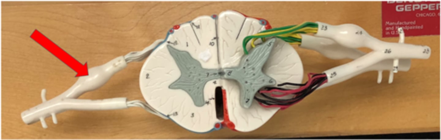
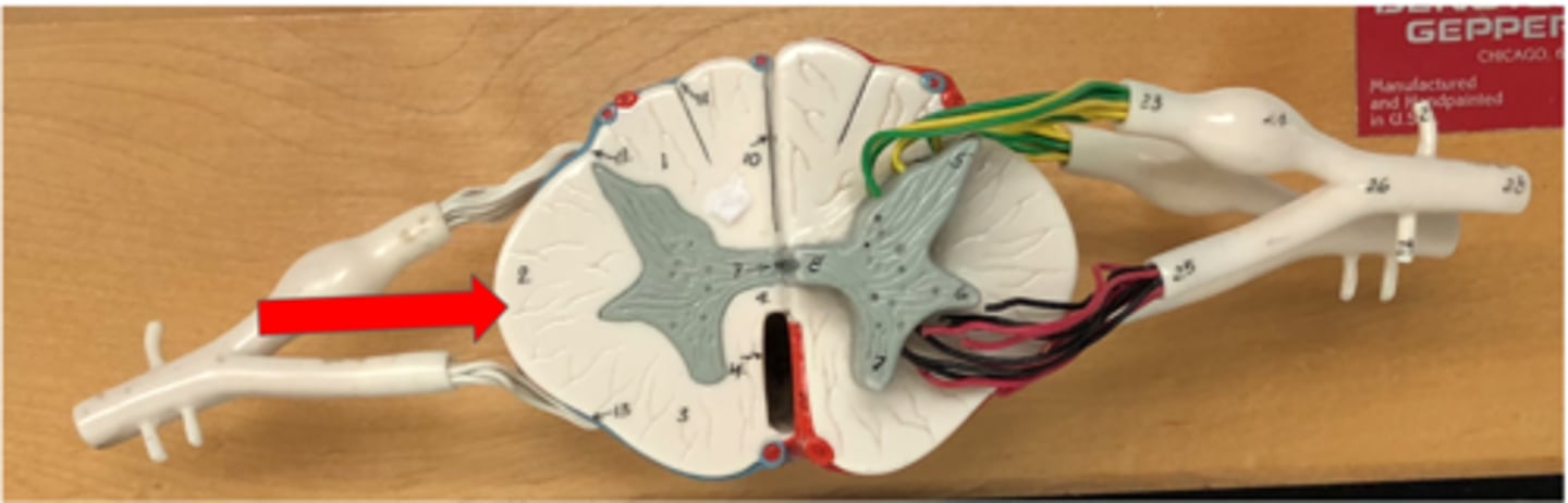
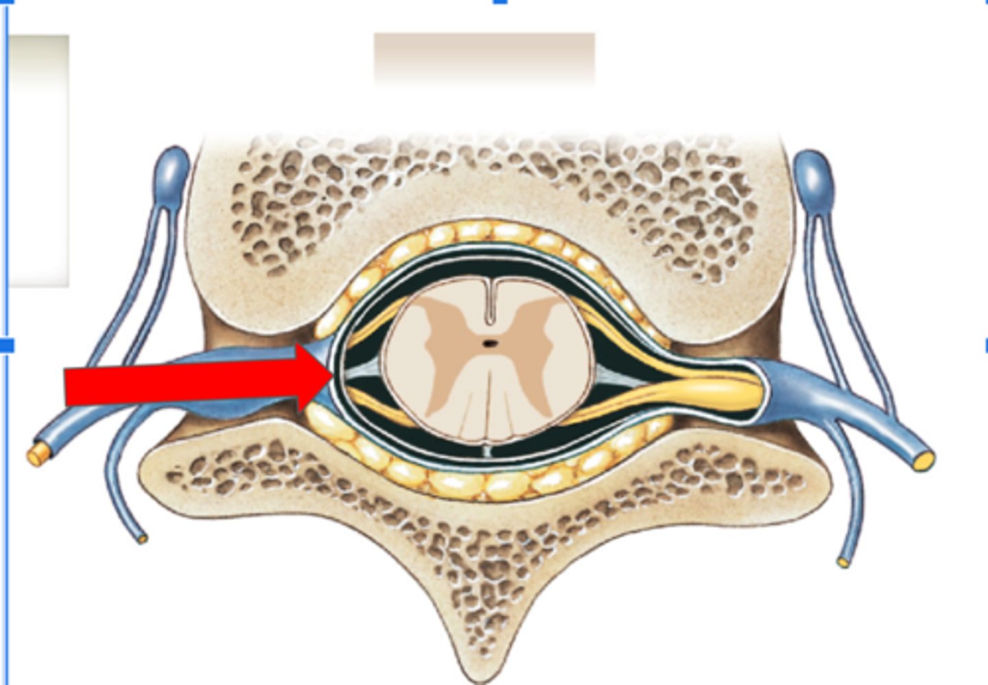
Spinal Nerve
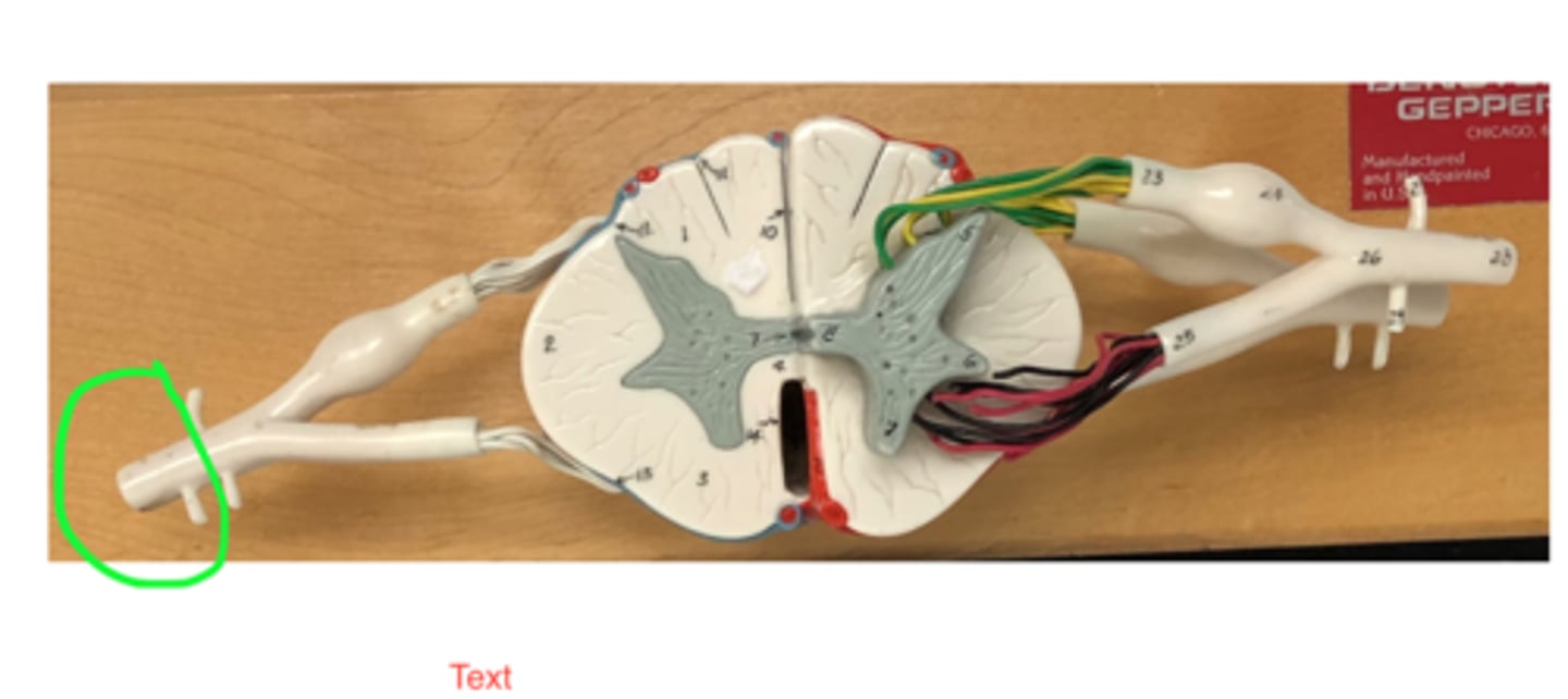
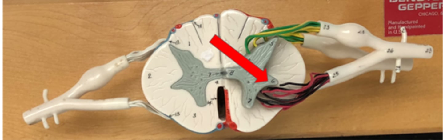
Anterior (ventral) root
Contains motor neurons
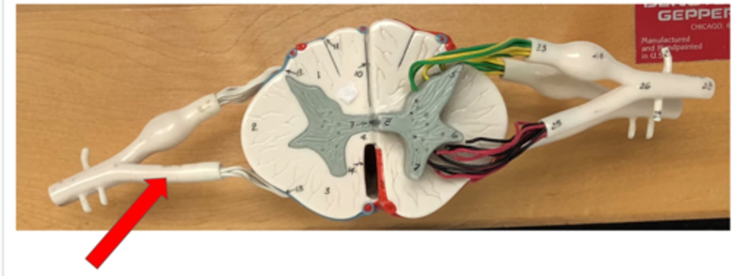
Posterior (dorsal) root
Contains sensory neurons
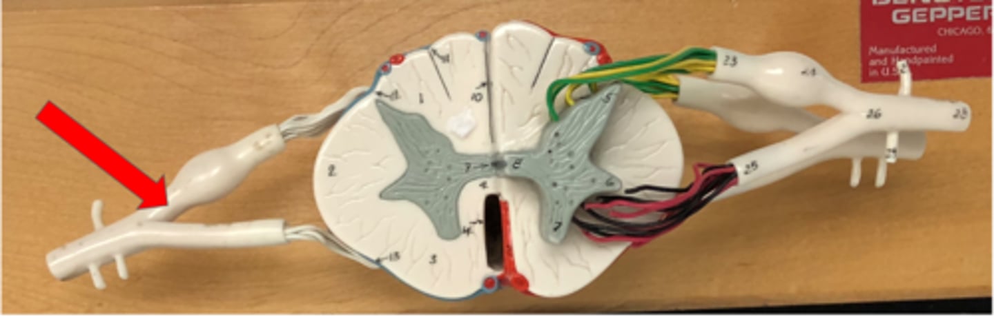
Posterior (dorsal) root ganglion
Contains cell bodies of sensory neurons
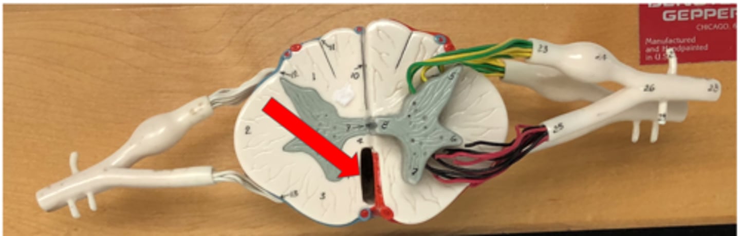
Anterior median fissure
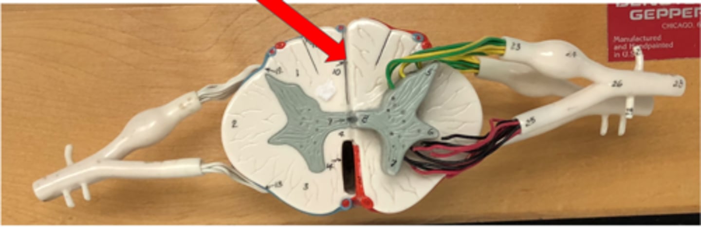
Posterior median sulcus
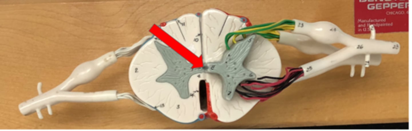
Central canal
Contains cerebrospinal fluid in the spinal cord
Gray matter
Inside of spinal cord, outside of brain
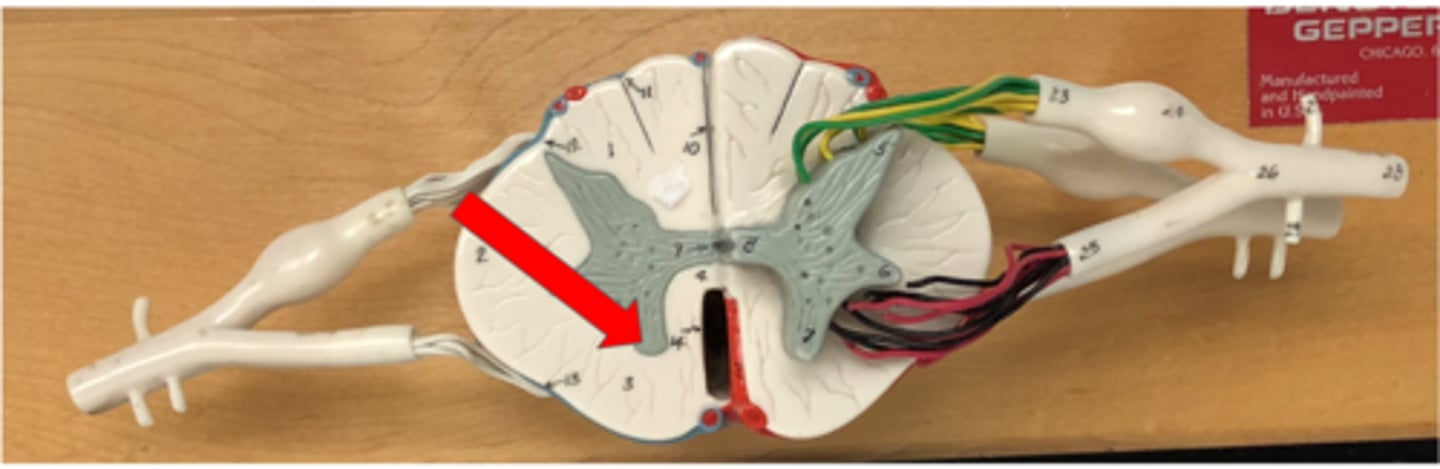
Anterior horn
Contains cell bodies of motor neurons in the spinal cord
Lateral horn
Contains cell bodies of autonomic motor neurons in the spinal cord
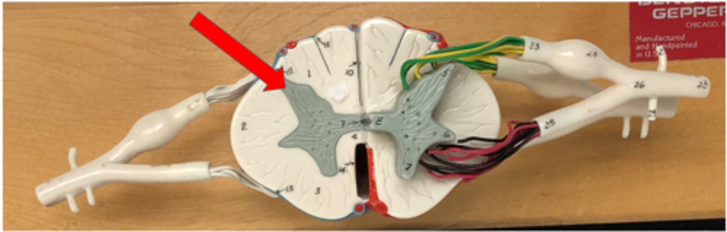
Posterior horn
Contains axons of sensory neurons in the spinal cord
White matter
outside of spine, inside of brain
Anterior white column (funiculus)

Posterior white column (funiculus)

Lateral white column (funiculus)
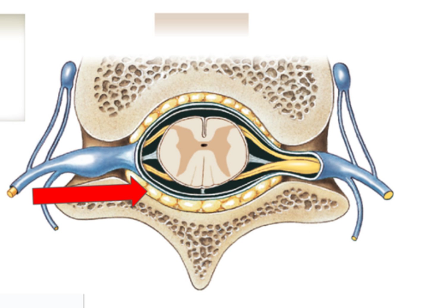
Pia mater (spine)
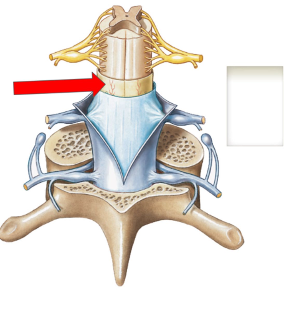
Subarachnoid space (spine)
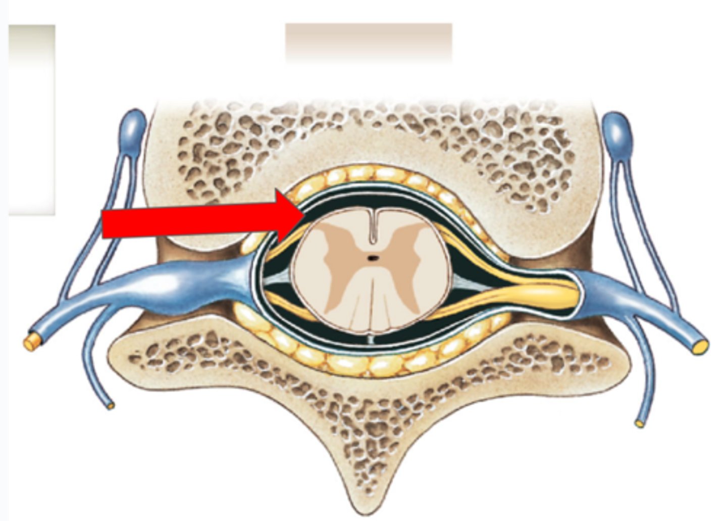
Arachnoid mater (spine)
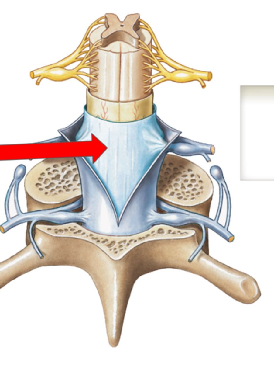
Subdural space (spine)
(space)
Dura mater (spine)
Outermost layer of the meninges in the spinal cord
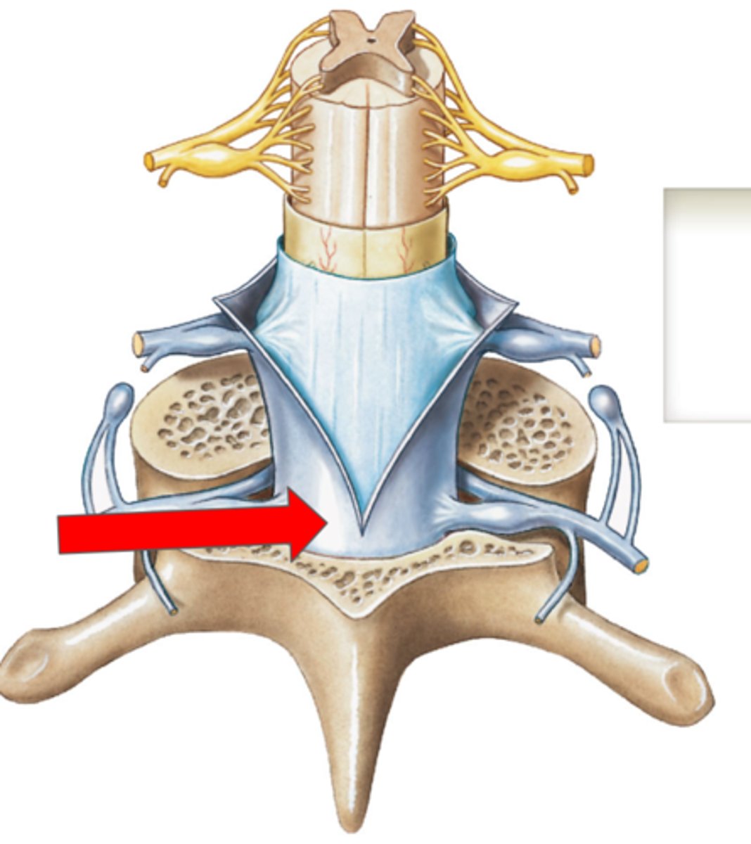
Epidural space (contains fat) (spine)
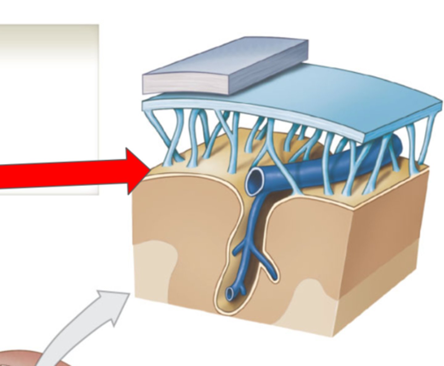
Cranial Meninges
Protective layers around the brain
Dura mater
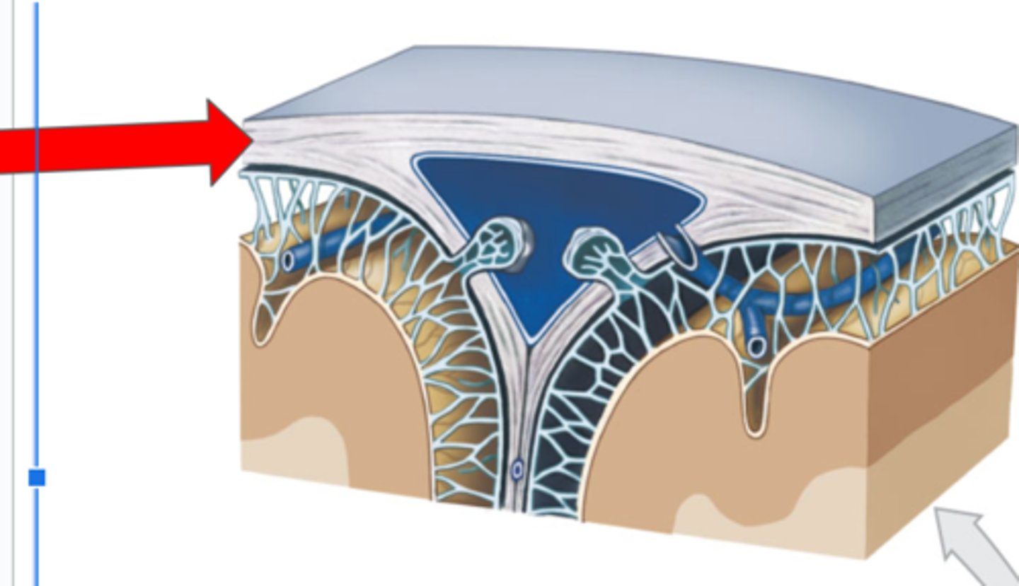
Arachnoid mater
Middle layer of the cranial meninges
Subdural space
(space)
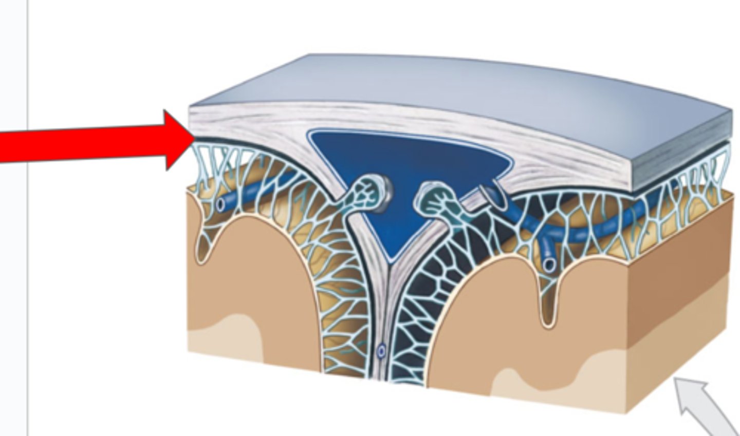
Subarachnoid mater
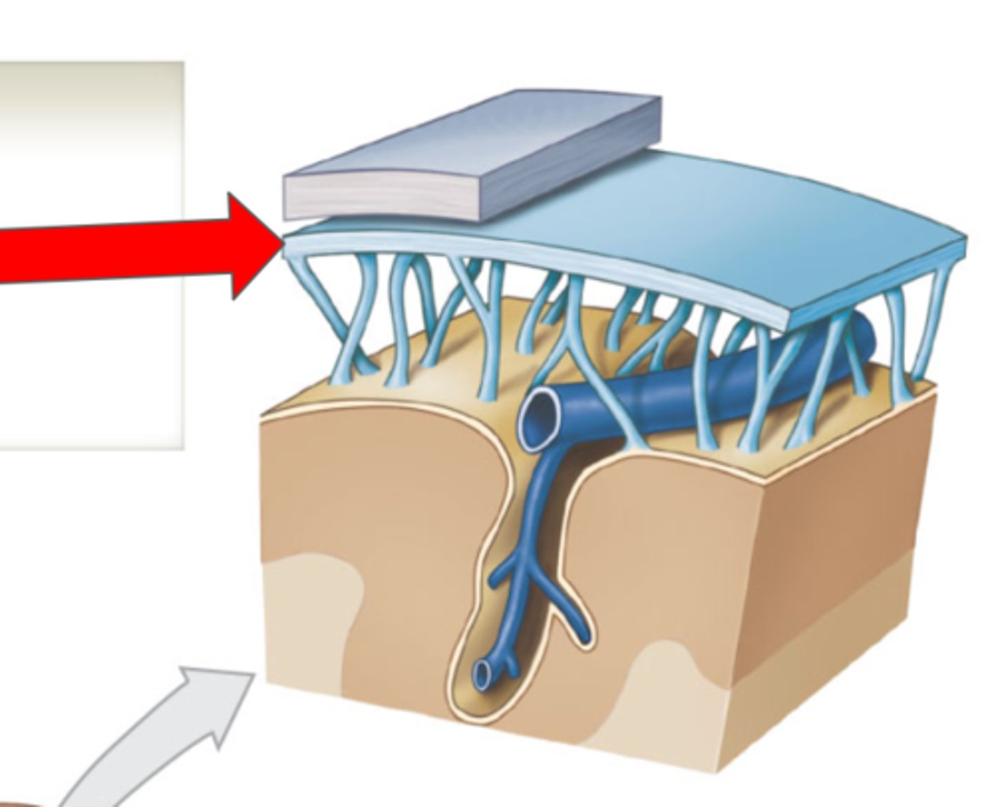
Arachnoid trabeculae

Pia mater
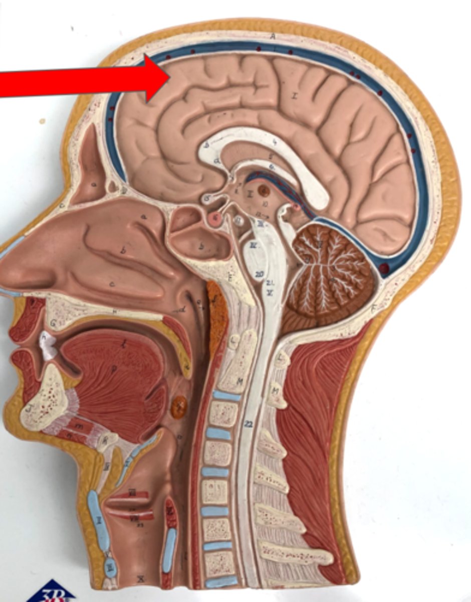
Cerebrum (Two Cerebral Hemispheres)
Cerebral cortex
Ventricles
Fluid-filled cavities in the brain
Lateral ventricles
Cerebral aqueduct
Third ventricle
Fourth ventricle
Choroid plexus
Produces cerebrospinal fluid
Fornix
Epithalamus
contains the pineal gland
Pineal gland
Produces melatonin
Thalamus
Interthalamic adhesion
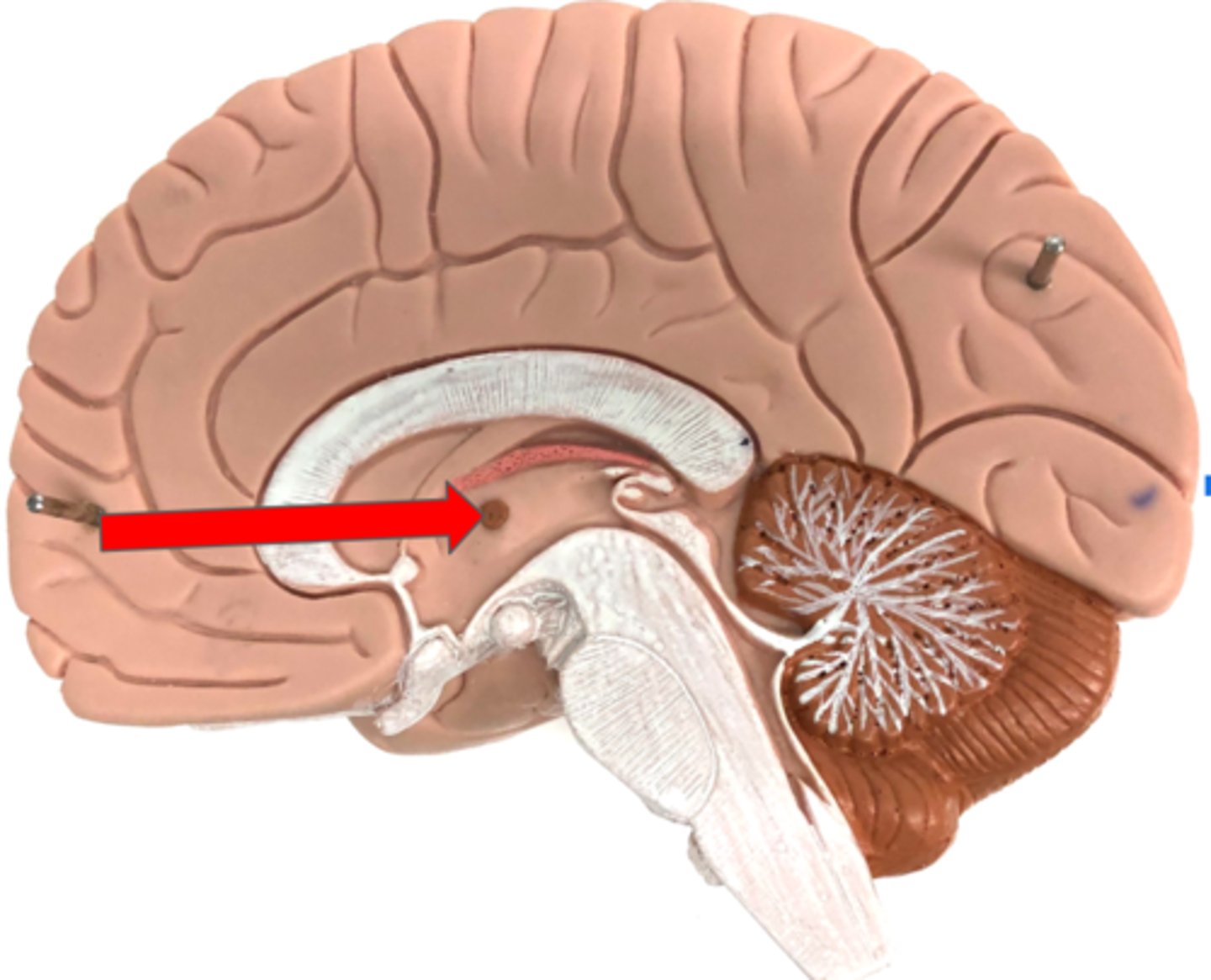
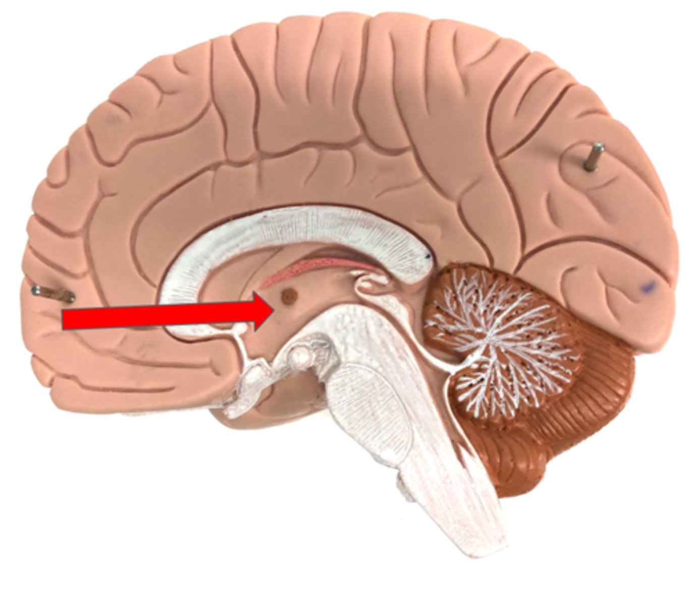
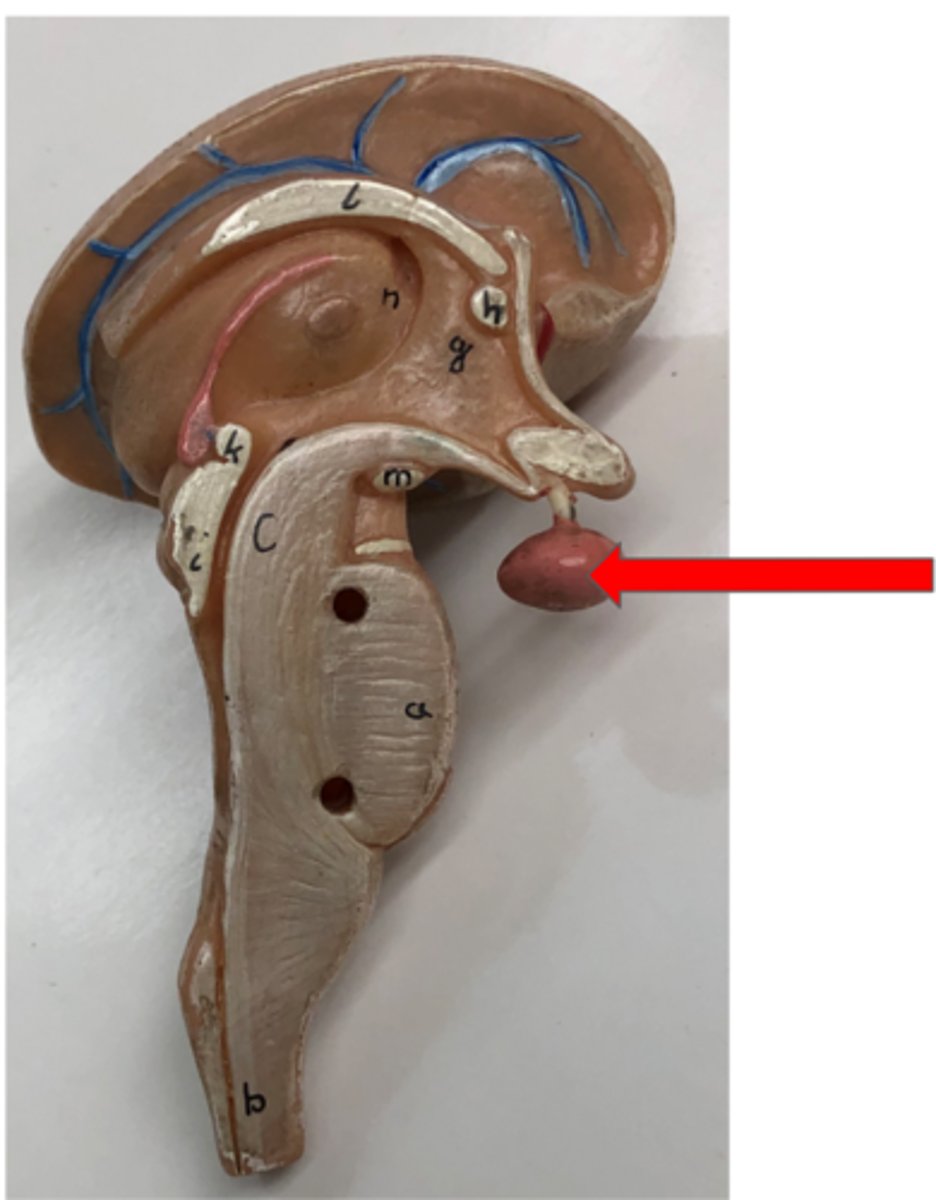
Hypothalamus
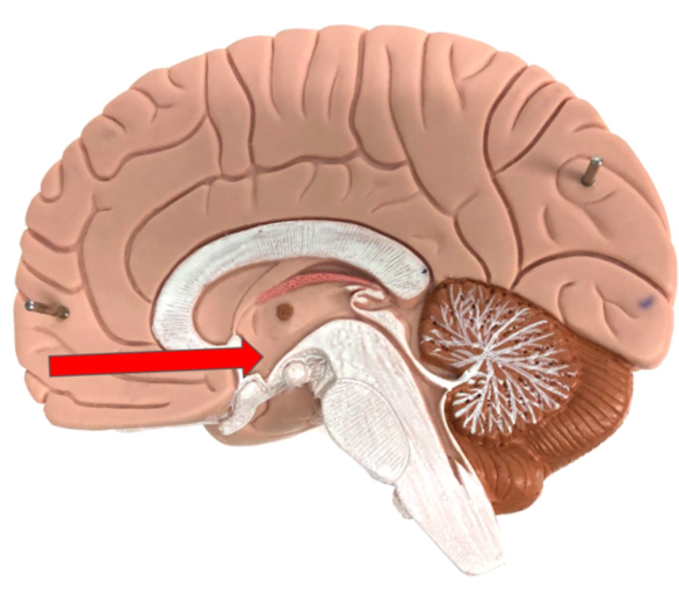
Infundibulum
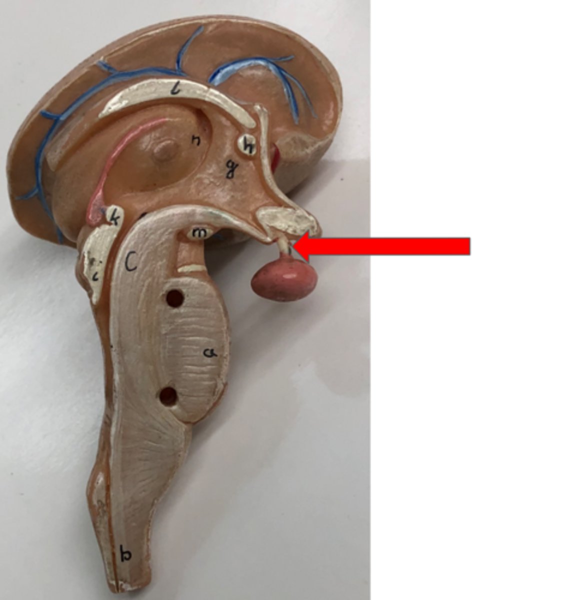
Pituitary gland
Produces and releases hormones
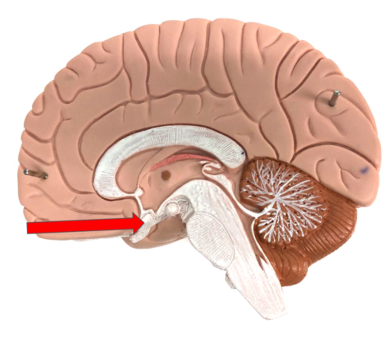
Optic chiasm
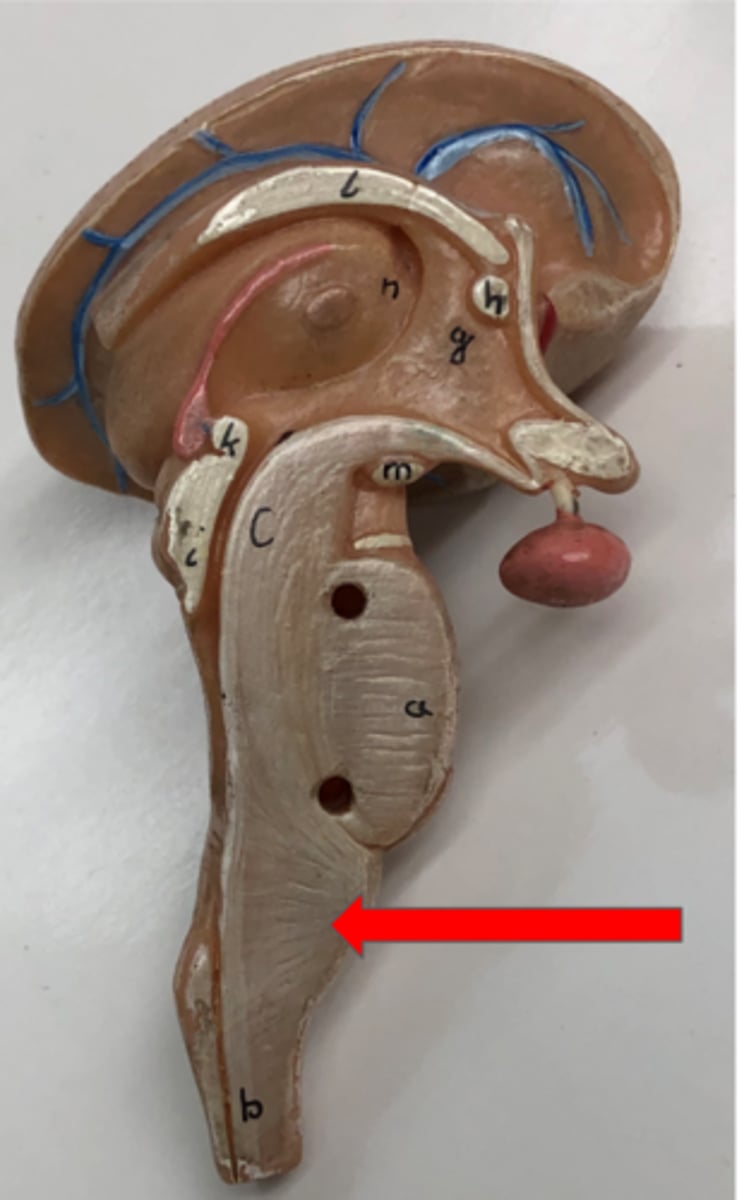
Brain Stem
Connects the brain and spinal cord
Pons
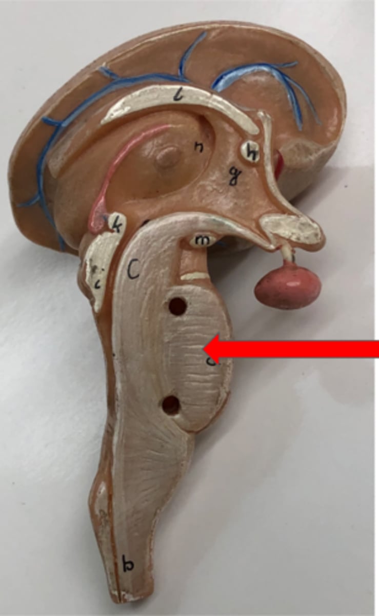
Medulla oblongata
Frontal lobe
Part of the cerebrum responsible for decision making and motor control
Parietal lobe
Part of the cerebrum responsible for sensory processing
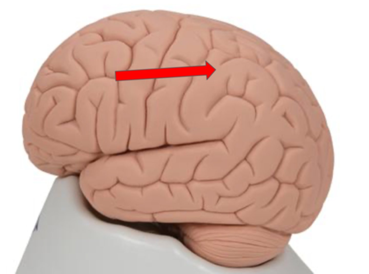
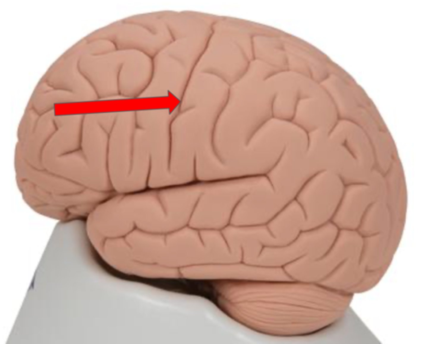
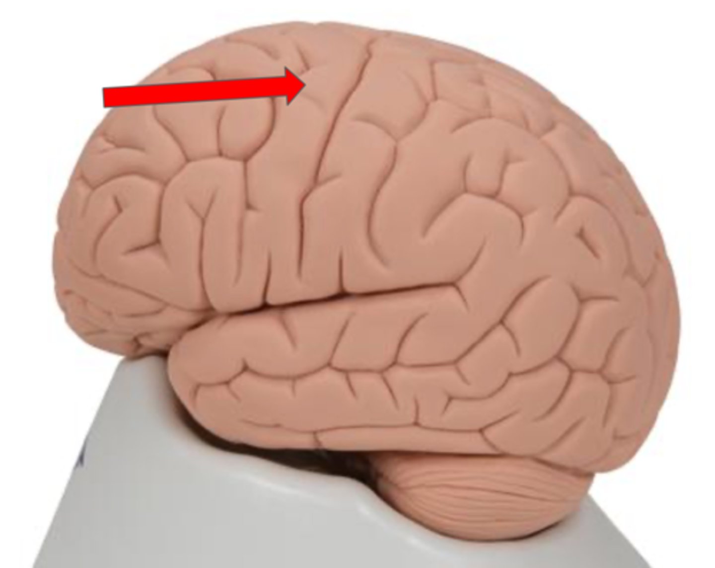
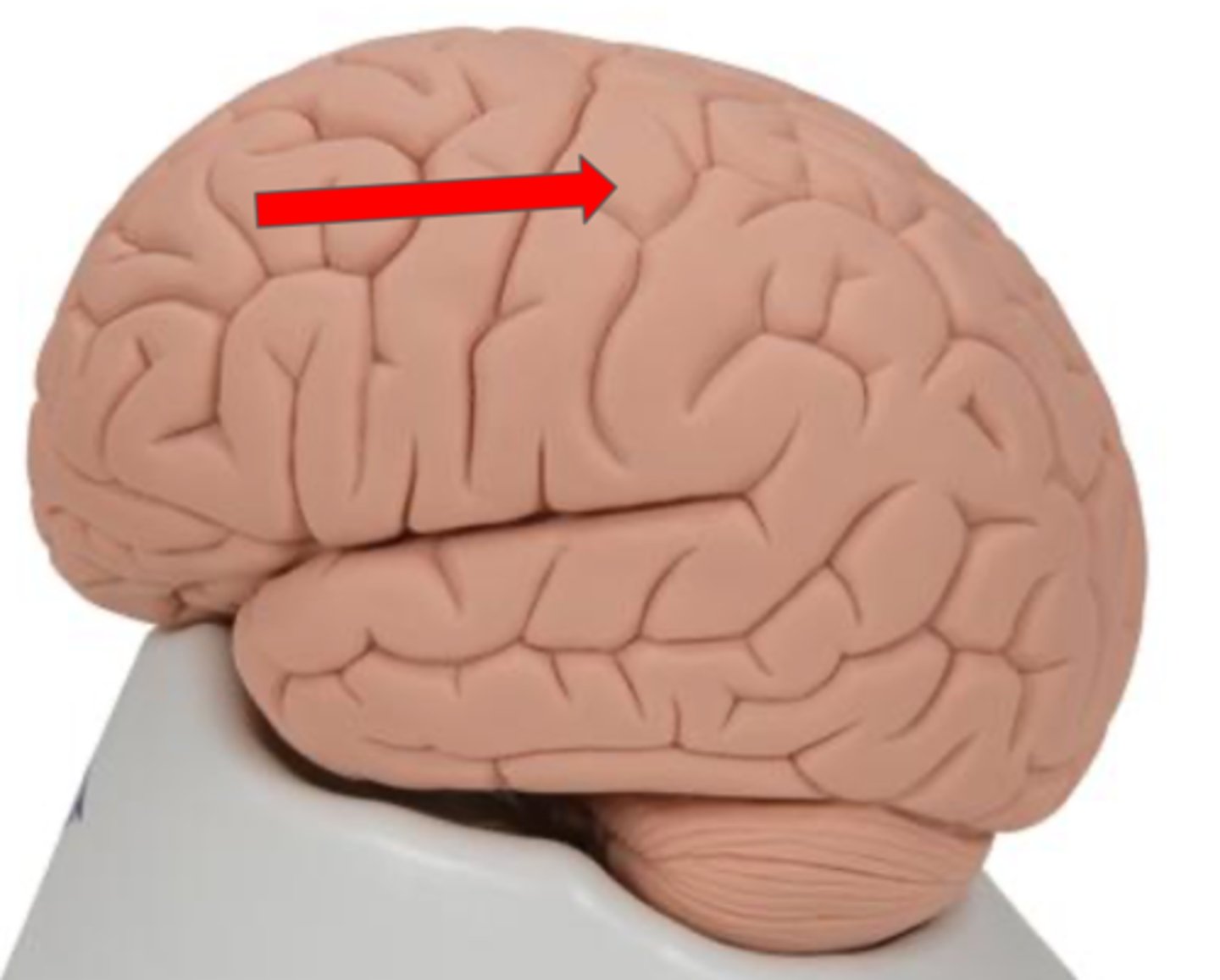
Central sulcus
Precentral gyrus
Primary motor cortex located in the frontal lobe
Postcentral gyrus
Primary sensory cortex located in the parietal lobe
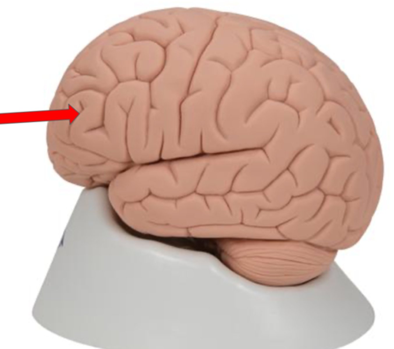
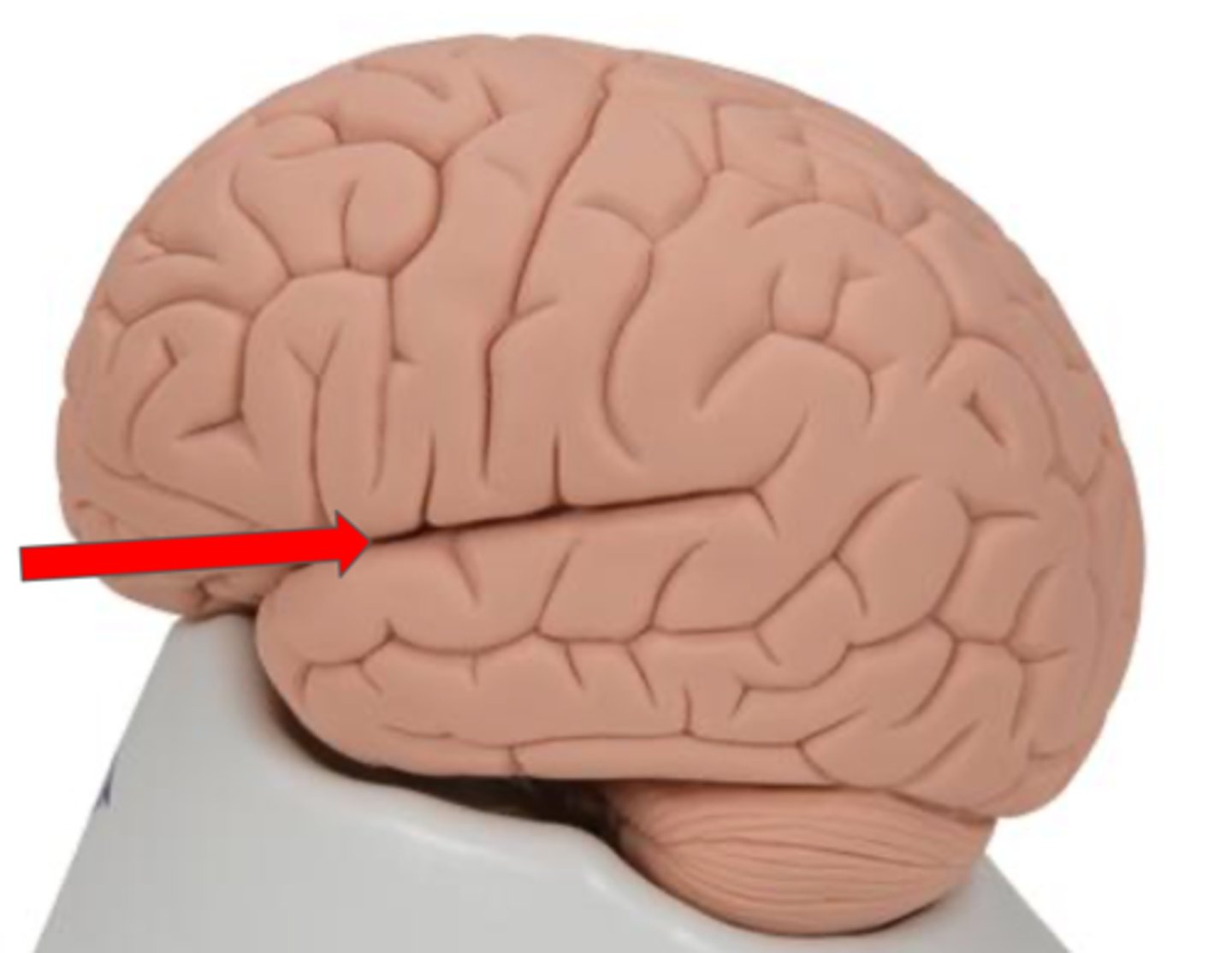
Lateral sulcus
Groove that separates the temporal lobe from the frontal and parietal lobes
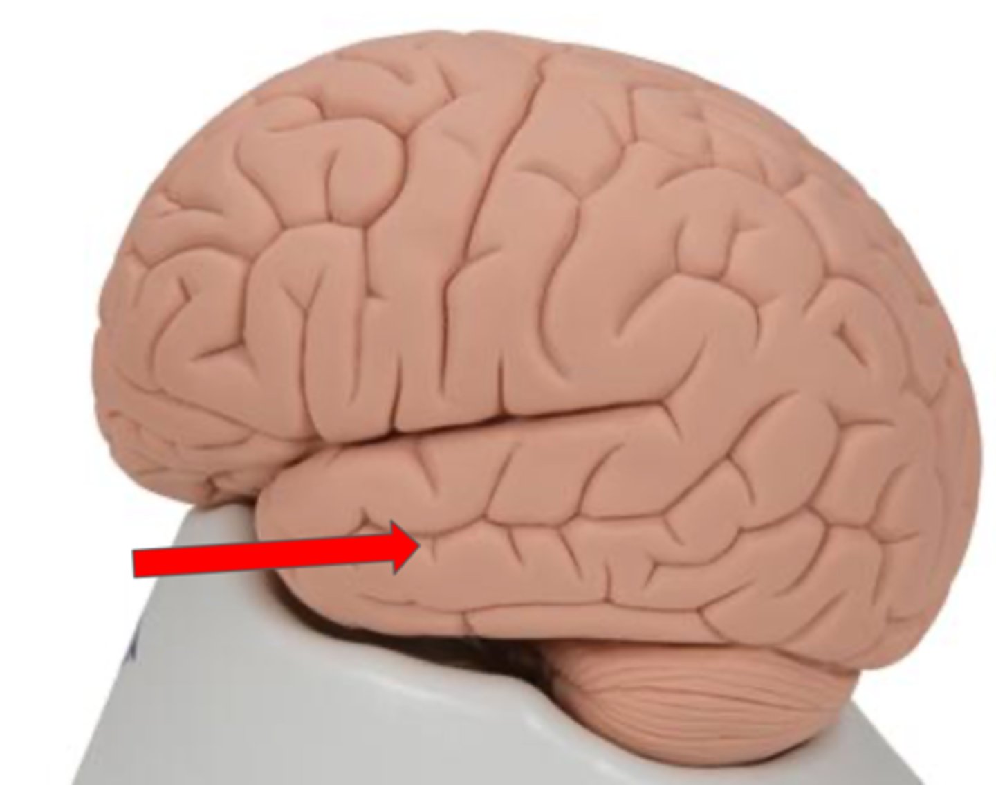
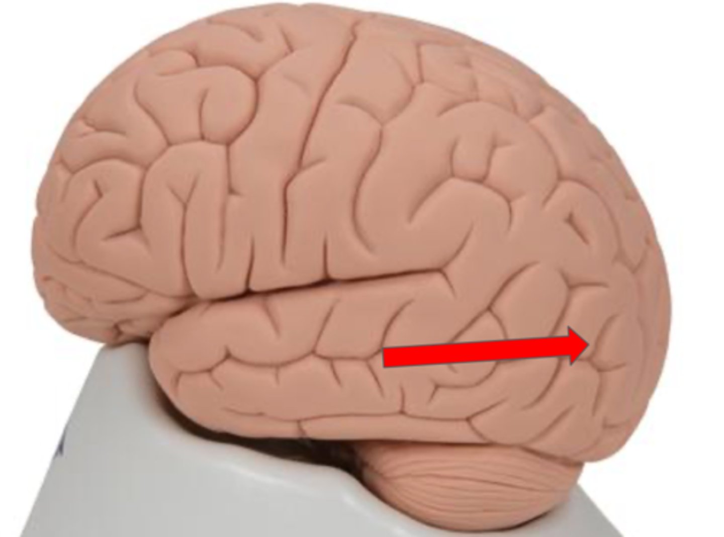
Temporal lobe
Part of the cerebrum responsible for auditory processing and memory
Occipital lobe
Part of the cerebrum responsible for visual processing
Insula
deep to temporal lobe
Corpus Callosum
Septum pellucidum
Thin membrane that separates the lateral ventricles
central canal (brain model)
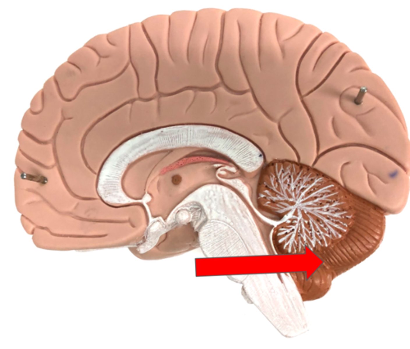
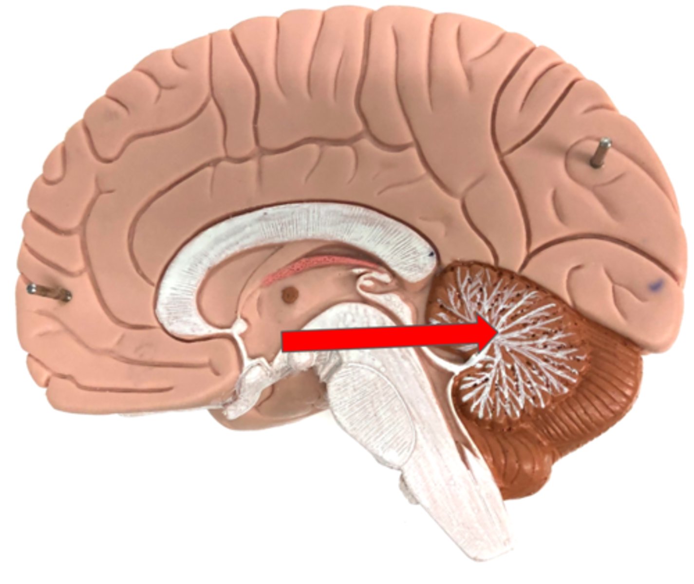
Cerebellum
Arbor vitae
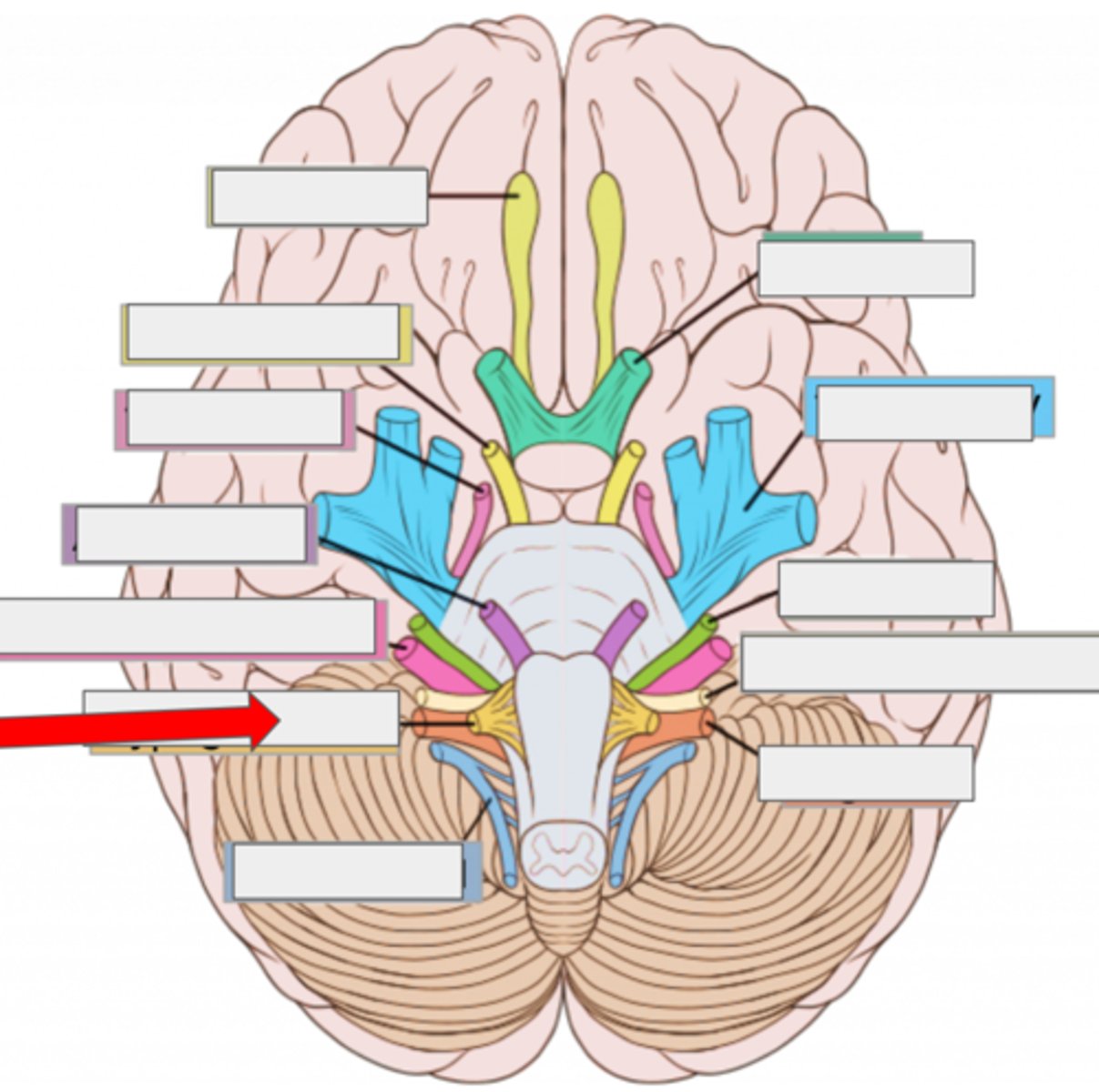
I olfactory- smell
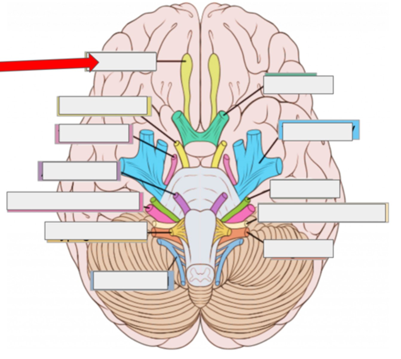
II optic- sight

III oculomotor- moves eye
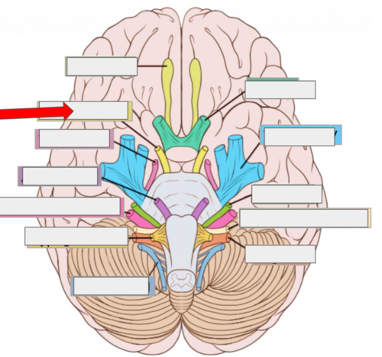
IV trochlear- moves eye
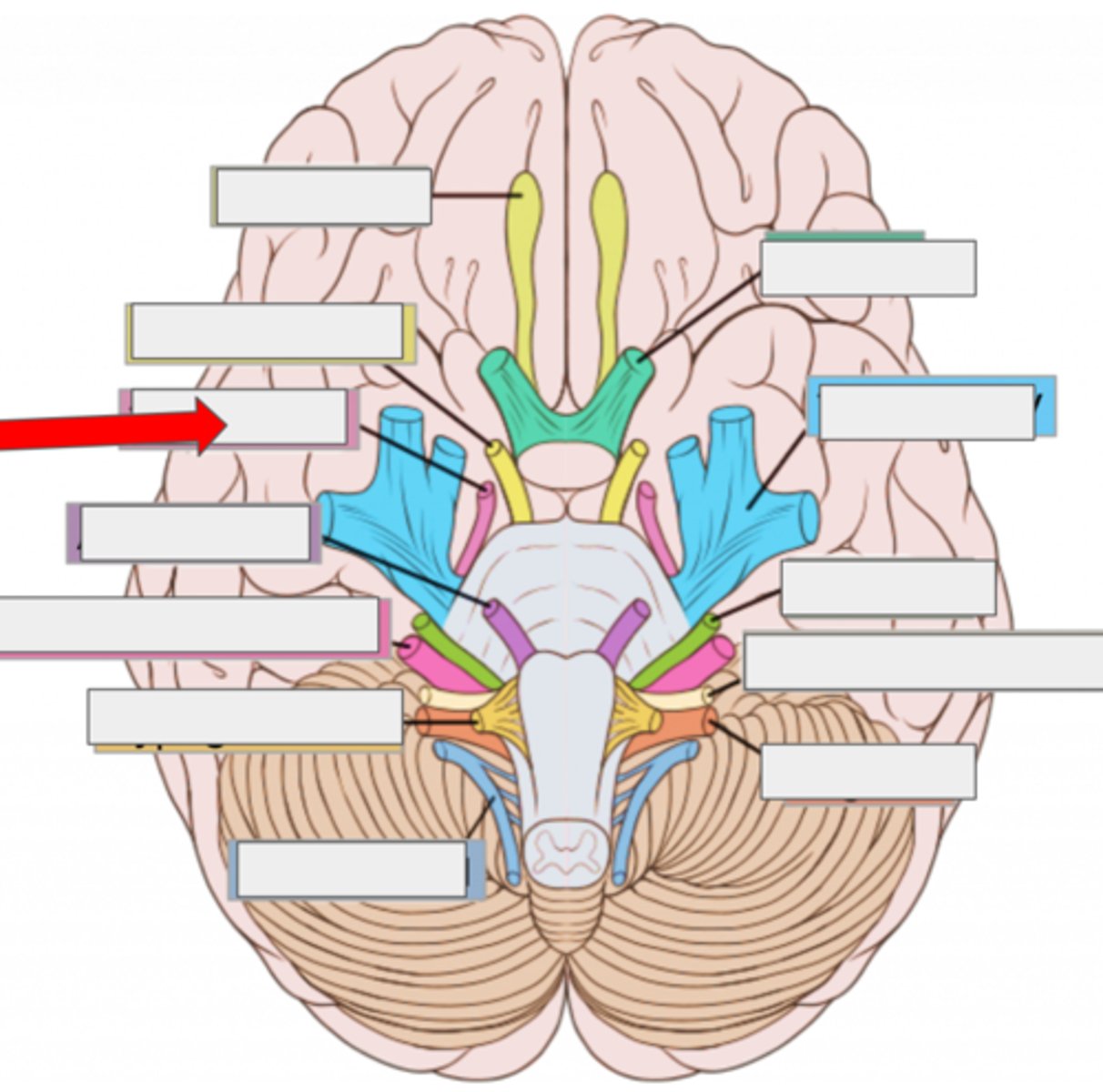
V trigeminal- face sensation
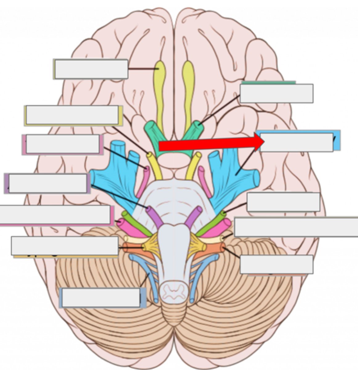
VI abducens- moves eye
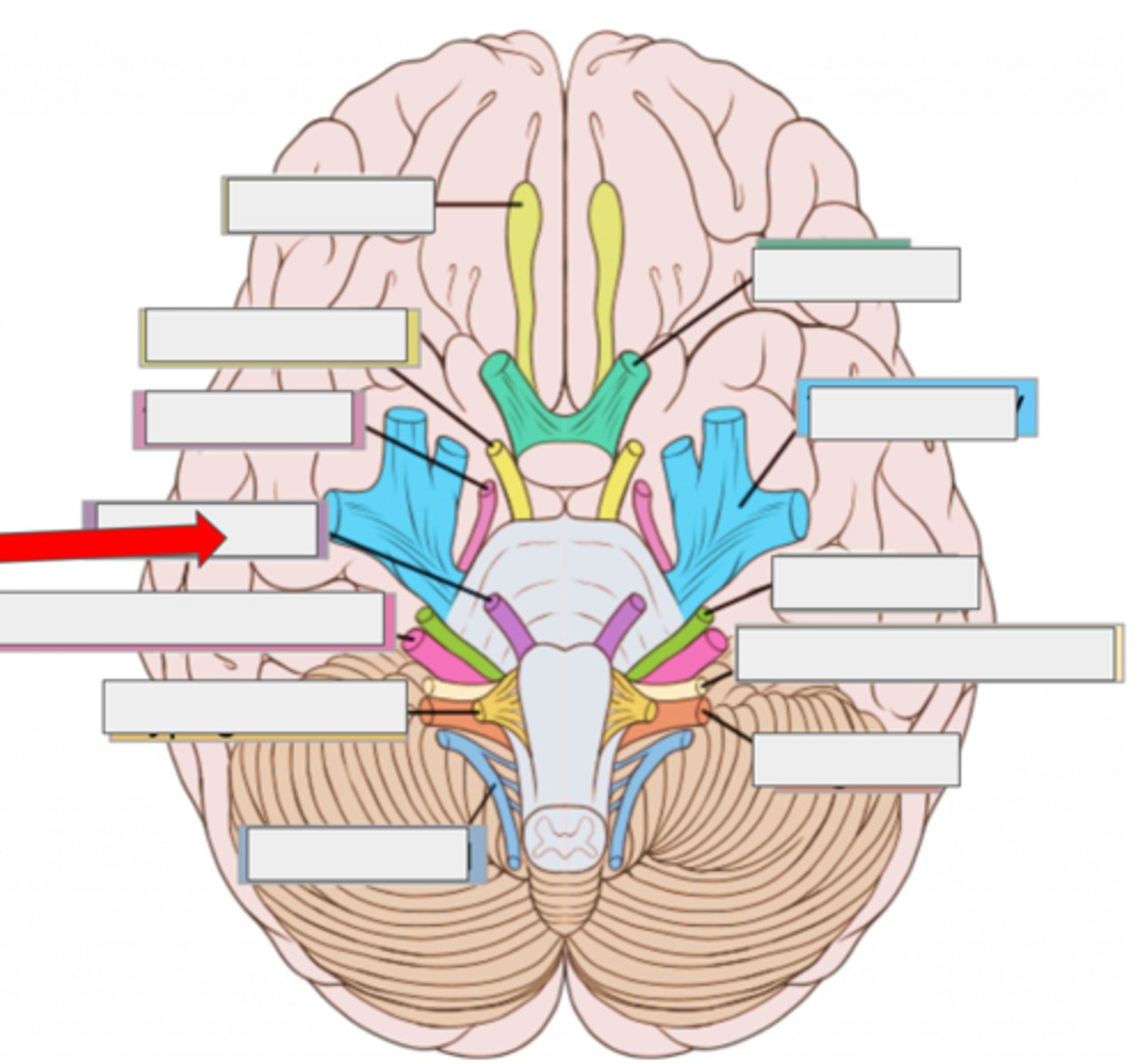
VII facial- moves face, salivate
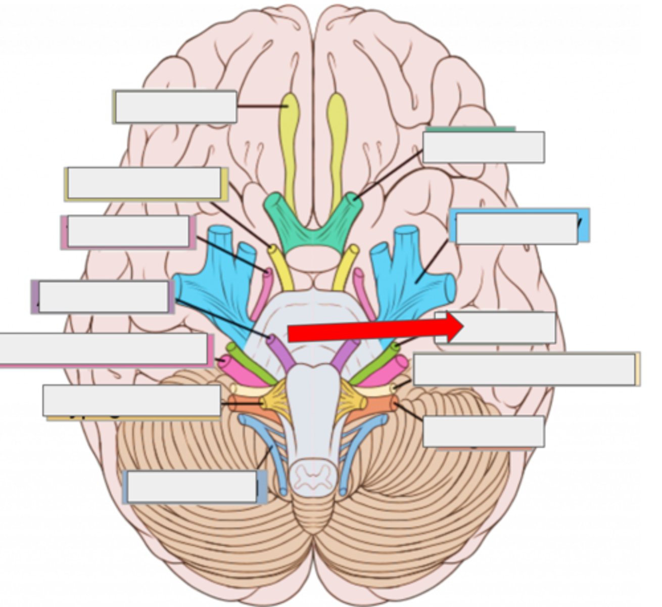
VIII vestibulocochlear- hearing, balance
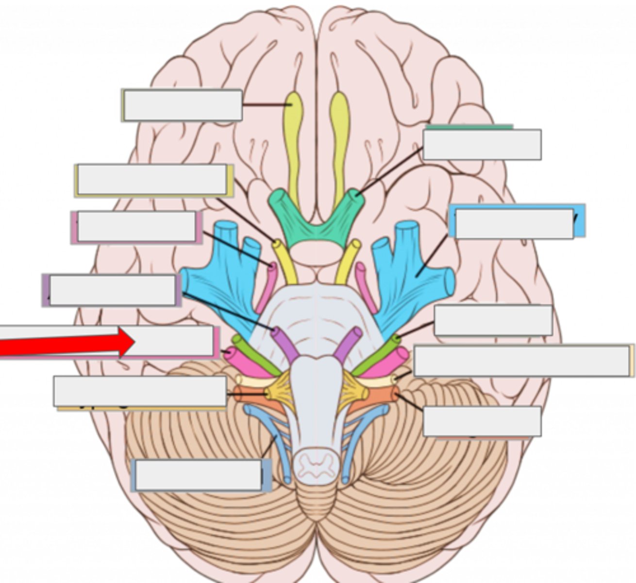
IX glossopharyngeal- taste, swallow
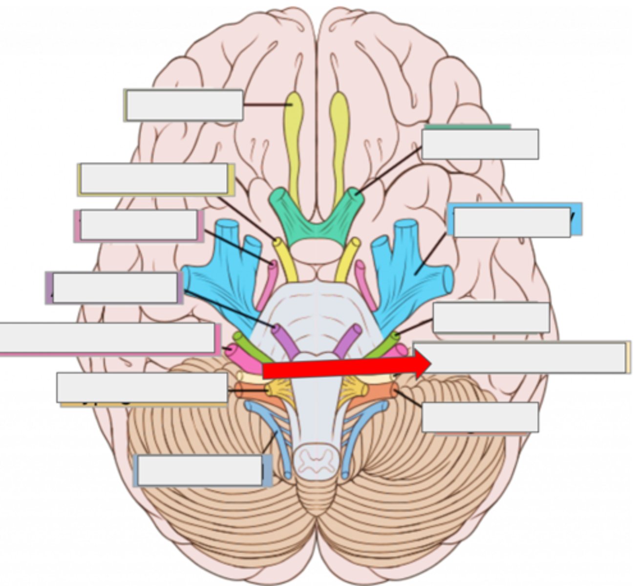
X vagus- heart rate, digestion
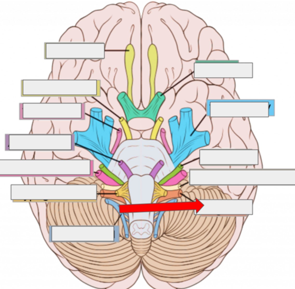
XI accessory- moves head
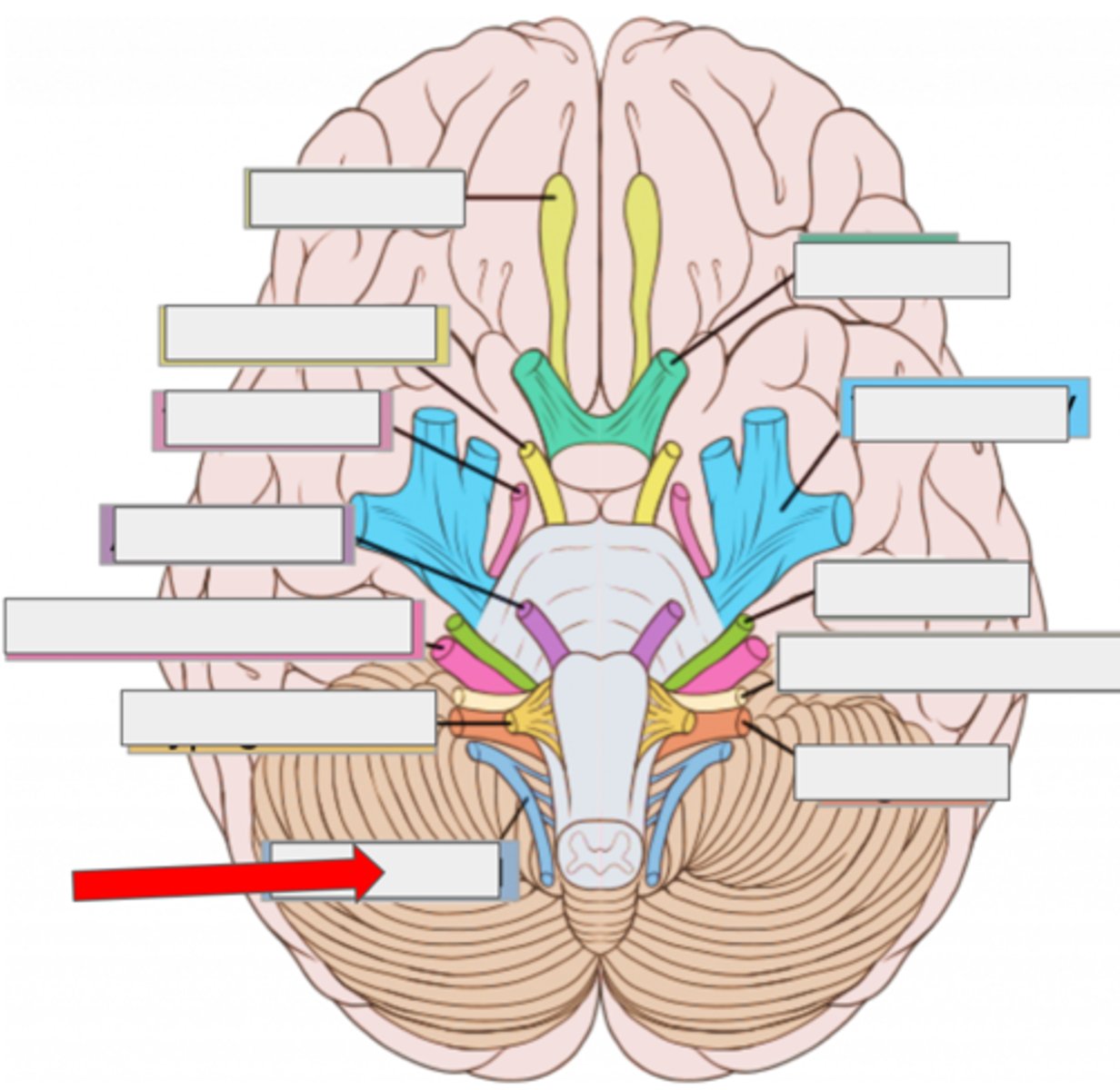
XII hypoglossal- moves tongue
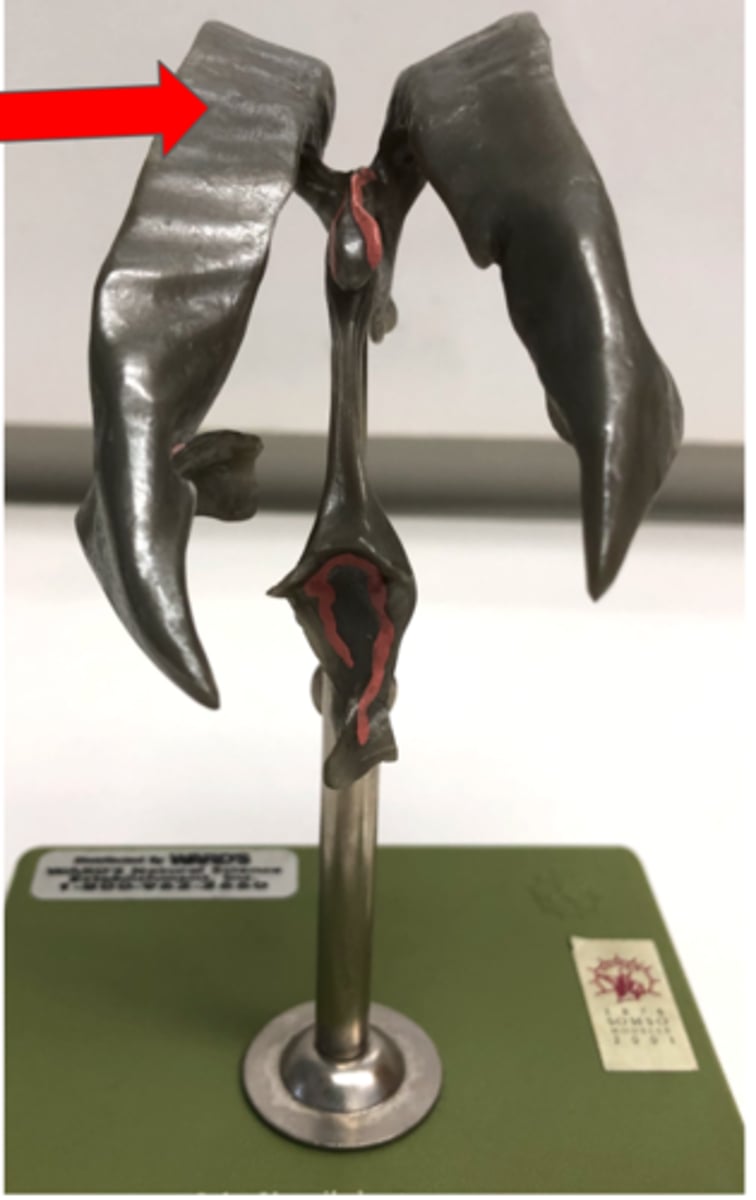
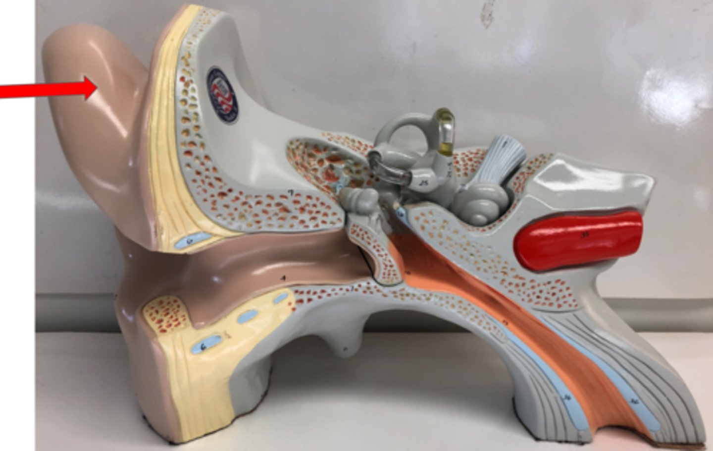
Auricle (OR pinna)
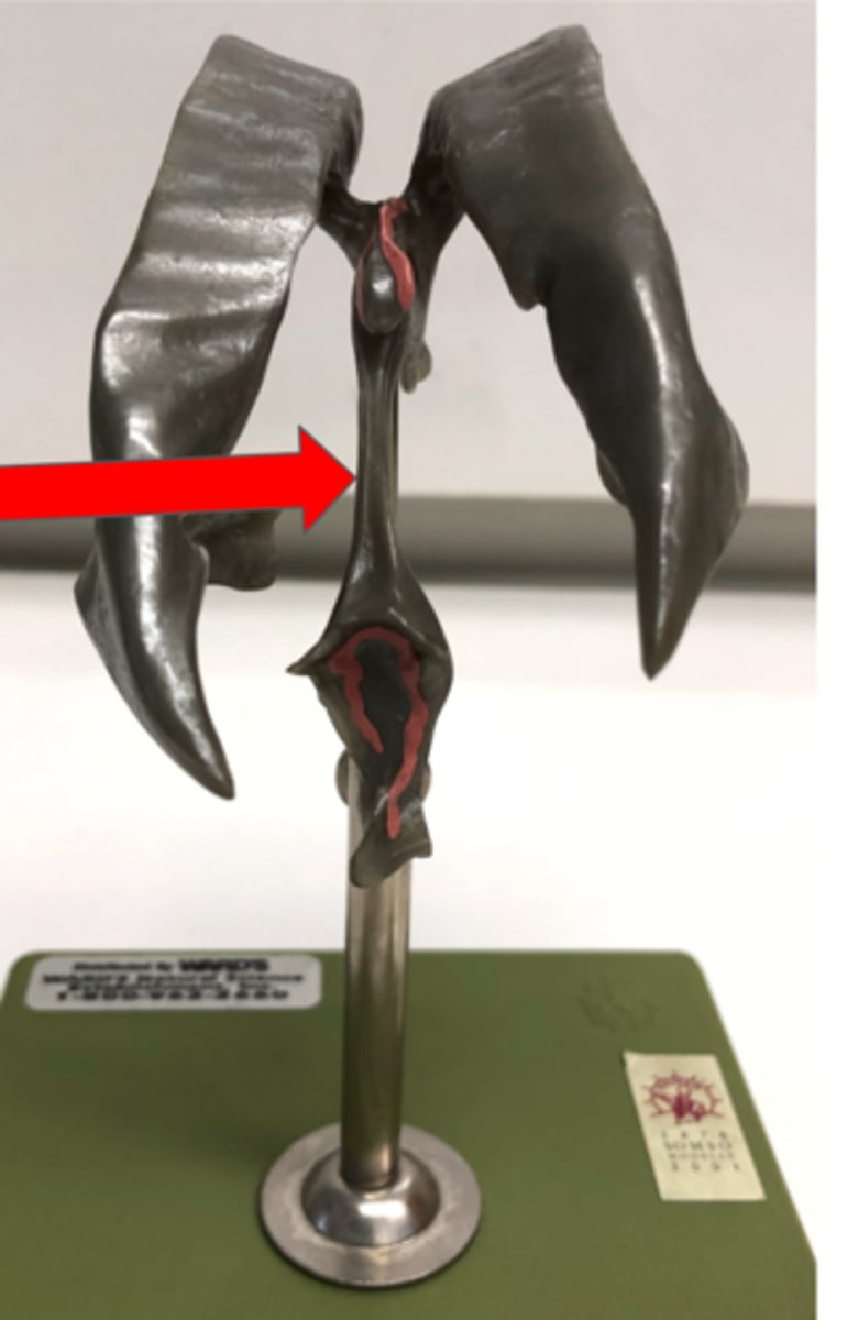
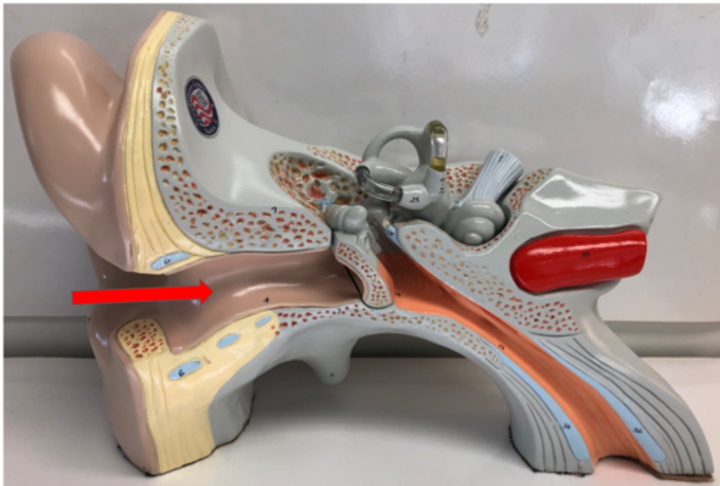
External auditory canal OR external acoustic meatus
Tympanic membrane
Middle ear
Cavity between the eardrum and inner ear
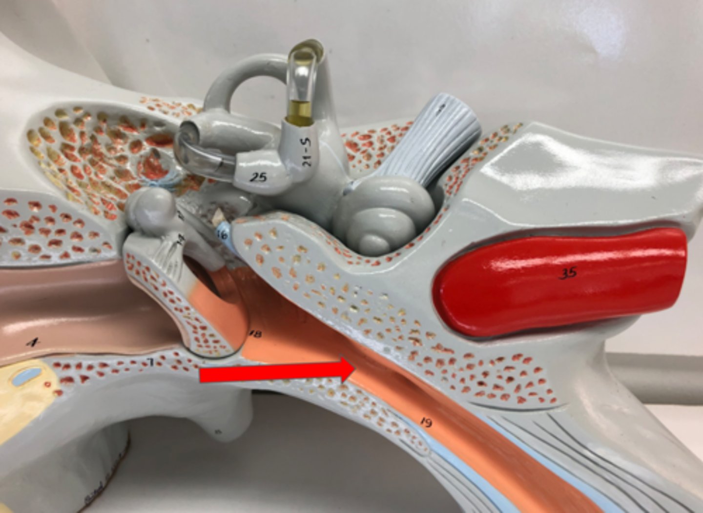
Pharyngotympanic tube
Connects middle eat to throat
Auditory Ossicles
Three small bones in the middle ear
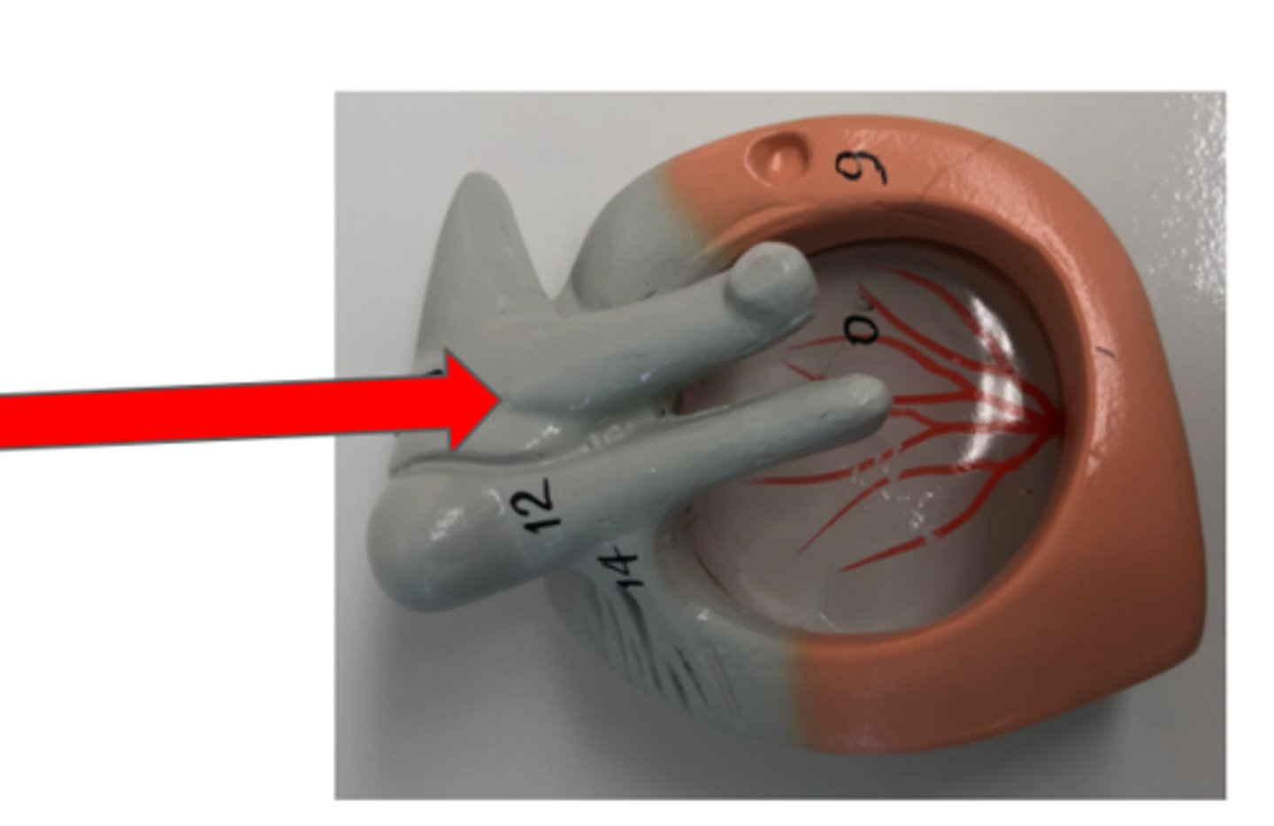
Malleus
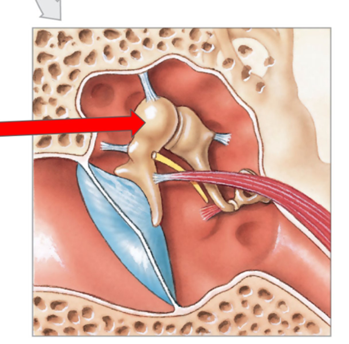
Incus
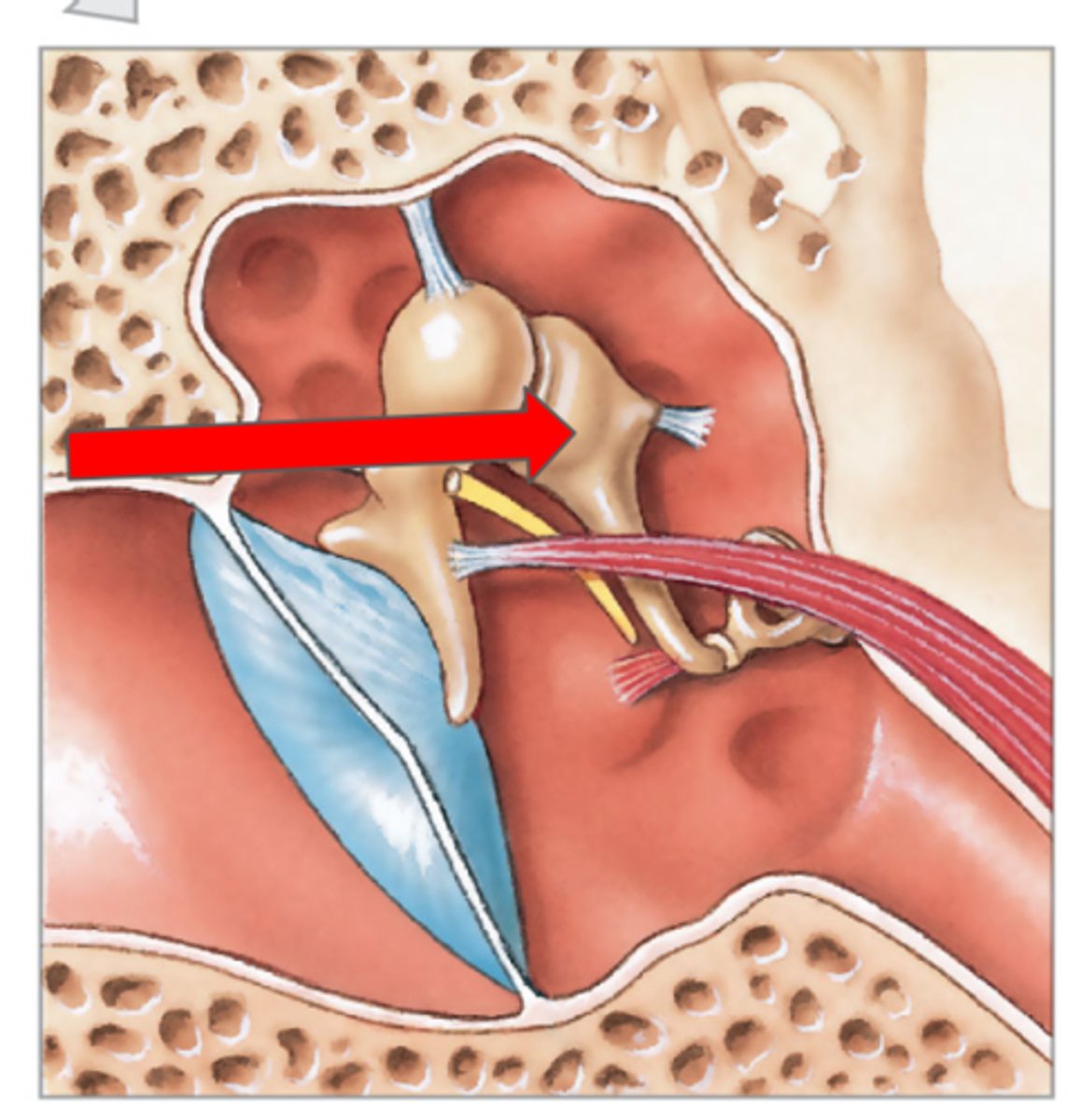
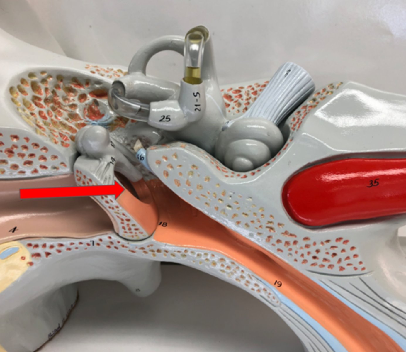
Stapes—connects to oval window
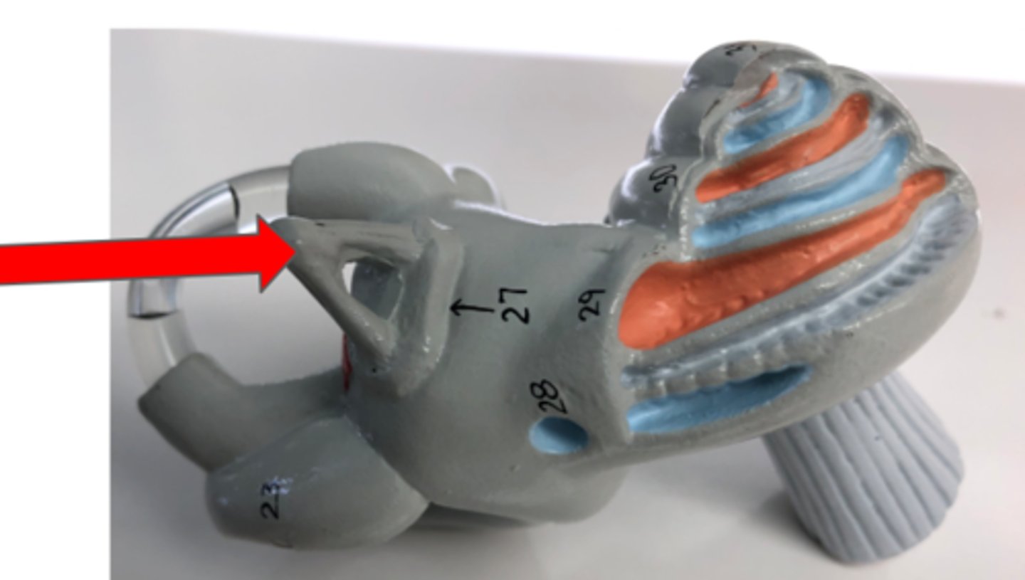
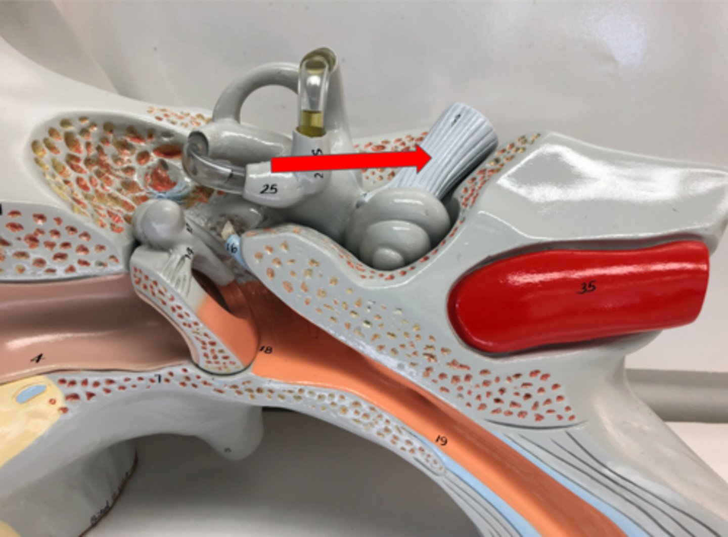
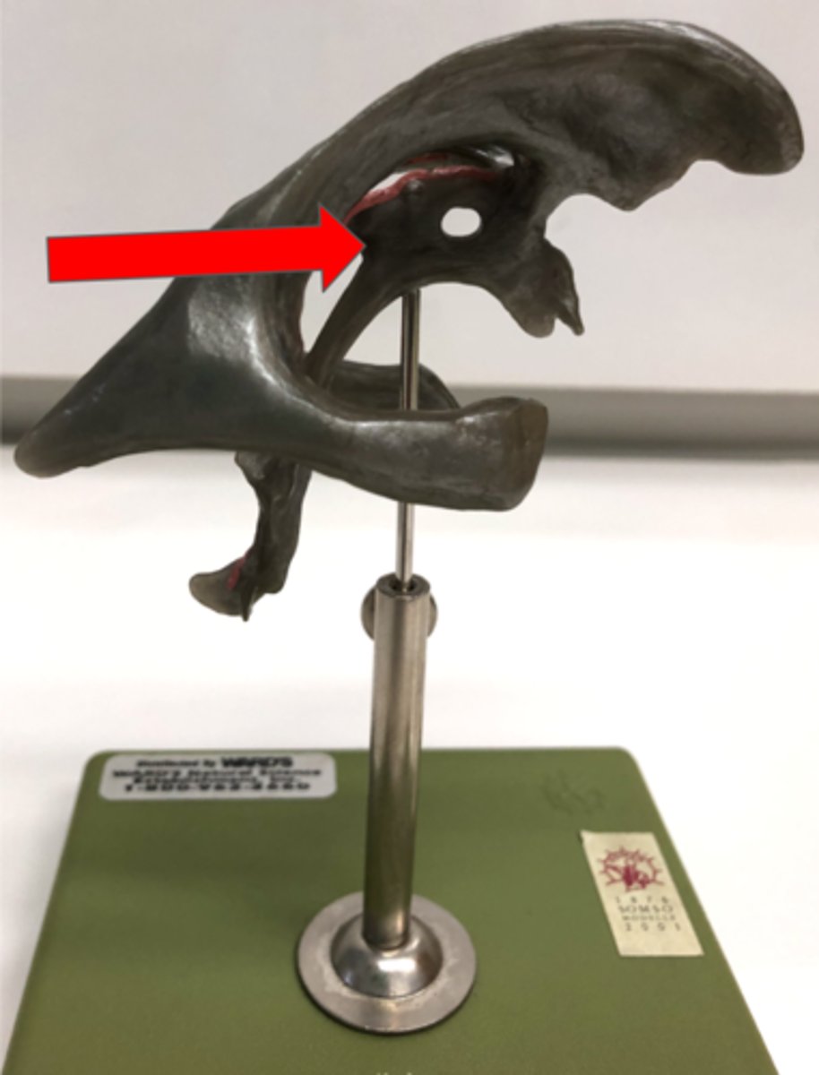
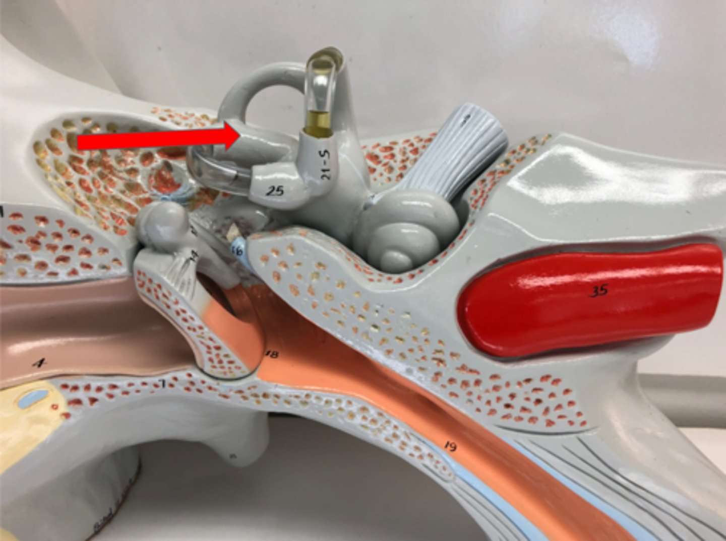
Semicircular canals
Fluid-filled canals in the inner ear for balance
Vestibule
Part of the inner ear for balance