ch 18 – modulation of movement by the basal ganglia (motor selection)
1/32
There's no tags or description
Looks like no tags are added yet.
Name | Mastery | Learn | Test | Matching | Spaced |
|---|
No study sessions yet.
33 Terms
basal ganglia and cerebellum
what regulates upper motor neurons?
false
true/false: basal ganglia and cerebellum regulate lower motor neurons
subcortical loop
formed by the basal ganglia, substantia nigra, and subthalamic nucleus
links motor cortex and upper motor circuits
neurons in these circuits change their activity at the beginning and end of voluntary movements
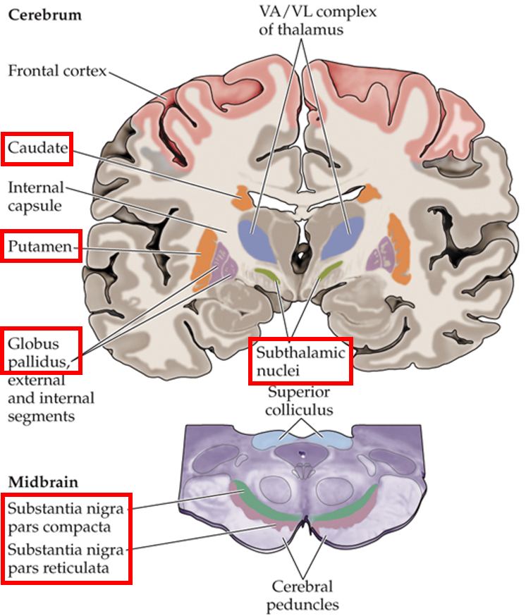
basal ganglia
striatum
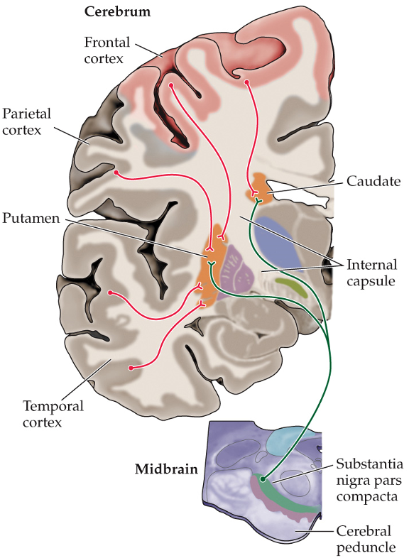
caudate
putamen
pallidum
globus pallidus
substantia nigra
pars reticulata
pars compacta
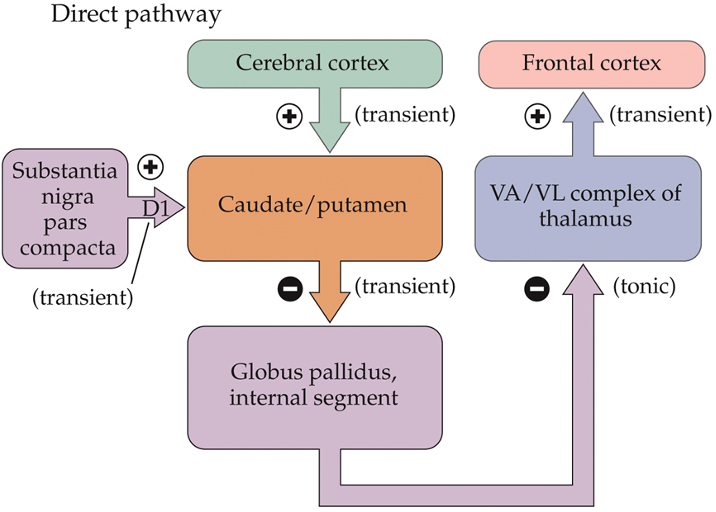
cortex → striatum → pallidum → thalamus
order of basal ganglia
projections to the basal ganglia
caudate
putamen
globus pallidus
substantia nigra
subthalamic nuclei
medium spiny neurons
where do cortical neurons and substantia nigra project their inputs onto?

caudate and putamen
where are the medium spiny neurons located?
dopaminergic; cortical
__________ inputs from substantia nigra synapse closely to ______ synapses, modulating them
medium spiny neurons
have spines where glutamatergic synapses form
have inward K+ rectifying currents (even at rest) → little spontaneous activity
require many excitatory inputs to fire
firing correlates with the occurrence of movement
fire seconds before movements and at termination of movement
firing helps select and initiate a movement
GABAergic (inhibitory)
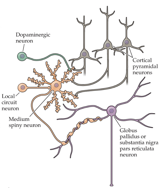
relationship between cortical neurons and medium spiny neurons
each cortical neuron synapses on one spine, but each medium spiny neuron receives many cortical inputs
allows for widespread information distribution

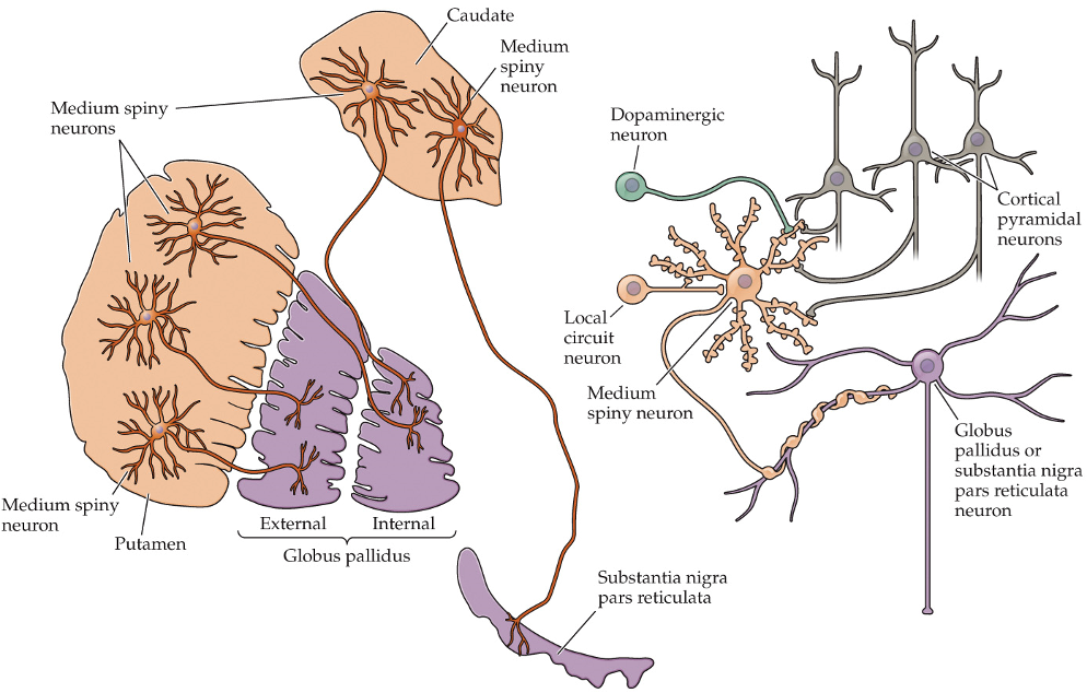
basal ganglia
one cortical neuron also connects with multiple _____ _____ neurons (telegraph poles)
firing patterns in caudate
medium spiny neurons fire in anticipation of eye movements

firing patterns in putamen
medium spiny neurons fire in anticipation of limb and trunk movements
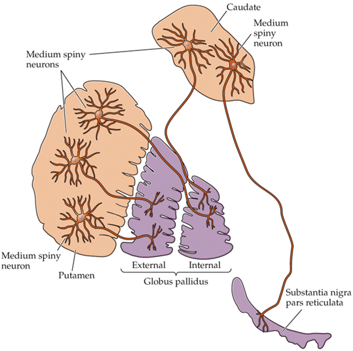
globus pallidus (GP) and substantia nigra pars reticulata (SN)
where do medium spiny neurons in the caudate and putamen project to?
GABA-ergic
are the neurons of the GP and SN GABA-ergic or glutamatergic?
degree of convergence
100 medium spiny neurons innervate each cell in the GP (funneling of information)
GP to cortex pathway
internal segment of GP → thalamus (ventral anterior and lateral muscle) → motor cortex (frontal lobe)
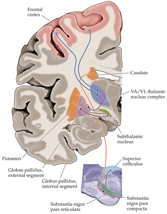
SN to superior colliculus pathway
SN → brainstem → superior colliculus
direct connection to upper motor neurons controlling eye and head movements

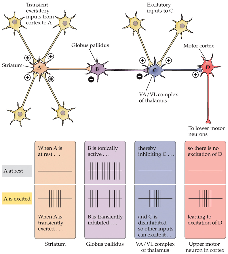
GP and SN inhibitory loop
inhibitory neurons of the striatum synapse onto inhibitory neurons of the GP and the SN
inhibition of inhibition → activation (disinhibition)
loop originates and ends in the cortex
GP and SN have high spontaneous firing to prevent unwanted movements
when striatum is activated, it inhibits the inhibitory neurons of the GP and SN → induces activation of thalamus and superior colliculus → initiation of movement

gate
basal ganglia acts as a ____ for the initatiation of movement
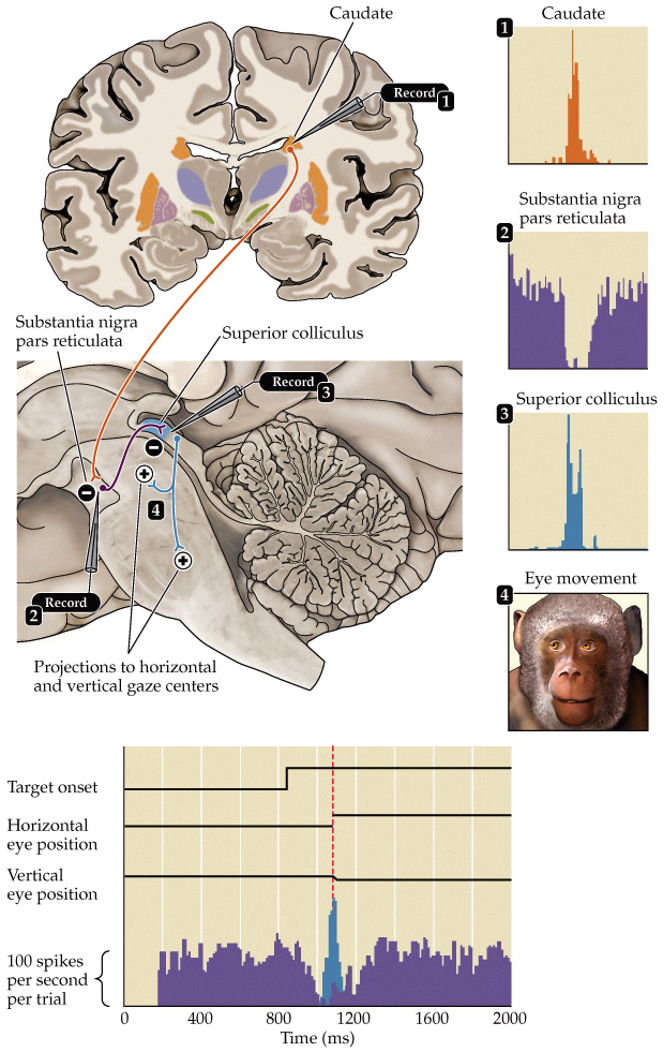
saccades
rapid eye movements
oculomotor loop
humans are foveating primates (spend a lot of time on moving eyes to focus on something)
when the eyes are fixated on a target, the upper motor neurons controlling the movement are inhibited by the SN (SN inhibits superior colliculus) → no saccades occur
before a saccade occurs: caudate inhibits SN activity via GABAergic input → upper motor neurons of superior colliculus are disinhibited → saccade occurs
direct pathway in basal ganglia
striatum to GP
facilitates voluntary movement initiation
indirect pathway in basal ganglia
some medium spiny neurons project to external segment of GP → external segment neurons project to internal segment and subthalamic nucleus
inhibits initiation of movement
increases inhibition provided by basal ganglia
cortex also projects to subthalamic nucleus
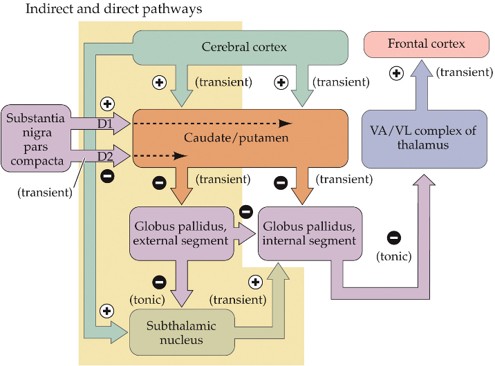
balance
output of the globus pallidus (GP) results from the _______ of the activity of both the direct and indirect pathway
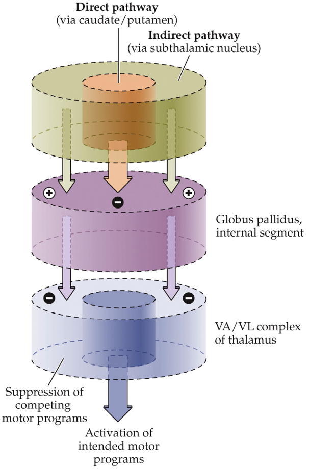
focused selection
created by GP output balance between direct and indirect pathways of basal ganglia
desired movement is allowed while unwanted movements are suppressed
enhances behavioral contrast (movement vs no movement)

hypokinesia
decrease in voluntary movement
ex. Parkinson’s disease
pars compacta; striatum
dopaminergic neurons of the SN ___ _______ send projection to the _______
D1 receptors
used by dopaminergic neurons in SN pars compacta to signal to the striatum
dopaminergic
on medium spiny neurons
located close to synapses between medium spiny neurons and cortical projections
increase in cAMP → enhance excitatory input
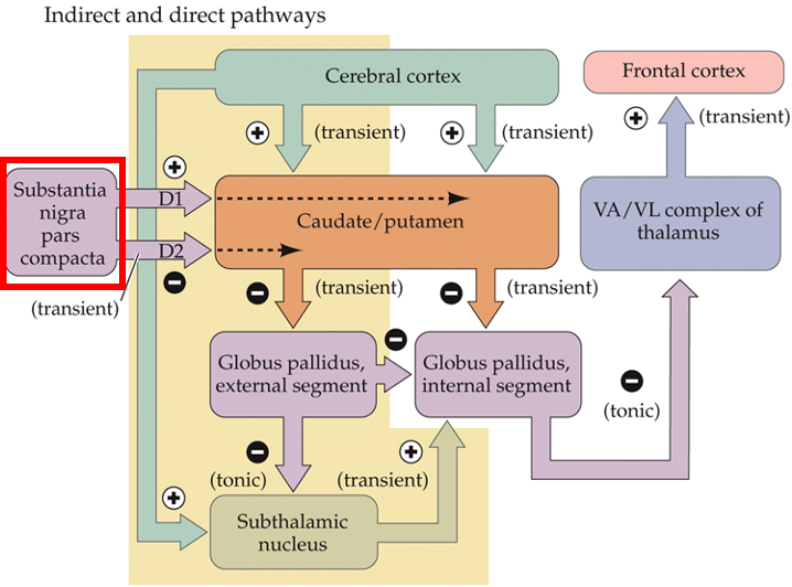
D2 receptors
used by dopaminergic neurons in SN pars compacta to signal to the striatum
dopaminergic
on medium spiny neurons
located close to synapses between medium spiny neurons and cortical projections
decrease in cAMP → inhibit excitatory input

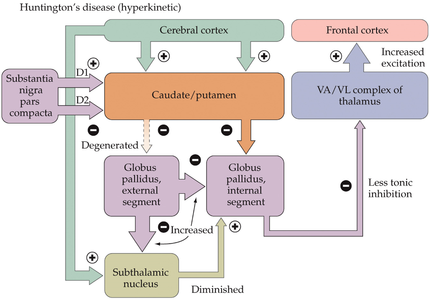
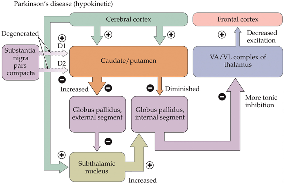
Parkinson’s disease
degeneration of dopaminergic neurons in SN pars compacta
loss of dopamine → inhibitory activity of basal ganglia is enhanced → reduces activation of upper motor neurons by thalamus → difficulty initatiating movement
loss of neuromelanin in SN
hyperkinesia
ex. Huntington’s disease
insufficient output from the pallidus → unwanted, jerky movements
atrophy of striatum
excitatory subthalamic nucleus can’t effectively oppose the direct pathway → cortex is more excited by the thalamus → excessive movement