Looks like no one added any tags here yet for you.
What is asthma?
The widespread narrowing of the airway
What physiological changes are caused by asthma?
Swelling of mucous membranes
Excess mucous secretions
Smooth muscle spasm
What is the radiographic appearance of asthma?
Can only be seen during an acute attack:
Bronchial narrowing
Hyperlucent lungs
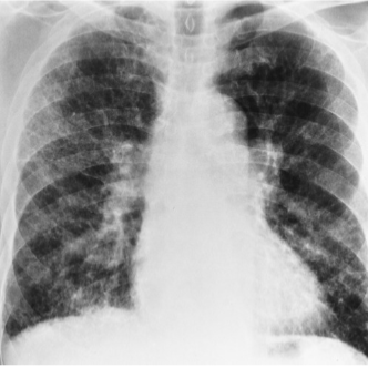
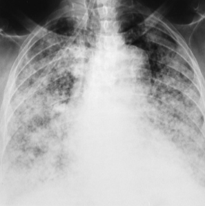
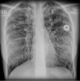
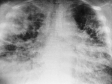
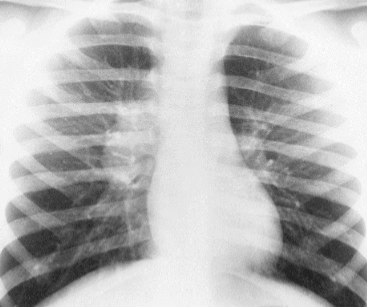
Name the pathology
Asthma
What are the diseases included in COPD?
Bronchitis
Emphysema
Asthma
Bronchiectasis
What is atelectasis?
Non-aeration collapse of part or whole lung (deflated alveoli)
What can cause atelectasis?
Anything that reduces air entry to alveoli including:
Pneumothorax
Excess secretions
Tumor
Abscess
Bronchial narrowing
ET tube misplacement
How does atelectasis appear on an xray?
Increased density in area of collapse following lobe fissures
Ipsilateral overinflation
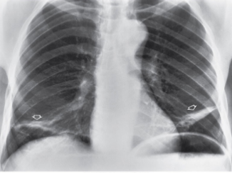
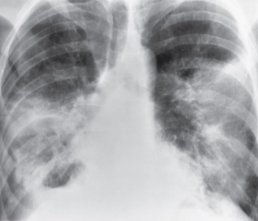
Name the pathology
Atelectasis
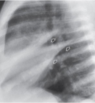
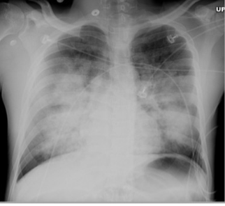
Name the pathology
Atelectasis
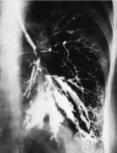
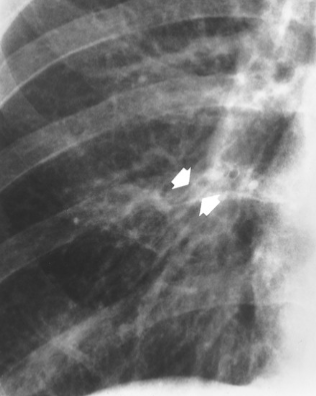
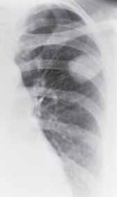
What is bronchiectasis?
Permanent dilation of one or more large bronchi due to destruction of muscular and elastic components
What can cause bronchiectasis?
Complications from bronchitis
What is the radiological appearance of bronchiectasis?
Loss of definition in interstitial markings
Oval or circular cysts
Interstitial fibrosis
Bronchiectasis radiographic sigh
Honeycomb pattern
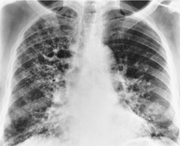
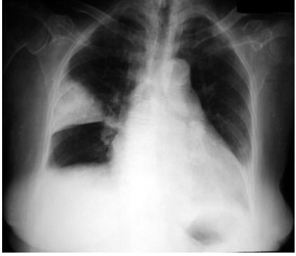
Name the pathology
Bronchiectasis
Name the pathology
Bronchiectasis
What is bronchitis?
Chronic inflammation of bronchi causing thickening and excessive mucus
What are the manifestations of bronchitis?
Blue bloaters - blue skin, deep breaths but still hypoxic
Severe coughing
Radiographic appearance of bronchitis
50% of patients have no changes on xray
Dirty chest - increased lower lung markings
Tram lines - parallel tubular lines
Name the pathology
Bronchitis
Bronchitis may eventually lead to what other pathology?
Emphysema
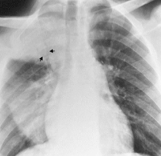
What are the three lung neoplasms?
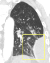
Solitary pulmonary nodule
Bronchogenic carcinoma
Pulmonary metastases
A solitary nodule is found on a cxr, what are the next steps?
CT to confirm location
PET scan
Fine needle aspiration biopsy
Name the pathology
Solitary pulmonary nodule
Name the pathology
Solitary pulmonary nodule
Should you use contrast in a scan for a pulmonary nodule?
If the nodule is smaller than 3 cm - no
If the nodule is greater than 3 cm (a mass) - yes
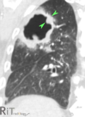
What is bronchogenic carcinoma?
Primary cancer of bronchial tree
What are the risk factors of bronchogenic carcinoma?
Carcinogen inhilation
How is bronchogenic carcinoma confirmed?
Bronchoscopy, biopsy
What is the radiographic appearance of bronchogenic carcinoma?
Mass off a main bronchus
May have air-fluid levels
Possible rib destruction
What cancers most commonly metastasize to the lungs?
Carcinomas of:
Breast
Prostate
Colon
Thyroid
Sarcomas of:
Muscle
Skeleton
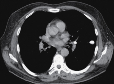
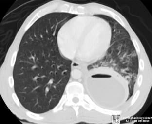
What modalities are used to assess pulmonary metastasis?
Gold star = CT
Assess metabolism (benign vs malignant) = PET
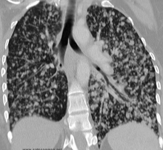
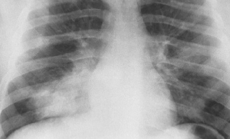
Name the pathology
Mets
Name the pathology
Mets
Name the pathology
Mets
What is COPD?
A category of processes which obstruct gas exchange
Another name for cystic fibrosis
Mucoviscidosis
What is cystic fibrosis?
Defect of chromosome 7 causing excessive production of mucous
What organs are affected by cystic fibrosis?
All organs with exocrine glands:
Sweat glands
Goblet cells of GI tract
Respiratory tract
Pancreas
What are the secondary pathologies caused by cystic fibrosis?
Lung collapse
Recurrent infections
Bronchiectasis
Cysts and abscesses
Failure to gain weight
Radiographic appearance of cystic fibrosis?
Hyperinflation of lungs
Irregular thickening of lung markings
Name the pathology
Cystic fibrosis
Name the pathology
Cystic fibrosis
What is croup?
Obstructive, inflammatory swelling og subglottic trachea
What symptoms are associated with croup?
Stridor
Barking cough
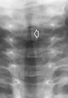
What image would best display croup? how would the image appear?
AP soft tissue neck with smooth, tapered hourglass shape
Name the pathology
Croup
What is epiglottitis
Acute infection of epiglottis and surrounding supraglottic pharyngeal structures
Demographic of epiglottitis?
Children
What is the usual cause of epiglottitis?
Haemophilus influenza
What image would best display epiglottitis? how would the image appear?
Lateral soft tissue nexk
Thumb print sign
What is emphysema?
Injured cilia
Mucosal inflammation
Bronchial narrowing
Air trapping
Bullae rupture
Blue bloaters and pink puffers?
Blue bloaters - Bronchitis
Pink puffers - Emphysema
Radiographic appearance of emphysema?
Overinflated barrel chest, flat diaphragms
Name the pathology
Emphysema
Name the pathology
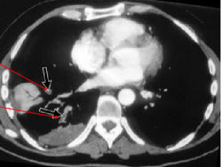
Empyema
Name the pathology
Empyema
Name the pathology
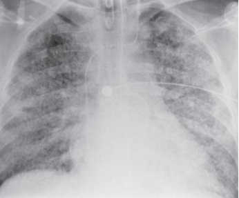
Lung abscess
Lung abscess
Necrotic area of pulmonary tissue with a fibrous outer wall
Common causes of lung abscess
Aspiration
Bacterial pneumonia
Bronchi obstruction
Foreign body
Common causes of pleural effusion:
CHF
PE
TB
Pleurisy
Common manifestations of pleural effusion
Chest pain
Non-productive cough
Dyspnea
Orthopnea
Alveolar pneumonia is also called
Air space pneumonia
Pneumococcal pneumonia
What is alveolar pneumonia
Inflammatory exudate replaces air in alveoli
Radiographic appearance of alveolar pneumonia
Homogenous consolidation with air bronchograms (hypodense bronchial tree through hyperdense area of lung)
Name the pathology
Alveolar pneumonia
What is bronchopneumonia
A staph infection originating in the bronchial tree causing patches of consolodation
What can happen if bronchopneumonia is not managed?
Airway obstruction
Atelectasis
Loss of lung volume
Name the pathology
Bronchopneumonia
Interstitial pneumonia
Viral and mycoplasmal infection of alveoli and interstitial tissues
Appearance of interstitial pneumonia
Linear / reticular pattern
Shaggy heart sign
Honeycomb lung
Name the pathology
Interstitial pneumonia
Name the pathology
Aspiration pneumonia
What os pulmonary edema?
Fluid buildup in pulmonary tissues (alveoli)
What can cause pulmonary edema?
Left sided heart failure = elevated pulmonary venous pressure
Poison
Infection
Medication
Renal failure
Exercise at high altitudes
Heart disease
Heart attack
Pulmonary edema manifestations
Coughing (blood)
Orthopnea
Doctunal dypsnea
Wheezing
Leg edema
Pale
LOC
Pulmonary edema radiographic appearance
Batwing or butterfly shaped hyperdensity
Decreased pulmonary markings
Perihilar haze from heart failure
Name the pathology
Pulmonary edema
What is the main cause of pulmonary embolism?
Lower limb venous stenosis
Trauma
Venipuncture
Indwelling catheter
Manifestations of pulmonary edema
Cheast pain
SOB
Cough
Shock
Cardiac arrest
Pulmonary infarct
Ischemic necrosis of lung tissue
What can cause a pulmonary infarct
PE
Radiographic appearance of pulmonary infarct
Wedge sign with apex towards hilum
Name the pathology
Pulmonary infarct
Name the pathology
Pulmonary infarct
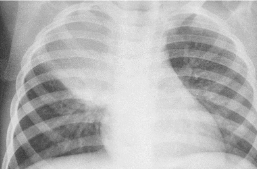
Respiratory distress syndrome
sudden life-threatening disorder caused by inhalation of toxins, trauma or overdose, the lung parenchyma dissolves
Respiratory distress syndrome is also called
ARDS
Shock lung
Radiographic appearance of Respiratory distress syndrome
Patchy, ill-defined consolidation between both lungs with normal heart shadow
Name the pathology
Acute respiratory distress syndrome
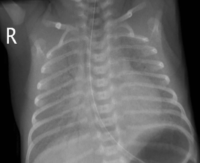
What is IRDS?
infant respiratory distress syndrome - hyaline membrane disease due to failure to produce surfactant
Name the pathology
IRDS
What is SARS?
Severe acute respiratory syndrome - a serious form of pneumonia in the coronavirus family
SARS symptoms
Cough
SOB
Fever
Chills
Aches
SARS radiographic appearance
Similar to pneumonia - patchy infiltrates proceed to consolidation and lack airspace
What is TB
A waxy coat mycobacterium
How is TB spread and how can it be prevented?
Airborn droplets spread through coughing. Can be killed by sunlight
What are the types of TB?
Primary - pulmonary
Secondary - reactivation of dormant disease
Radiographic appearance of primary TB
Well-defined consolidation
Hilar enlargement
Pleural effusion
Ghon lesion
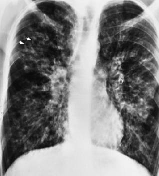
Name the pathology
TB
Name the pathology
TB