Fascial Spaces and Deep Neck Infections
1/44
There's no tags or description
Looks like no tags are added yet.
Name | Mastery | Learn | Test | Matching | Spaced | Call with Kai |
|---|
No analytics yet
Send a link to your students to track their progress
45 Terms
Deep Neck Infection
Infectious and inflammatory conditions of the upper aerodigestive tract are the primary cause of
Dental Infections
most common cause of DNI in adults.
Oropharyngeal Infections
most common cause of DNI in children.
Staphylococcus aureus
more common in the nose and throat and may participate in mixed odontogenic infections.
Actinomyces
endogenous saprophytic organisms of the oral cavity and tonsil.
Actinomyces
A granulomatous reaction with central abscess formation and necrosis with “sulfur granules” is characteristic.
TUBERCULOUS AND NONTUBERCULOUS INFECTIONS OF THE HEAD AND NECK
most commonly come to medical attention with cervical lymphadenopathy.
Histopathologically, caseating necrotizing granulomatous inflammation is present.
CAT SCRATCH DISEASE
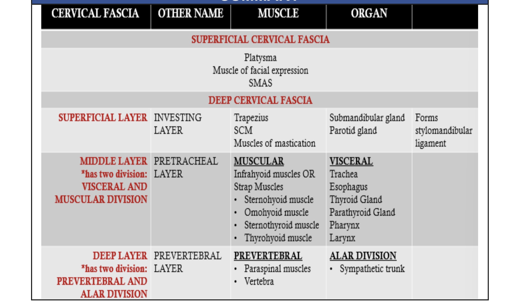
Carotid Sheath
Condensation of fascia of ALL THREE LAYERS OF DEEP CERVICAL FASCIA
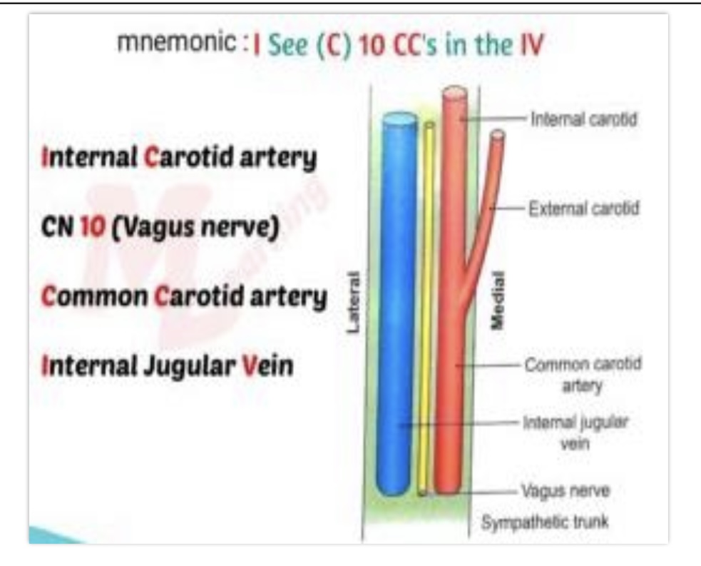
Common Carotid
What structure is the most medial?
Internal Jugular Vein
What is the most lateral part?
Vagus Nerve (CN X)
What nerve is in between the IJV and Common carotid?
D
CAROTID SHEATH REPRESENTS THE CONFLUENCE OF
WHAT LAYERS OF CERVICAL FASCIA?
a. Superficial and deep
b. Investing and pretracheal
c. Pretracheal and prevertenral
d. all three layers of deep cervical fascia
Canine Spave
Buccal Space
2 Maxillary Space
Canine Space
becomes infected almost exclusively as a result of apical infection of the root of the maxillary canine tooth.
Canine Space
located between the anterior surface of the maxilla and the levator labii superioris.
BUCCAL SPACE
an ovoid space below the zygomatic arch and above the inferior border of the mandible.
SUBMENTAL SPACE
lies between the anterior bellies of the digastric muscles and between the mylohyoid muscle and the skin.
SUBLINGUAL AND SUBMANDIBULAR SPACE
Both spaces exit on the medial aspect of the mandible.
SUBLINGUAL SPACE
lies between the lingual oral mucosa and the mylohyoid muscle.
Its posterior boundary is open, so it communicates freely with the submandibular space and the secondary spaces located more posteriorly and superiorly.
SUBMANDIBULAR SPACE
lies between the mylohyoid muscle and the skin.
When this space becomes infected, the swelling begins at the inferior lateral border of the mandible and extends medially to the digastric area and posteriorly to the hyoid bone.
Ludwig’s Angina
If all three primary mandibular spaces bilaterally become infected, the infection is known as
Trismus
Drooling
Dysphonia
Dysphagia
Ludwig Angina Presentation (4)
Ludwig Angina
Tense and swollen floor of the mouth, submandibular area
• Incision and Drainage
• Tracheostomy
• Antibiotics
Ludwig Angina Management (3)
Mediastinitis
most common major complication of DNSI
Peritonsillar abscess (PTA)
AKA QUINSY
Localized deep neck infection that develops between the tonsil and its capsule.
Peritonsillar abscess (PTA)
most common deep neck space infection.
Peritonsillar cellulitis
inflammatory reaction of the tissue between the capsule of the palatine tonsil
and the pharyngeal muscles that is caused by infection
Peritonsillar abscess
collection of pus located between the capsule of the palatine tonsil and the
pharyngeal muscles.
Streptococcus pyogenes
Most common aerobic organisms
Fusobacterium
Most common anaerobic organisms
Peritonsillar Space
Consist of loose alveolar tissue overlying the palatine tonsils.
Tonsillar Fossa
Symptoms:
• Fever
• Malaise
• Severe sore throat (worse on one side) –UNILATERAL
• Dysphagia
• Otalgia (ipsilateral) – HYPOGLOSSAL NERVE
Tonsillar Fossa
• Erythematous, swollen soft palate with uvula deviation to contralateral side and enlarged tonsil
• Trismus - irritation and reflex spasm of the medial pterygoid muscle
• Drooling or Pooling of saliva
• Muffled voice (“hot potato voice”)
• Needle Aspiration
• Incision and Drainage
• Antimicrobial Therapy
• Tonsillectomy
Tonsillar Fossa Management (4)
Lemierre Syndrome
rare thrombophlebitis of the internal jugular vein most often caused by the anaerobic, gram-negative bacillus Fusobacterium necrophorum.
Lemierre Syndrome
period of pharyngitis before progressing to fever, lethargy, lateral neck tenderness and edema, occasional trismus, and septic emboli
bilateral
nodular infiltrates on chest radiography or as septic arthritis
IV β-lactamase
Lemierre Syndrome Management (1)
CAVERNOUS SINUS THROMBOSIS
retrograde spread of infection from the upper dentition or paranasal sinuses via the valveless ophthalmic venous system to the cavernous sinus
Mediastinitis
Relatively rare complication of DNI caused by spread of infection along the retropharyngeal and prevertebral planes of the neck into the upper mediastinum
Necrotizing Fasciitis
Severe form of DNI that occurs more often in
Older adults (age >60 years)
Immunocompromised patients
Poorly controlled diabetes.
Necrotizing Fasciitis
PITTING NECK EDEMA
ORANGE-PEEL APPEARANCE from obstructed dermal lymphatics
with or without subcutaneous CREPITUS