TAP lab 7 short list
1/14
There's no tags or description
Looks like no tags are added yet.
Name | Mastery | Learn | Test | Matching | Spaced |
|---|
No study sessions yet.
15 Terms
Bovine lungs
Left lung
cranial lobe: cranial and caudal parts
caudal lobe
Right lung
cranial lobe: cranial & caudal parts
tracheal bronchus
middle lobe
accessory lobe
caudal lobe
Equine lungs
Left lung
cranial lobe
caudal lobe
Right lung
cranial lobe: cranial & caudal parts
accessory lobe
caudal lobe
Canine lungs
Left lung
cranial lobe: cranial and caudal parts
caudal lobe
Right lung
cranial lobe
middle lobe
accessory lobe
caudal lobe
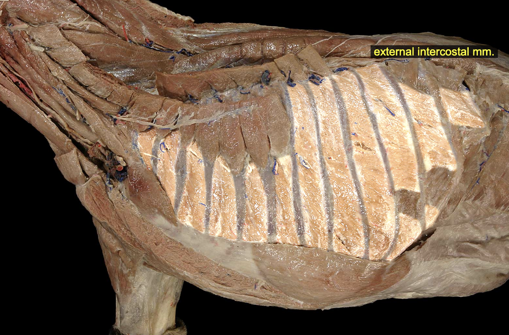
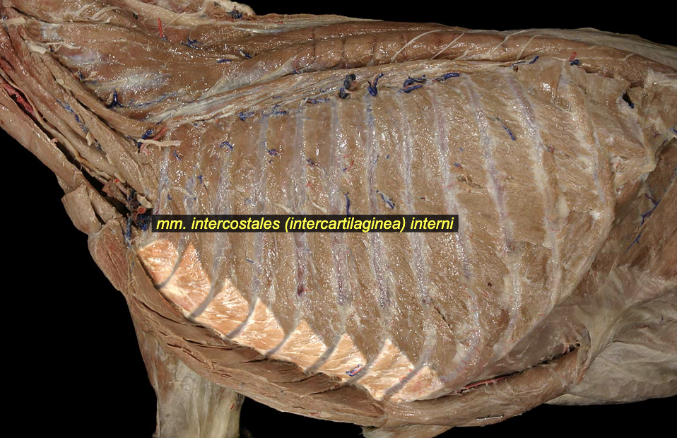
External & Internal Intercostal mm.
The muscles run periperpendicular to one another
External intercostal m. Superficial
craniodorsad → caudoventrad
O: caudal border of ribs 1 – 12
I: cranial border of ribs 2 – 13
A: draw ribs closer together; contraction produces a lifting of the thoracic cage, associated with inspiration
N: intercostal nn.
Internal intercostal m. Deep
cranioventrad → caudodorsad
O: caudal border of ribs 1 – 12
I: cranial border of rib 2 – 13
A: respiration (exact role debated), fixation of ribcage during locomotion
N: intercostal nn.
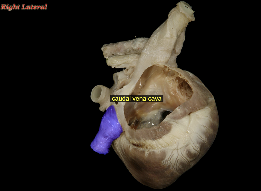
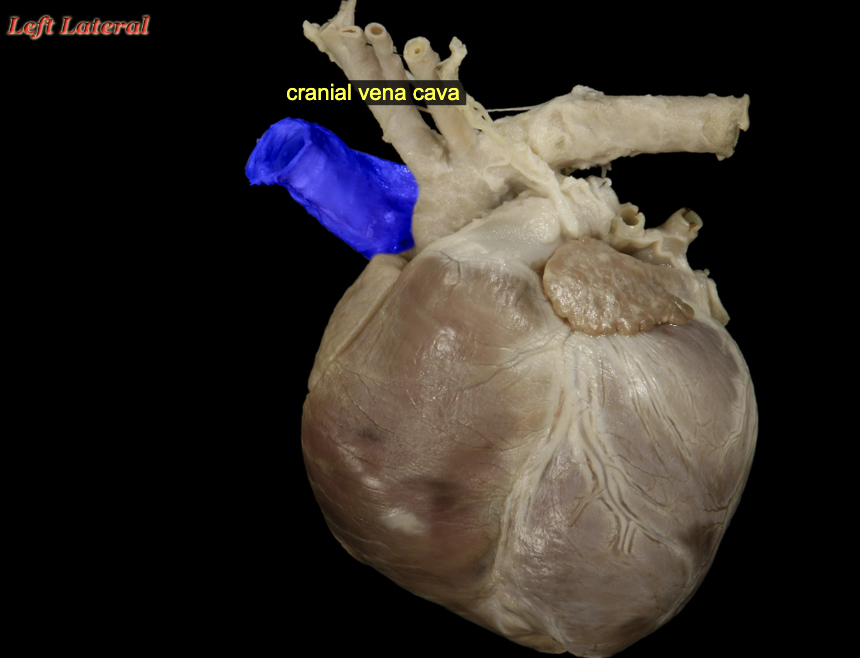
Cranial vena cava & Caudal vena cava
Cranial vena cava
The right and left brachiocephalic veins join just inside the thoracic inlet to create the cranial vena cava which passes caudad ventral to the trachea and to the right of the esophagus to terminate at the right atrium. The cranial vena cava also receives venous blood from the azygos, costocervical, caudal thyroid, and internal thoracic veins.
Caudal vena cava
The caudal vena cava arises at about the level of lumbar vertebra six where the two common iliac veins merge. In its cranial course, it receives venous blood from somatic veins draining the body wall and trunk and from the renal arteries. The caudal vena cava passes dorsal to the liver, receiving hepatic venous blood just before passing through the caval foramen of the diaphragm.
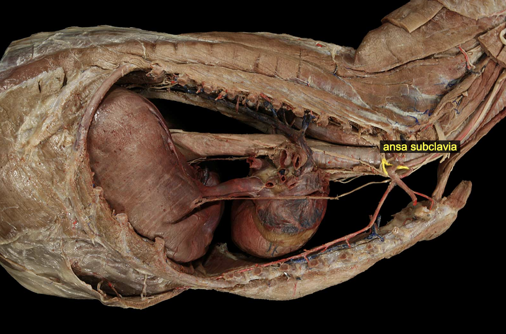
Ansa Subclavia
Motor: sympathetic preganglionic fibers (visceral motor autonomics)
Sensory: general visceral afferent fibers from thoracic viscera
Origin: cervicothoracic ganglion
Insertion: middle cervical ganglion
The ansa subclavia is a loop of preganglionic sympathetic fibers that encircles the subclavian artery; it is present bilaterally in the cranial thorax. Fibers in the ansa subclavia have passed first through the cervicothoracic ganglion and may subsequently either synapse in the middle cervical ganglion (ventral to the ansa) or follow the vagosympathetic trunk to the head.
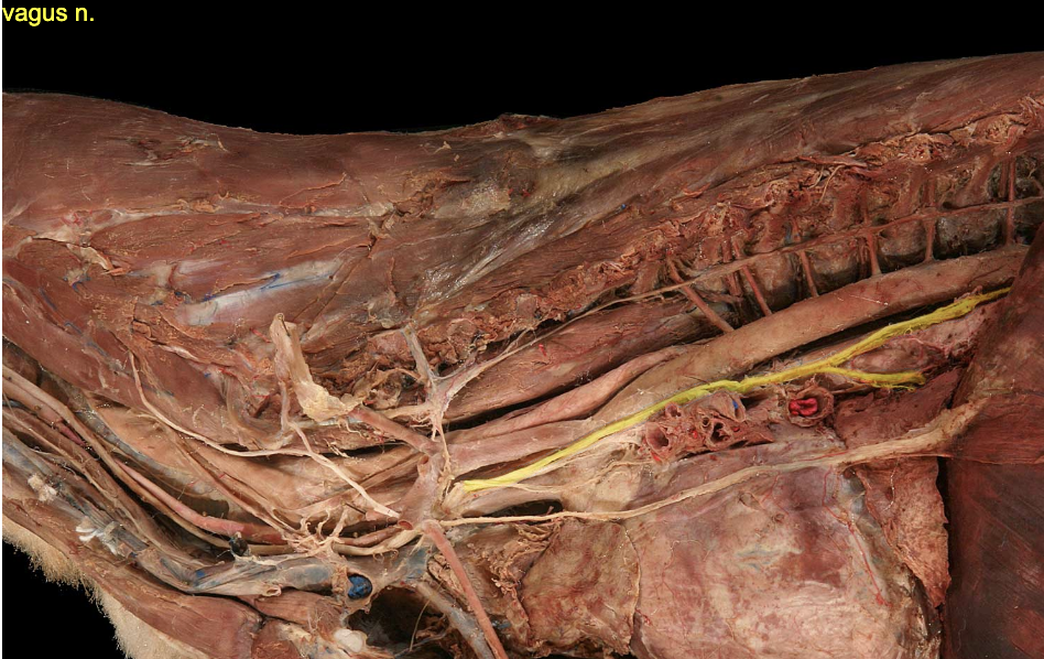
Dorsal vagal trunk
Vagus nn.
Motor (somatic): laryngeal, pharyngeal, esophageal muscles
Parasympathetic: all thoracic viscera, most abdominal viscera
Sensory: laryngeal vestibule, all thoracic viscera, most abdominal viscera
Dorsal vagal trunk
The left and right vagus branches of the vagus n. join and make a dorsal vagal trunk before the esophageal hiatus.
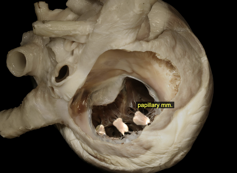
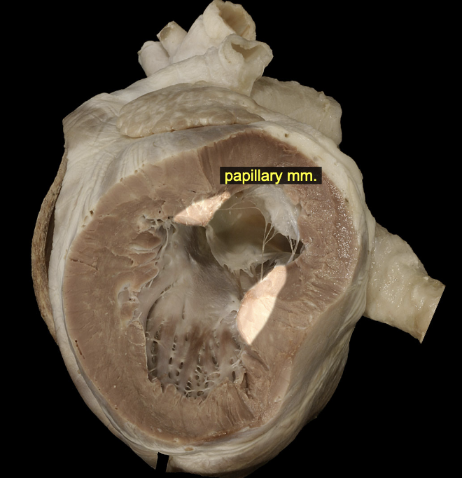
Papillary muscles
The papillary muscles are raised, blunt projections of the interior ventricular wall. The right ventricle of the dog typically possesses three main papillary muscles, although variations (fusions, splitting, and duplications) are common. The left ventricle more consistently possesses two papillary muscles. The collagenous chordae tendineae that tether the valve leaflets of the atrioventricular valves are anchored in the papillary muscles.
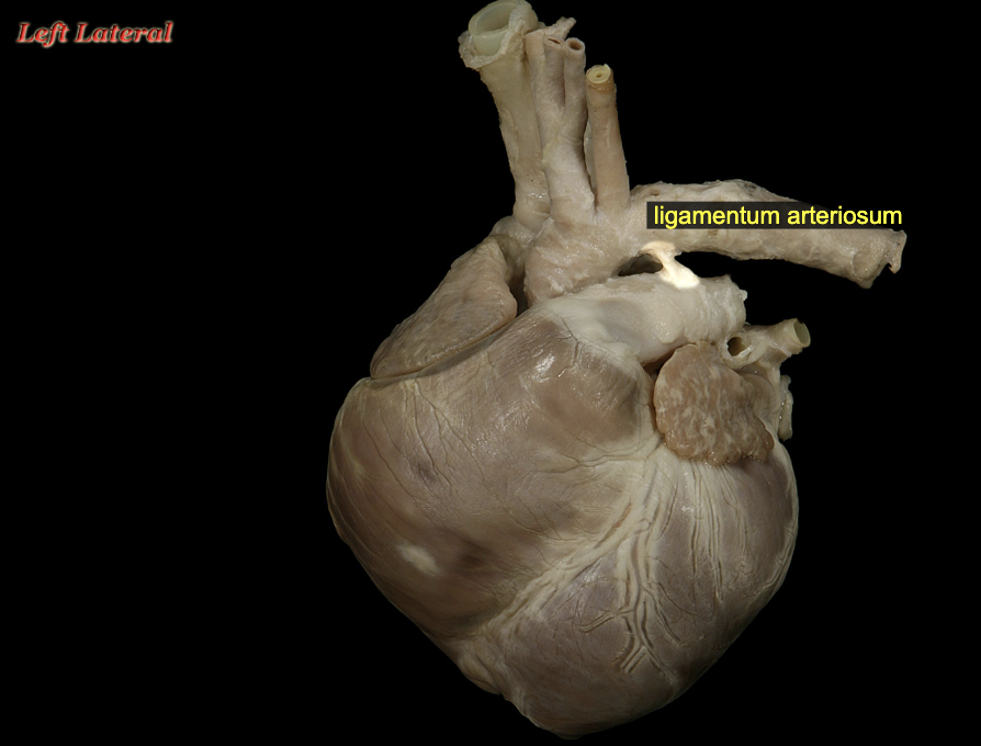
Ligamentum arteriosum
During fetal life, pulmonary trunk and aorta are connected by a large anastomosis, the ductus arteriosus, which permits blood in the higher pressure pulmonary trunk to pass to the lower pressure aorta, bypassing the unventilated lungs. Immediately at birth, dramatic changes in pulmonary circulation of oxygen saturations produces a muscular constriction of the ductus arteriosus, which achieves a complete obliteration of its lumen over the course of the first week of extrauterine life. The ligamentous remnant of the ductus, now the ligamentum arteriosum, remains as a fibrous attachment between the pulmonary trunk and aorta.
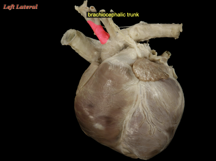
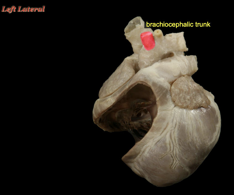
Brachiocephalic trunk
The first branch from the aortic arch is the brachiocephalic trunk, which inclines to the right side of midline, passing ventral to the trachea. It will give rise to the two common carotid arteries and the right subclavian artery, as well as a variable number of small arteries that supply nearby structures of the cranial mediastinum.
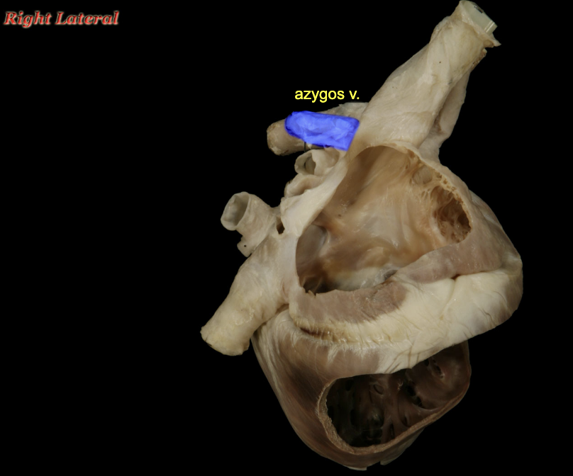
Azygos v. (ca)
The azygos vein is an unpaired vessel arising between the crura of the diaphragm at the level of L3 vertebral body. It passes craniad through the aortic hiatus and within the thorax inclines to the right side of midline, running between vertebral bodies and thoracic aorta. The azygos vein terminates by emptying into the cranial vena cava close to the latter's termination in the right atrium. Lumbar, intercostal, costoabdominal, and bronchoesophageal veins all drain to the azygos vein, making it the primary venous drainage for thoracic and abdominal body walls.
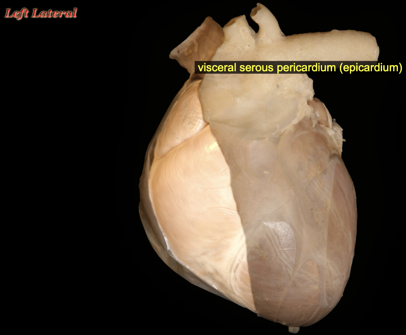
Visceral vs. Parietal serous membranes (pericardium)
Visceral pericardium (epicardium) Deep
in mediastinum
heart is directly covered in this serous membrane
The visceral pericardium faces the potential space of the pericardial cavity, and reflects to become the parietal pericardium around the base of the heart
Parietal pericardium (epicardium) & Fibrous pericardium Superficial
Deep to the fibrous pericardium and firmly attached to it is a serous membrane, the parietal layer of the serous pericardium, often simply referred to as the parietal pericardium. The parietal pericardium faces the potential space of the pericardial cavity. The fibrous pericardium blends into the adventitia of the great vessels at the base of the heart; the apex of the heart is unattached inside the pericardium so that the heart is afforded frictionless movement within the pericardial cavity.
Pericardial sac or heart sac is the potential space b/w fibrous and parietal pericardium
Visceral vs. Parietal serous membranes (pleura)
Parietal pleura
Costal pleura
Pleura is the serosal investment associated with the thoracic viscera. It is a simple squamous epithelium and is underlain by a fibrous subserosal layer called endothoracic fascia. Individual regions of the parietal pleura are named for the body parts with which they are associated. The parietal pleura on the deep side of the thoracic wall is accordingly referred to as costal pleura, a specific subcategory of the parietal pluera that lines the walls of the thoracic cavity and mediastinum.
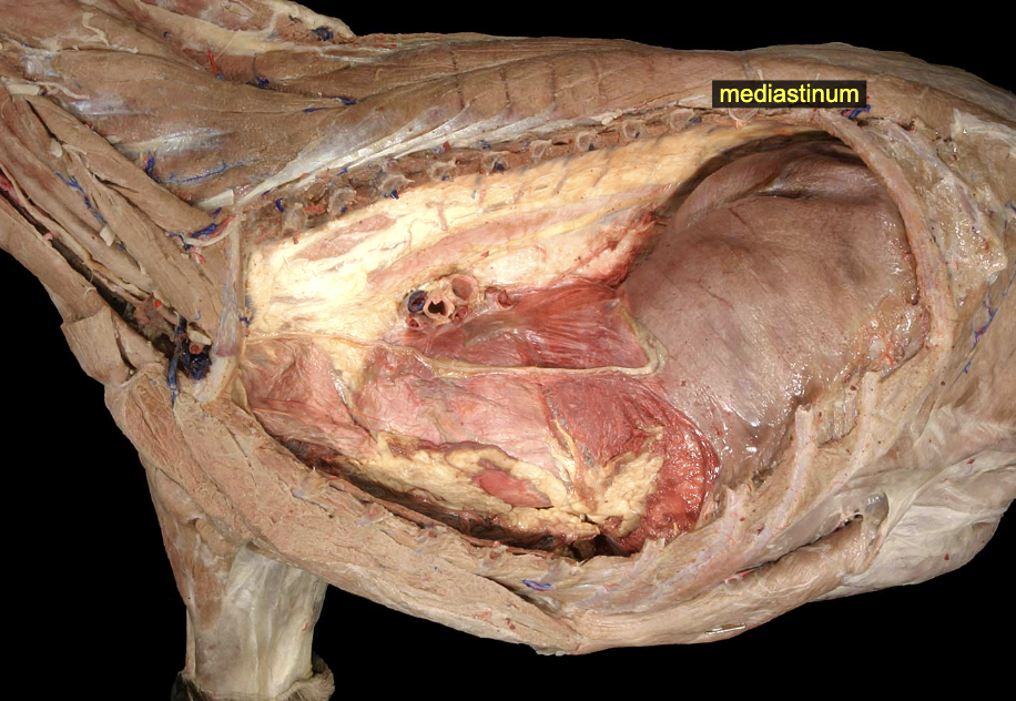
Mediastinum
The thoracic cavity is divided into right and left sides, each associated with a separate pleural cavity, by the presences of the tissues and organs near the midline of the body. These structures (which include the trachea; esophagus; heart; thymus; and many nerves, vessels, lymphatic organs, and fat) are covered by parietal pleura on right and left sides of the thorax. Collectively, all of these with their pleural coverings create an incomplete barrier between the right and left pleural cavities. This collective barrier is the mediastinum.
Diaphragmatic pleura
Pleura is the serosal investment associated with the thoracic viscera. It is a simple squamous epithelium and is underlain by a fibrous subserosal layer called endothoracic fascia. Individual regions of the pleura are named for the body parts with which they are associated. The pleural on the thoracic side of the diaphragm is accordingly referred to as diaphragmatic pleural, a specific subcategory of the parietal pleura that surrounds the lungs.
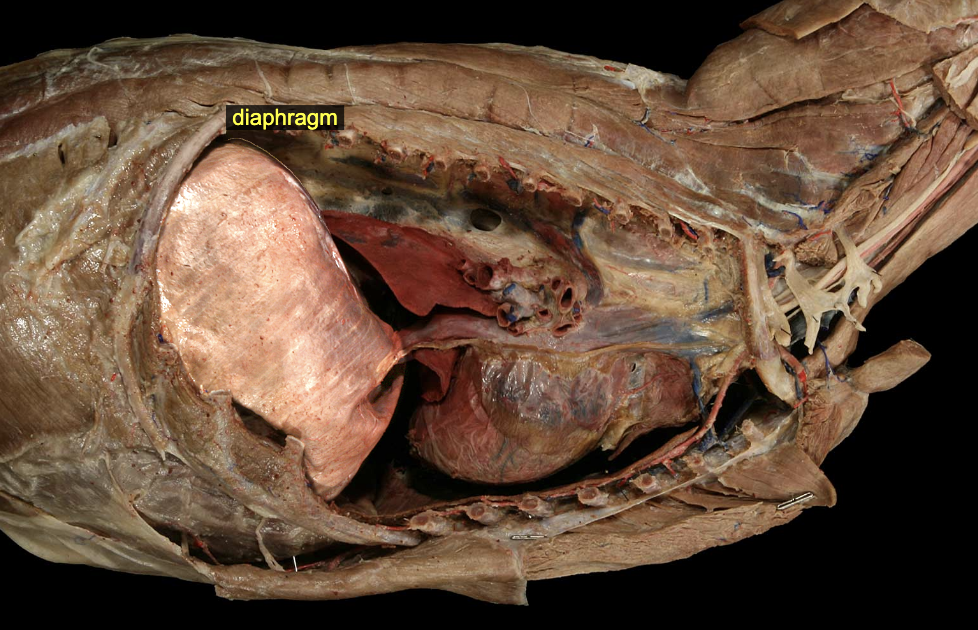
Diaphragm
The diaphragm is the musculotendinous sheet that divides the embryonic coelom into thoracic and abdominal cavities. The muscle of the diaphragm is striated, innervated by somatic motor fibers in the phrenic nerves. It is related intrathoracically to the lungs and heart and intraabdominally to the liver. It forms a deep dome, projecting well into the thorax from its attachments on the costal arches, lumbar vertebrae, and sternum. Within the thorax, the diaphragm is covered with diaphragmatic pleura, a specific subcategory of the parietal pleura that surrounds the lungs.
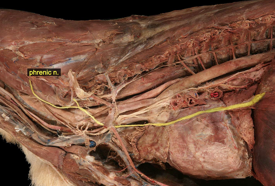
Phrenic nn.
Motor: diaphragm
Sensory: diaphragm
The phrenic nerve arises as individual nerve branches in the caudal cervical region which pass caudad next to the external jugular vein and become a single identifiable phrenic nerve as they enter the thoracic inlet. Inside the thorax, the nerve passes ventral to the subclavian artery in the cranial mediastinum, across the heart in a fold of pleura, and terminates by arborizing in the muscular part of the diaphragm.