Forming a 4 Chambered Heart (Cardiac Looping)
1/19
There's no tags or description
Looks like no tags are added yet.
Name | Mastery | Learn | Test | Matching | Spaced |
|---|
No study sessions yet.
20 Terms
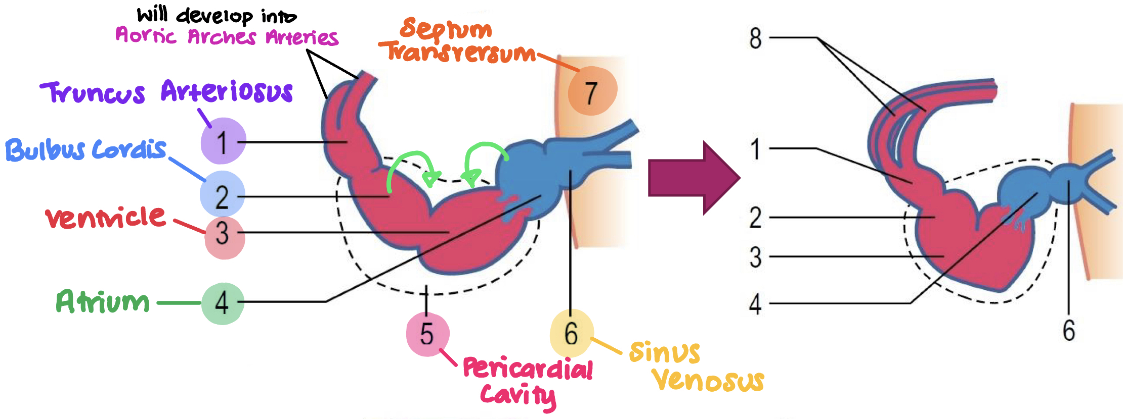
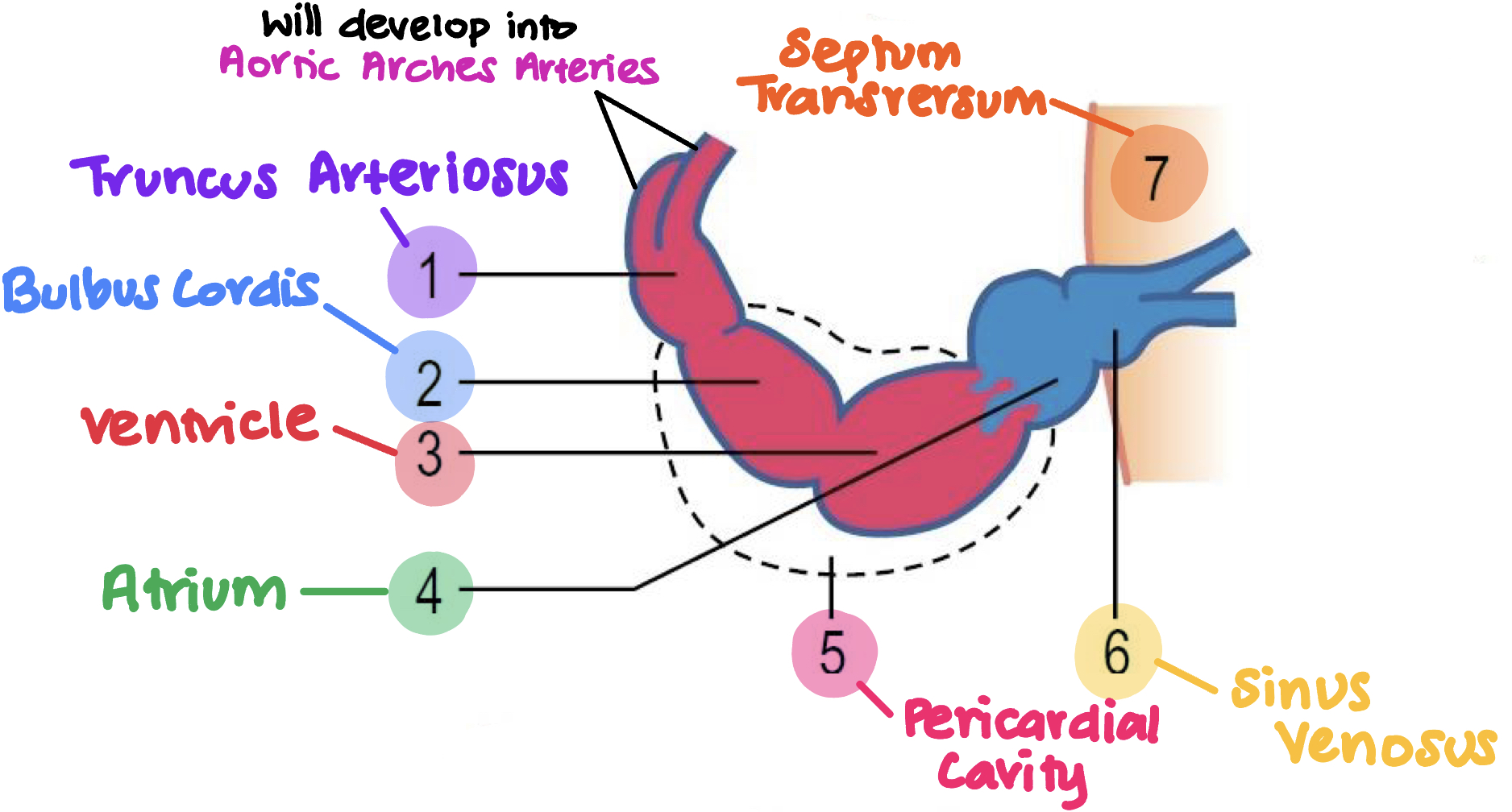
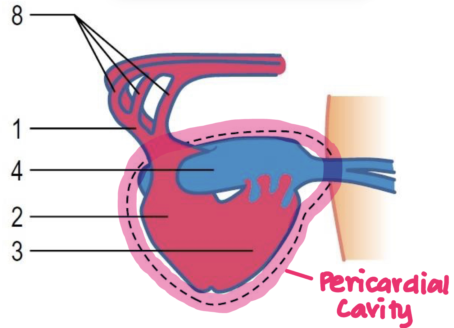
Recap: What are the regions of the straight heart tube?
Truncus arteriosus
Bulbus cordis
Ventricle
Atrium
Pericardial cavity
Sinus venosus
Septum transversum
Aortic arches arteries

What first occurs in the looping process?
Bulbus cordis (2) moves besides ventricle (3)
Atrium (4) moves dorsally (above) ventricle (3)
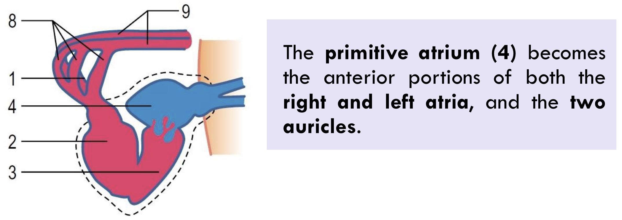
Due to the movement of atrium, it develops into?
Atrium (4) becomes anterior portions of
Both right and left atria
2 auricles
During cardiac looping what occurs to the sinus venosus (6)?
Develops in to the posterior portion of the
Right atrium
Sinoatrial nod
Coronary sinus
During cardiac looping what occurs to the pericardial cavity?
Envelopes the atrium and sinus venosus
Atrio-Ventricular Partitioning: Initially what does the AV canal look like?
Initially the heart tube = single structure which contain common AV canal
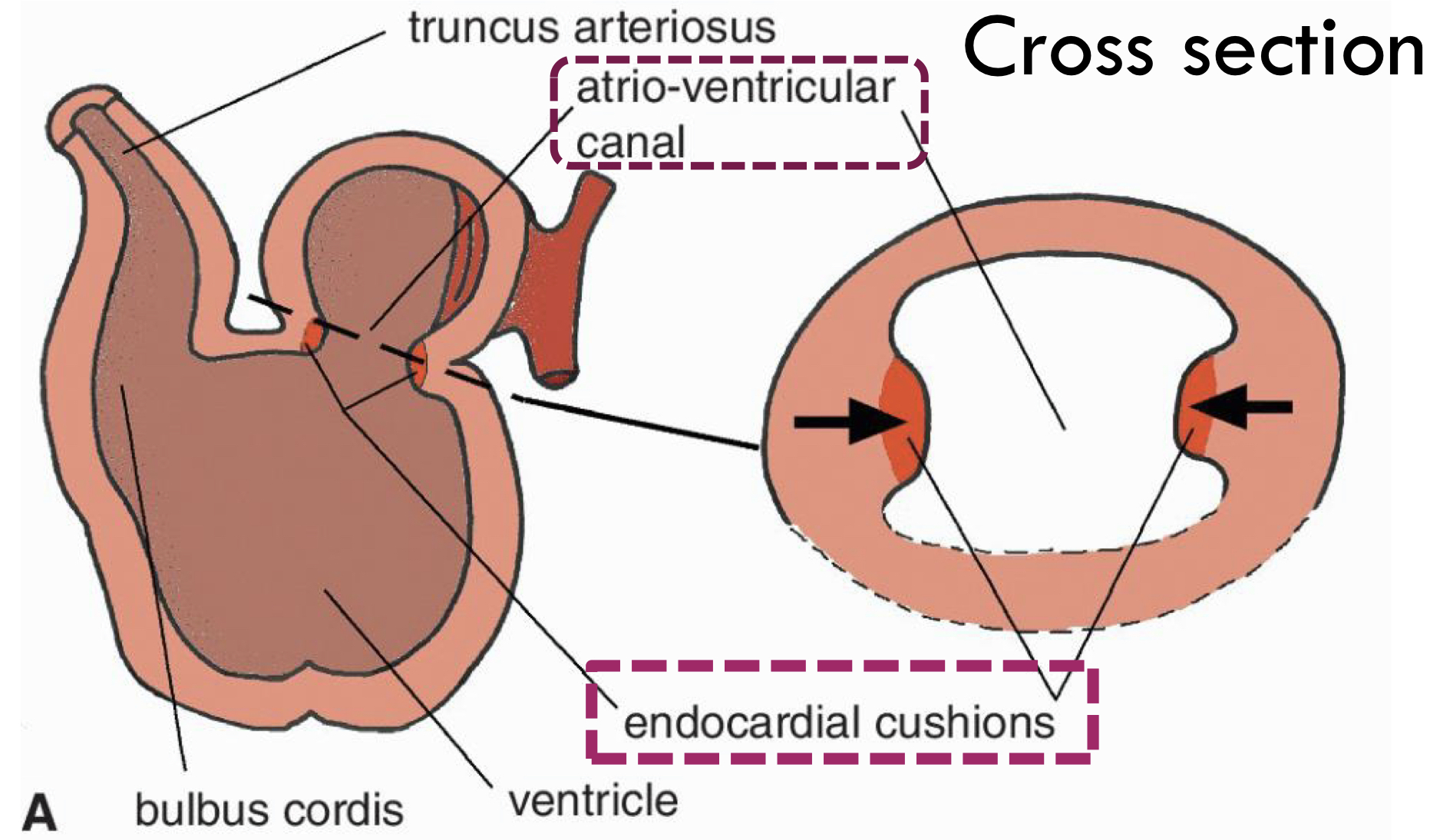
Atrio-Ventricular Partitioning: Therefore how does the AV canal become separate?
Due to the development of endocardial cushions:
They grow until they fuse in the middle of AV canal
Dividing the canal into right and left atrio-ventricular channels
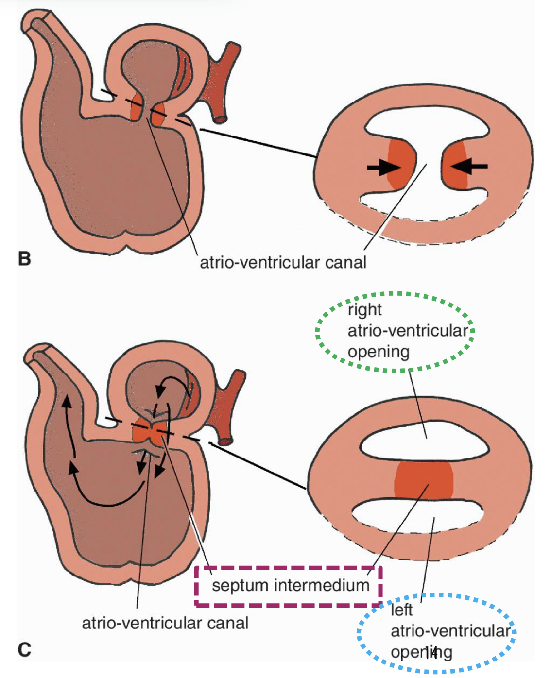
Atrio-Ventricular Partitioning: The fusion of the endocardial cushions formed what?
Septum intermedium (AV septum)
(the septum in the middle, separating atriums and ventricles)


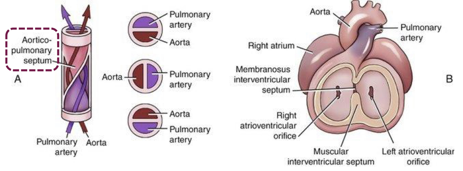
Septum formation in the ventricles: How does the interventricular septum develop?
From the ventricle walls that grows dorsally (upwards) towards the septum intermedium

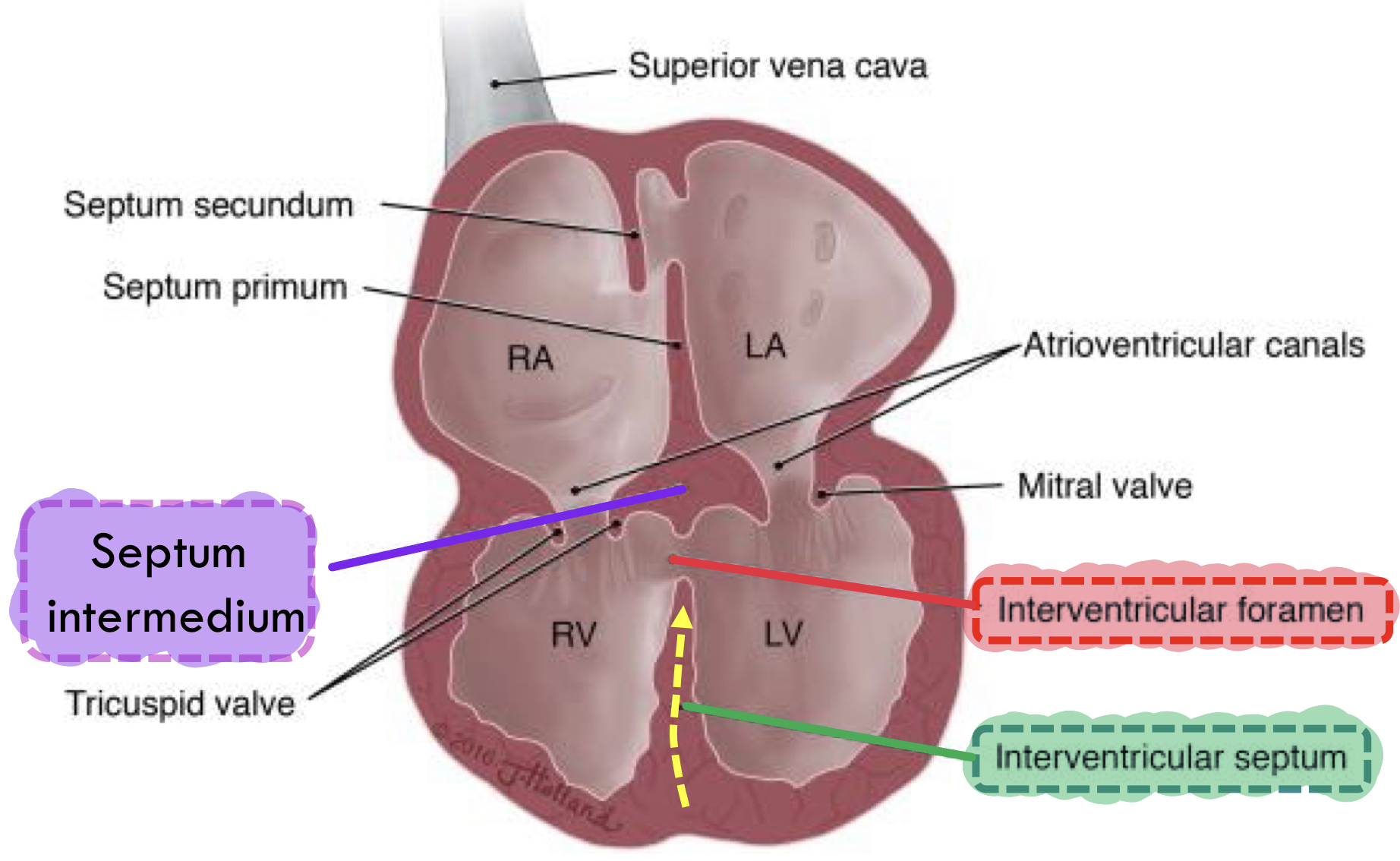
Septum formation in the ventricles: What about the interventricular foramen?
It persist for some time, but it eventually closes by the membranous part of interventricular septum

Septum formation in the ventricles: Therefore, what does the development of the interventricular septum and closure of the interventricular foramen form?
Forms the right and left ventricles
Clinical relevance: How could incomplete closure of the interventricular septum cause a disease?
It causes ventricular septal defect (VSD)
Common congenital disease
Leads to cardiac insufficiency
Because allows oxygenated and deoxygenated blood to mix
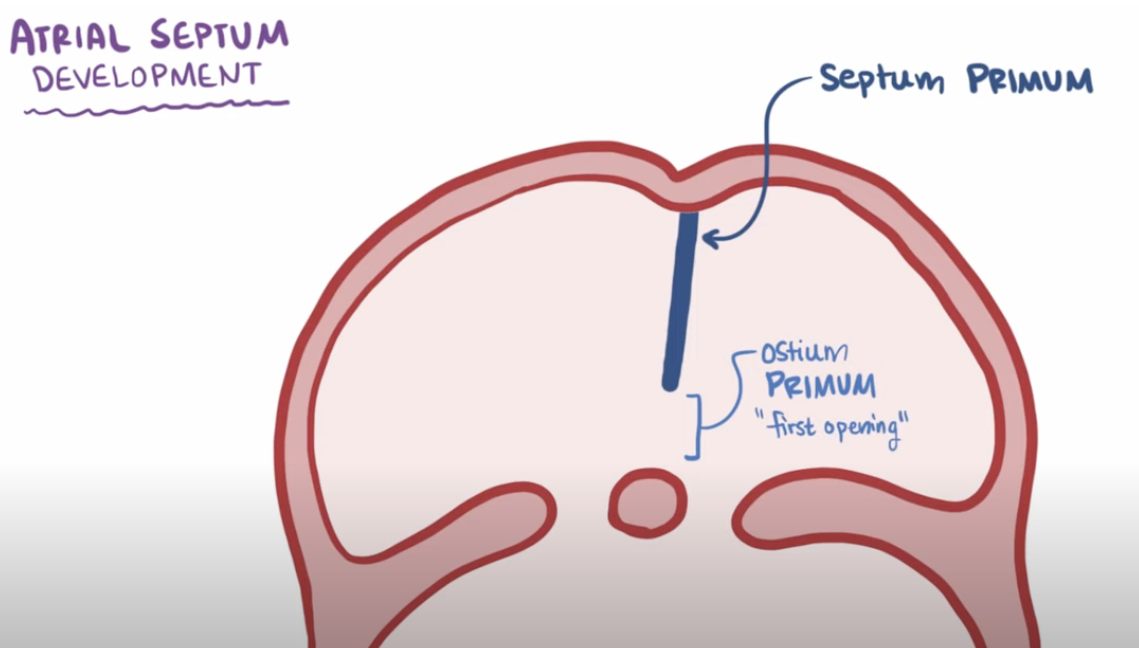
Formation of the Right and Left Atria: What is the septum primum and how is it formed?
What: First structure that makes up the atrial septum to grow
Forms:
Begins as a thin wall extending downwards from the roof of atrium —> endocardial cushions
This separates left and right atria
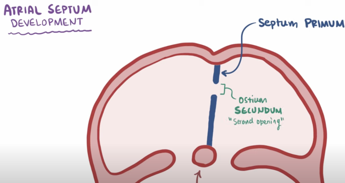
Formation of the Right and Left Atria: As the septum primum grows it leaves an opening called ostium primum, how does it close?
Eventually the septum primum continues to grow and then fuses with the endocardial cushions, closing the opening
Formation of the Right and Left Atria: However, there is still blood flow between right and left atria, how?
Because a second opening called the ostium secundum develops on a higher position
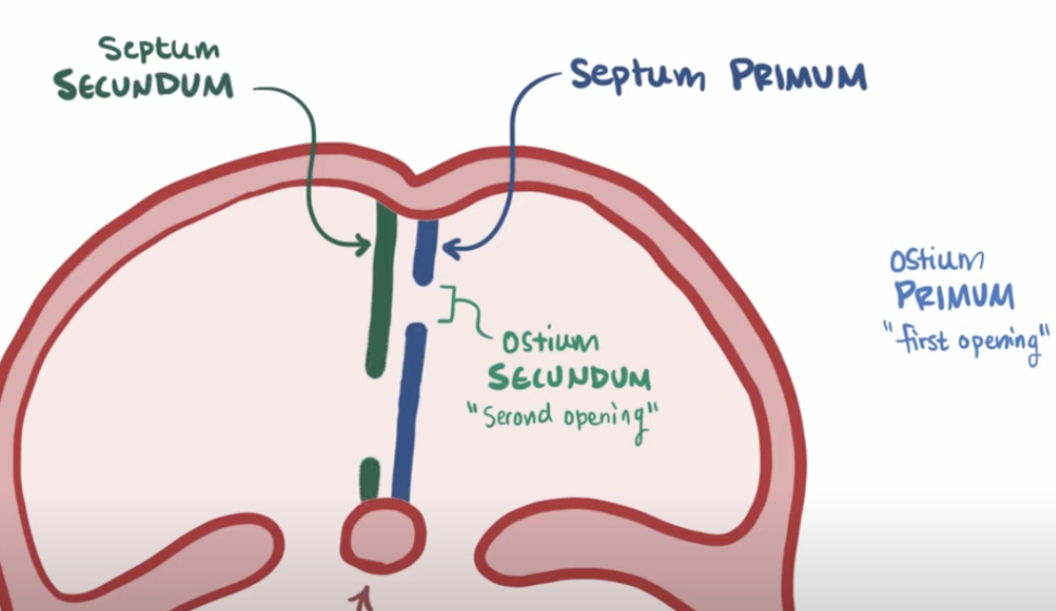
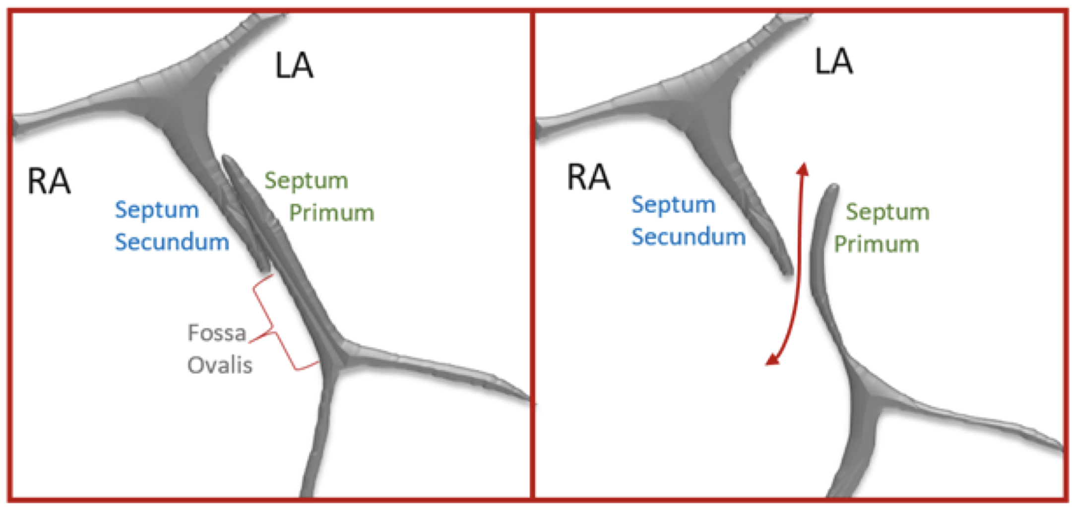
Formation of the Right and Left Atria: What is the septum secundum?
A septum that grows to the right side of the septum primum, extending downwards, covering the ostium secundum like a curtain but NOT entirely closed
Formation of the Right and Left Atria: Since the septum secundum doesn’t completely close, what opening does it form?
Foramen ovale
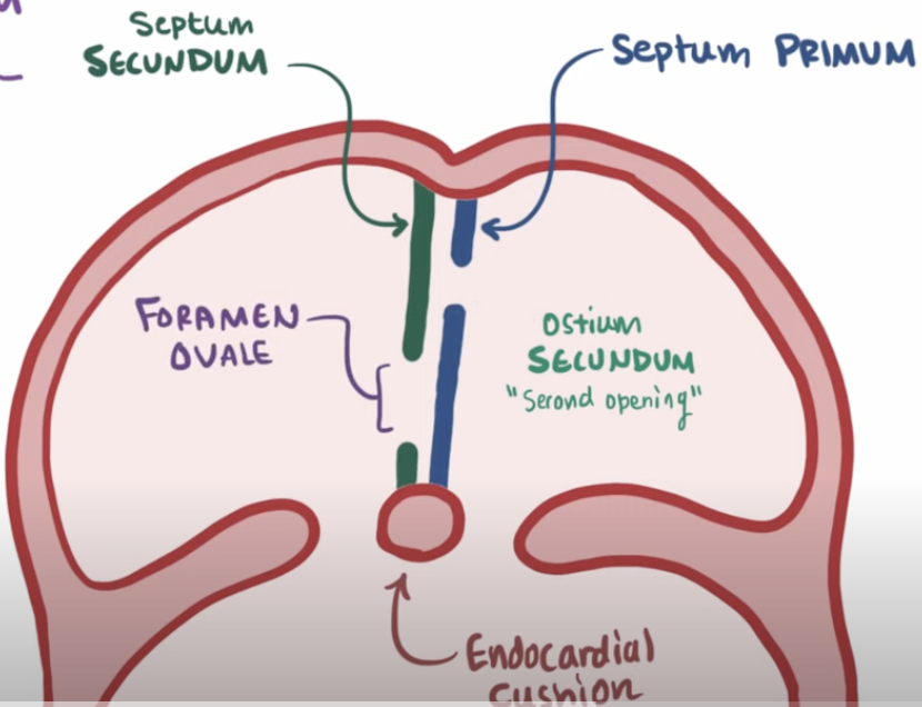
Formation of the Right and Left Atria: What is the function of the foramen ovale?
Allows oxygenated blood from the right atrium to bypass non functional fetal lungs and flow directly to into the left atrium to enter the systemic circulation
Formation of the Right and Left Atria: How does the foramen ovale close?
First breath after birth = lungs expand
Establishes pulmonary circulation
Increases O2 supply
Decreases pulmonary vascular resistance
Pressure in left atrium increases
Pressure in right atrium decreases (due to the cutting of umbilical cord)
Pressure difference forces the septum primum against the septum secundum
This closes the foramen ovale
Development of the Aorticopulmonary Septum: How does this septum develop and what does it separates?
Develops: Grows downwards, in a twisting pattern
Separates: Aorta and pulmonary trunk