Sternum and Ribs (2/12/2024 & 2/14/2024)
1/38
There's no tags or description
Looks like no tags are added yet.
Name | Mastery | Learn | Test | Matching | Spaced |
|---|
No study sessions yet.
39 Terms
What is Aurora’s sternum routine?
lateral and RAO
What is a common indication for a sternum x-ray?
MVA
What size IR is used for sternum images?
10 × 12 LW (with grid)
What is SID for the RAO sternum image?
40”
Explain patient and CR position for the RAO sternum image
patient
prone or erect
rotate 15-20o into RAO
places sternum to the left of the spine
large chest needs less rotation
small chest needs more rotation
CR
perpendicular to mid sternum
1 inch to the left of the spine
top of IR 1½ inches above jugular notch
Why do we do an RAO instead of an LAO?
to put the sternum into the uniform density heart shadow
What are breathing instructions for an RAO sternum image? Why?
shallow breathing to utilize long exposure time (3 sec) to blur lung markings and ribs
(on expiration if pt is unable to do shallow breathing)
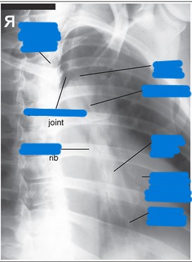
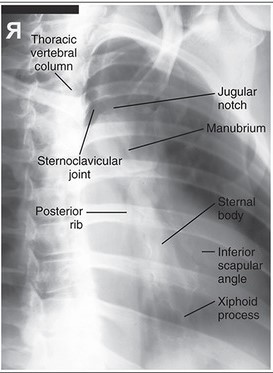
What needs to be demonstrated on an RAO sternum image (film eval)?
slight oblique of sternum utilizing heart shadow
any fractures or inflammatory processes
collimation side to side
marker (L because the right side of the body is not even on the image)
oriented as if person is standing in front of you
What is done in place of an RAO sternum for a trauma patient?
trauma oblique sternum
patient supine
15-20o tube angle from right to left or LPO position
angled tube: center at sternum
LPO: centered 1 inch lateral to sternum on left side (side down)
mark left side
Explain patient and CR positioning for a lateral sternum
patient
erect
lateral position (R or L)
seated or standing
shoulders and arms drawn backward
recumbent
lateral position (R or L)
true lateral
arms raised above head, shoulders back
CR
perpendicular to mid-sternum entering lateral border
top of IR 1½ inches above jugular notch
Explain the trauma lateral sternum
recumbent
dorsal decubitus position
raise arms above head (if possible)
What are breathing instructions for a lateral sternum? Why?
full inspiration; to get high contrast between posterior surface of sternum and lungs
What is SID for lateral sternum images?
Erect: 72”
Recumbent: 40”
Trauma shoot-thru: 40-72”
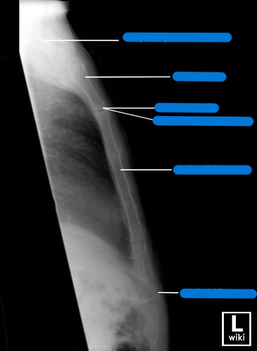
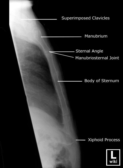
What needs to be demonstrated on a lateral sternum image (film eval)?
lateral view of entire sternum
any fractures or inflammatory processes
marker placed anterior for side down (usually L)
collimation anterior to posterior
oriented how you took the image
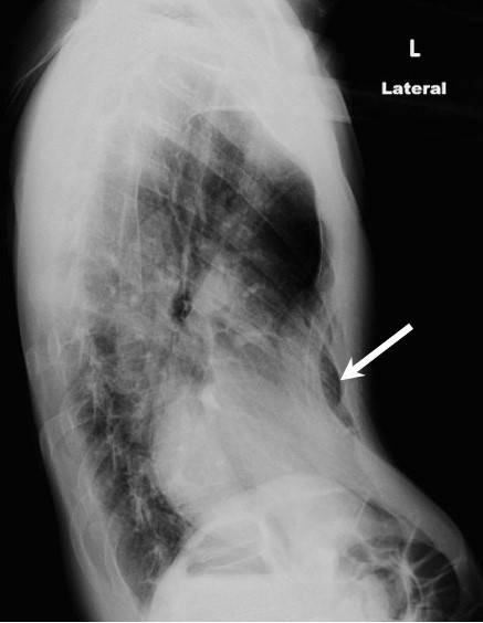
What pathology is shown?
pectus excavatum
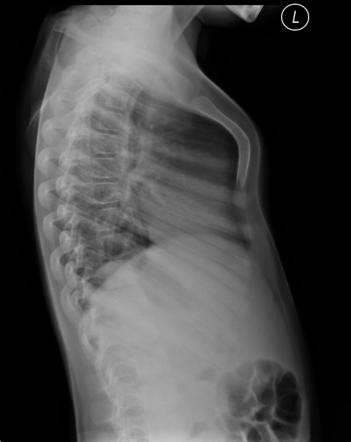
What pathology is shown?
pectus carinatum
What SID is used for ribs?
40”
What is the Aurora routine for ribs?
AP/PA upper
AP/PA lower
AP/PA oblique upper
AP/PA oblique lower
What size IR is used for ribs?
upper: 14×17 LW
lower: 11×14 LW
What are some complications that can occur with rib fractures?
pneumothorax
hemothorax
atelectasis
What determines whether ribs are done AP or PA?
location of the pain (area in pain is closer to IR)
Explain patient and CR position for AP/PA upper ribs
patient
AP or PA
no rotation
erect (preferred)
to lower the diaphragm
supine if patient cannot stand
raise chin
abduct affected arm
CR
perpendicular 3-4 inches below jugular notch
midway between lateral rib cage and mid-sagittal plane
top of IR 1½ inches above shoulder
What are respiration instructions for AP/PA upper ribs?
full inspiration (to lower diaphragm)
What needs to be demonstrated on an AP/PA upper rib image?
ribs 1-10
any fractures, neoplastic processes, pathologies
posterior ribs in detail
marker on affected side
collimate (make sure to include past jugular notch/spine)
oriented as if person is standing in front of you
Explain patient and CR position for AP/PA lower ribs
patient
AP or PA
supine recumbent preferred
raises diaphragm
abduct arm
CR
perpendicular to level of T12
midway between xiphoid and lower rib cage
midway between lateral rib cage and MSP
bottom of IR at crest
What are respiration instructions for AP/PA lower ribs?
on expiration
What needs to be demonstrated on an AP/PA lower rib image?
ribs 8-12
any fractures, neoplastic processes, other pathologies
posterior ribs in detail
marker on affected side
collimate (make sure to include all the way through the spine)
oriented as if person is standing in front of you
Explain patient and CR position for oblique upper ribs
patient
AP or PA
erect preferred
to lower diaphragm
otherwise supine recumbent
rotate 45o oblique
AP - affected side down
PA - affected side up
abduct affected arm
CR
perpendicular 3-4 inches below jugular notch
top of IR 1½ inches above shoulder
midway between spine and lateral border of rib cage
What are respiration instructions for oblique upper ribs?
on full inspiration
What needs to be demonstrated on an oblique upper rib image?
axillary portion of ribs (not seen on AP or PA)
any fractures, neoplastic processes
ribs 1-10
marker placed on affected side
oriented as if person is standing in front of you
collimate (make sure to include spine)
Explain patient and CR position for oblique lower ribs
patient
AP or PA
supine recumbent preferred
to raise diaphragm
45o oblique
AP - affected side down
PA - affected side up
abduct arm
CR
perpendicular to level of T12
midway between xiphoid and lower rib cage
midway between spine and lateral border of rib cage
bottom of IR slightly below rib cage or at top of crest
What are respiration instructions for oblique lower ribs?
on expiration
What needs to be demonstrated on an oblique lower rib image?
axillary ribs (not seen on AP or PA)
any fractures or neoplastic processes
ribs 8-12
marker on affected side
oriented as if person is standing in front of you
collimate (make sure to include spine)
What are techniques for an RAO sternum image?
80 kVp @ 16-20 mAs (breathing technique)
What are techniques for a lateral sternum image?
85-90 kVp @ 25-32 mAs
What are techniques for upper ribs?
AP: 70 kVp @ 16-20 mAs
Obl: 70 kVp @ 25-32 mAs
What are techniques for lower ribs?
AP: 80 kVp @ 20-25 mAs
Obl: 80 kVp @ 25-32 mAs