The Hip Joint, Joints and Ligaments of the Pelvis
1/24
There's no tags or description
Looks like no tags are added yet.
Name | Mastery | Learn | Test | Matching | Spaced |
|---|
No study sessions yet.
25 Terms
There are 3 compartments of the lower limbs…
There is thick fascia separating groups of muscles divided into an anterior compartment, a posterior compartment, and a medial compartment.
Compartments are important for two things: muscle ID, and compartment syndrome.
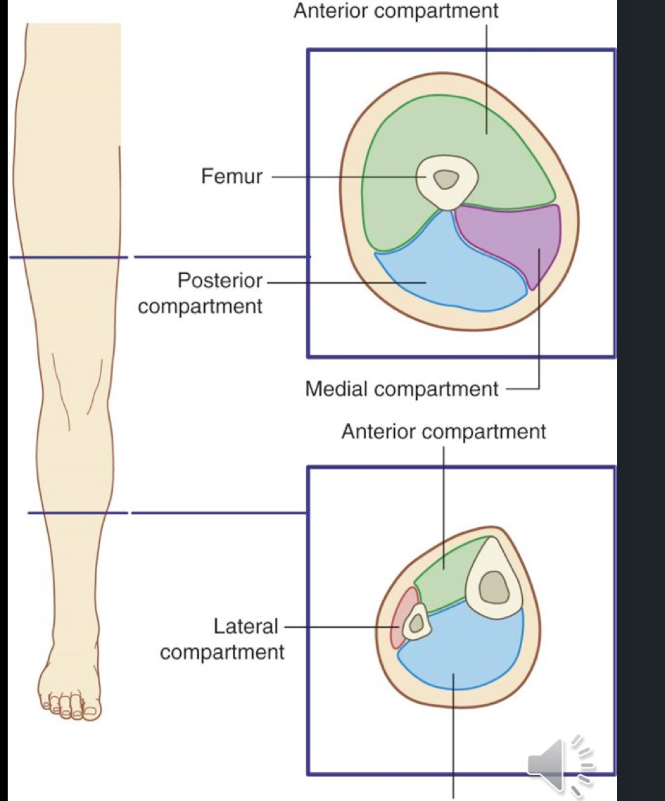
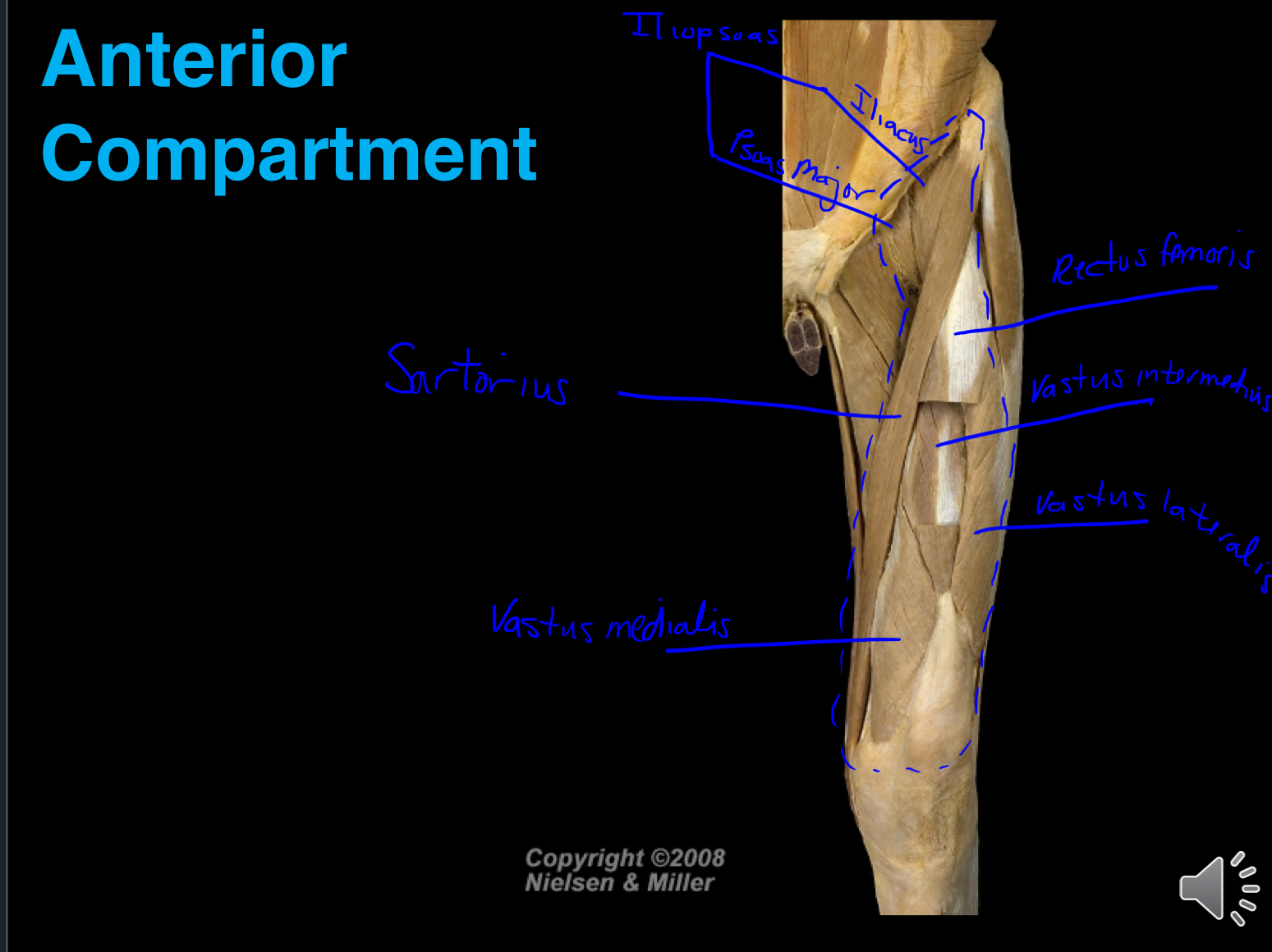
The anterior compartment of the thigh consists of…
Quadriceps Femoris Muscles
(RF, VL, VI, VM)
Sartorius
Psoas major and iliacus (terminal ends)
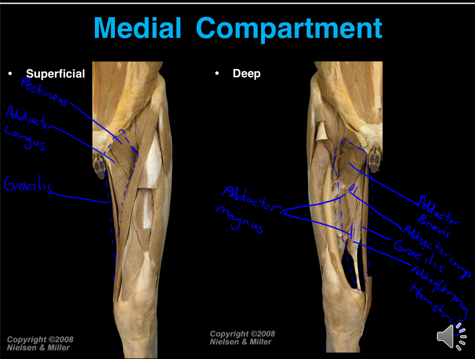
The medial compartment of the thigh consists of…
Gracilis
Pectineus
Adductor longus, brevis
Adductor magnus
Obturator externus
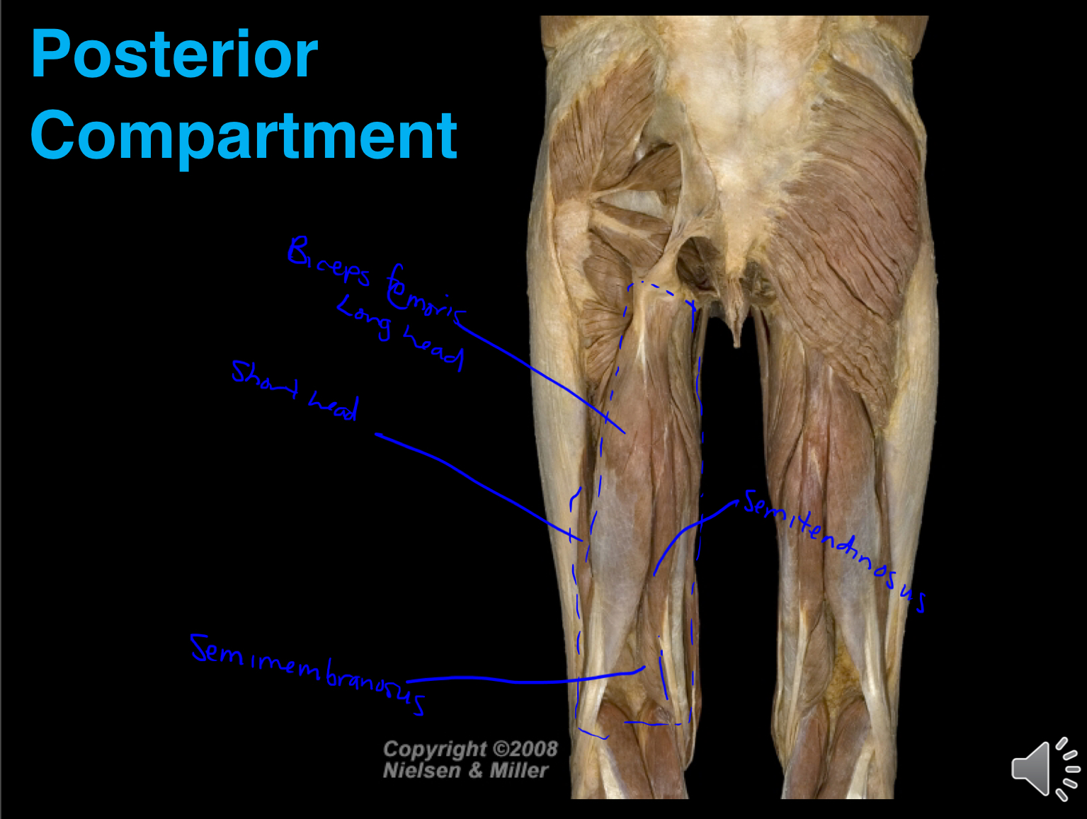
The posterior compartment of the thigh consists of…
Biceps femoris
Semitendinosus
Semimembranosus
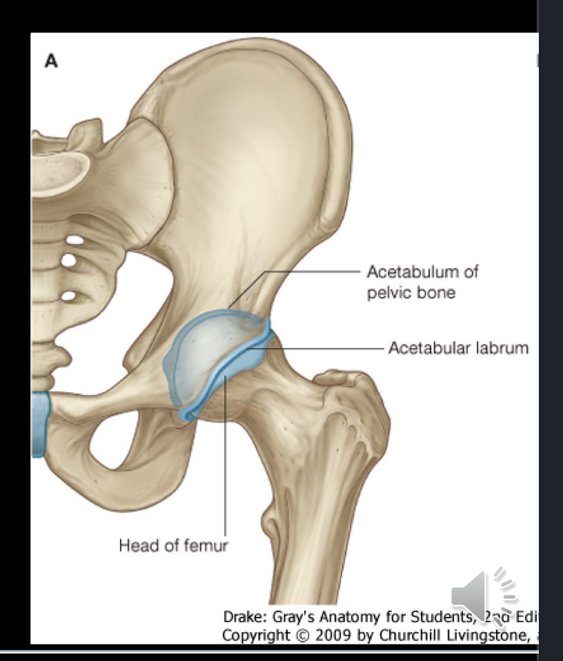
The hip joint…
Ball and socket synovial joint which is formed between the head of the femur and the acetabulum of os coxae. (Multi-Axial, meaning not as mobile as GH)
Consists of a very deep socket
Can withstand immense weight bearing responsibility and possesses good stability at the cost of mobility
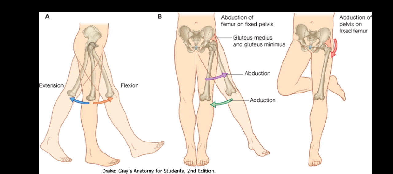
What are the movements at the hip joint?
Flexion
Extension
Abduction
Adduction
Internal/External Rotation
Circumduction
How is the hip joint more stable than the glenohumeral joint?
This is because nearly the entire head of the femur is within the acetabulum, in comparison with the humerus which is almost floating in the glenoid cavity within a mild labrum.
The articular surface of the acetabulum is made by the…
Lunate surface + articular cartilage, which forms a semi-circular shape around the acetabulum, providing a smooth surface for joint movement.
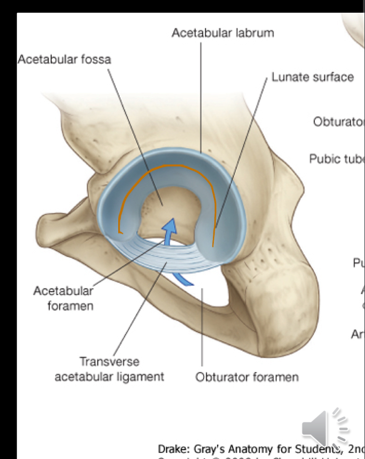
The acetabular labrum crosses the acetabular notch becoming…
The transverse acetabular ligament which bridges the notch, providing stability to the hip joint.

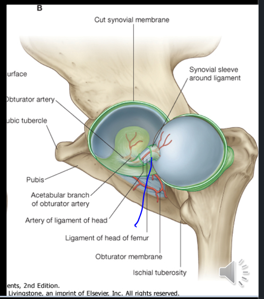
What ligament arises from the fovea capitis and connects it to the acetabular fossa and transverse acetabular ligament? What blood vessel runs through this ligament?
The ligamentum teres, which contains the artery of the ligamentum teres supplying blood to the head of the femur.
Another name for ligamentum teres is “ligament of the head of the femur”
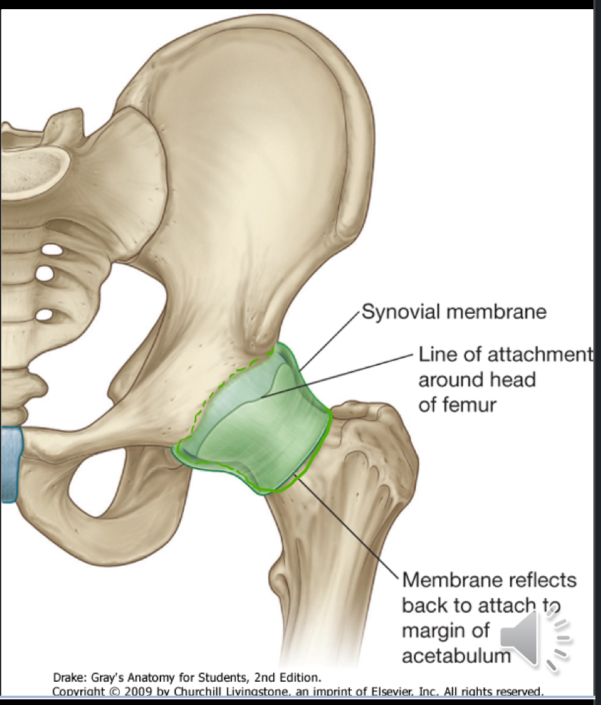
What does the synovial membrane of the hip joint attach to? What does it cover?
The synovial membrane of the hip attached to the articular surfaces of the femur and acetabulum.
It covers the neck of the femur
What are the fibrous joint capsule articulations for the hip joint?
Covers synovial membrane
Very strong and thick
Articulates with: Margin of acetabulum, transverse acetabular ligament, margin of obturator foramen, to the neck of the femur
What are the three stabilizing ligaments of the hip joint? What are the orientation of their fibers and how does this assist the hip joint/movement?
Iliofemoral, Pubofemoral, and Ischiofemoral ligaments
The fibers are oriented in spirals, which means that when the joint is extended the fibers become taut and firm. This helps to stabilize the joint and reduce the amount of energy/effort from muscles to maintain a standing position.
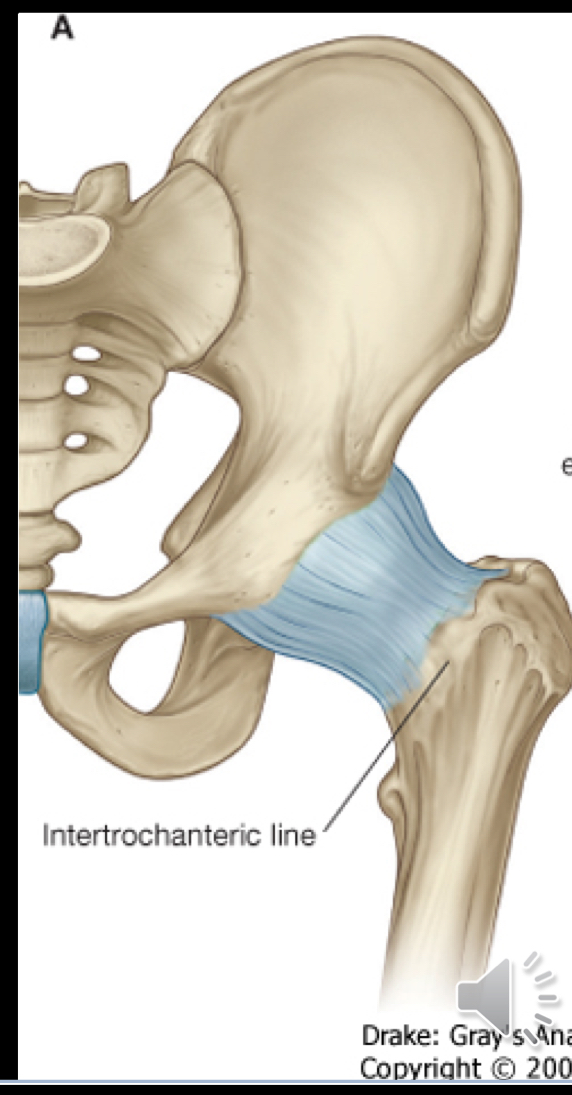
Iliofemoral Ligament
A strong ligament connecting the ilium to the femur, it helps stabilize the hip joint and limits hyperextension.
Anterior
Between the anterior inferior iliac spine and margin and the base of the acetabulum to the intertrochanteric line of femur
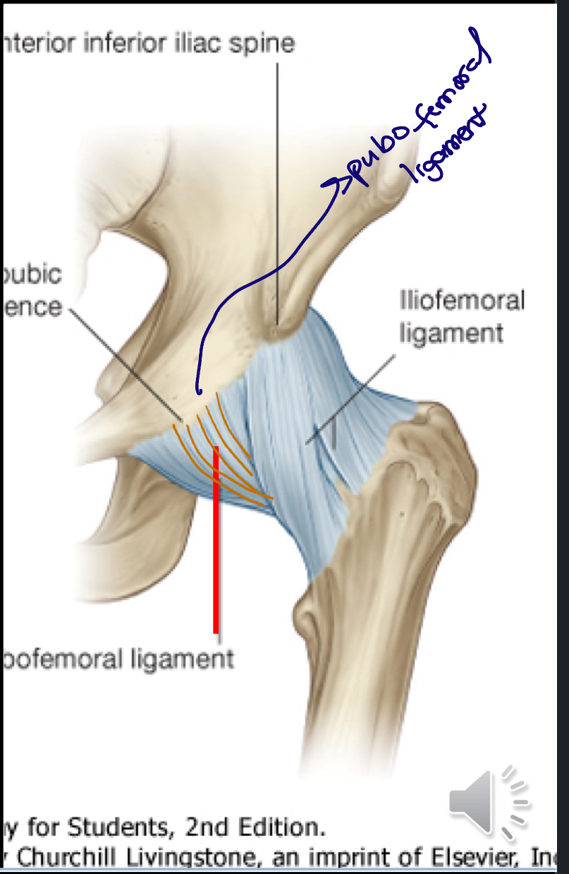
Pubofemoral Ligament
A ligament connecting the pubis to the femur, it aids in stabilizing the hip joint and preventing excessive abduction and extension. It runs from the pubis to the femoral neck.

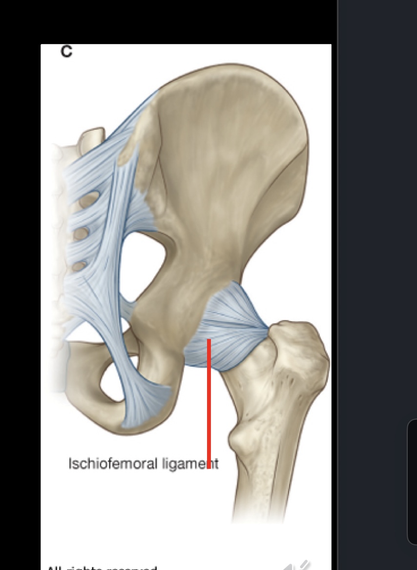
Ischiofemoral Ligament
A ligament that connects the ischium to the femur, it stabilizes the hip joint and limits internal rotation and adduction. It runs from the ischial part of the acetabulum to the femoral neck.
Posterior to the hip joint
From ischium posterioinferior to acetabulum
Goes to greater trochanter as well
What are the two main joints of the pelvic girdle?
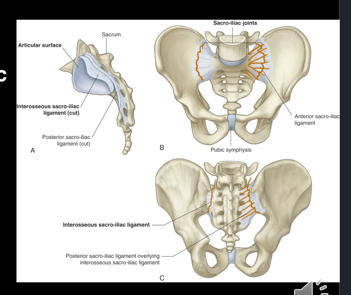
Sacroiliac joints
Between sacrum and ilium of os coxae
Transmits forces from lower limbs to axial skeleton
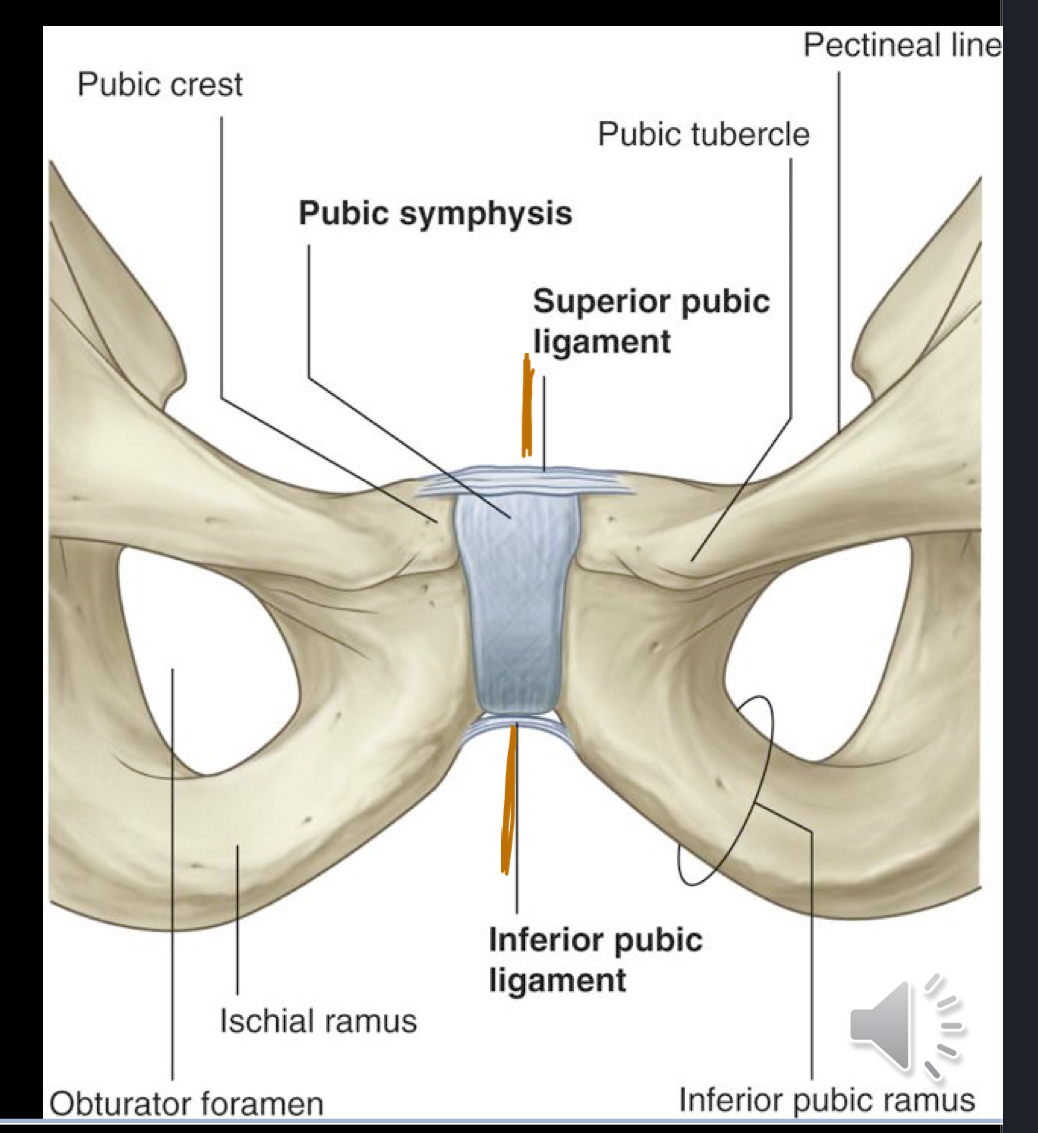
Pubic Symphysis

What are the stabilizing ligaments of the sacroiliac joints?
The anterior and posterior sacroiliac ligaments provide stability to the sacroiliac joints, supporting movement between the sacrum and ilium.
What are the stabilizing ligaments of the pubic symphysis?
Superior pubic ligament
Inferior pubic ligament
These ligaments connect the pubic bones at the pubic symphysis, providing stability and limiting separation during movement.
What are the stabilizing ligaments of the pelvic wall?
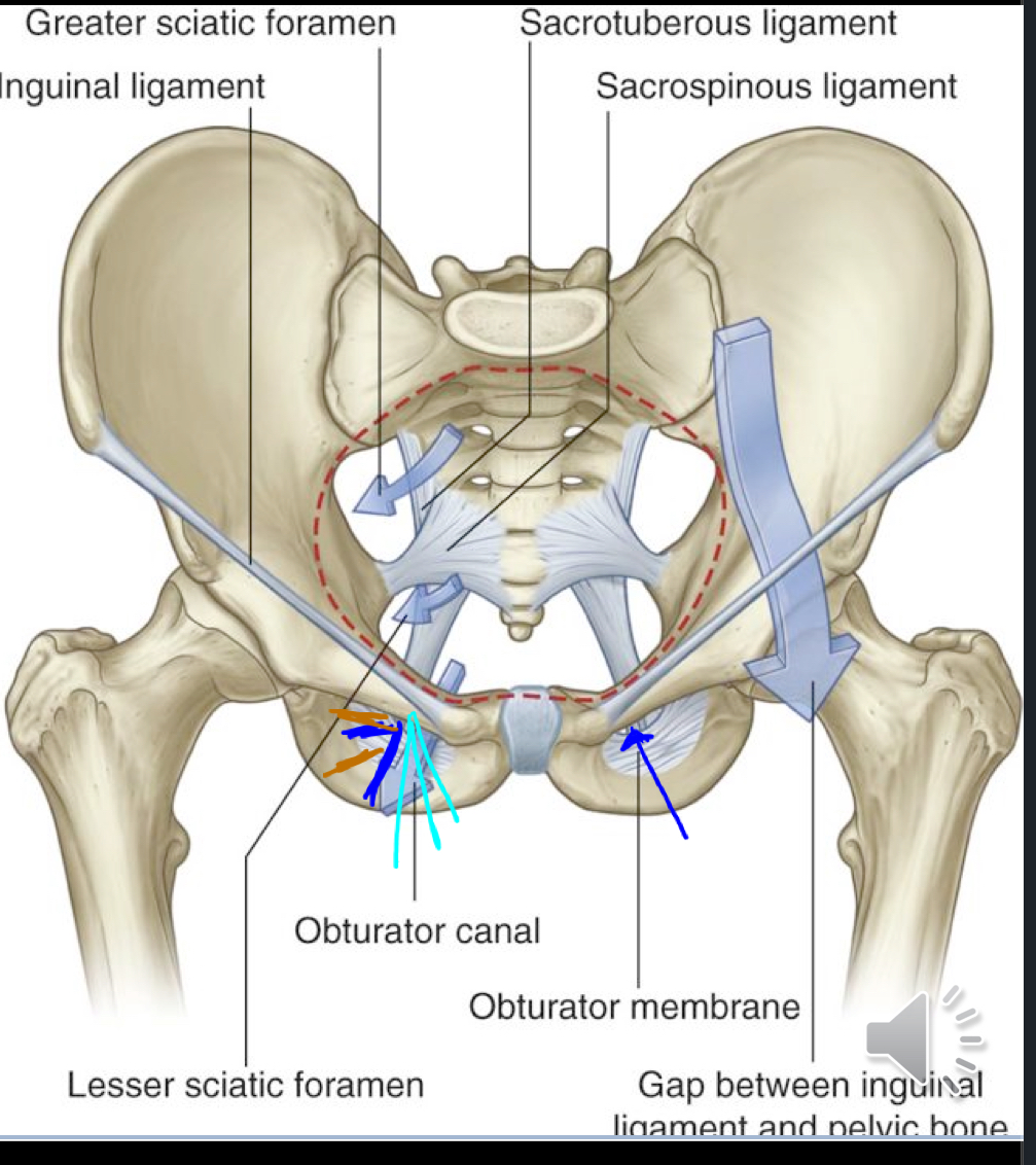
Sacrospinous ligaments
Ischial spine to sacrum & coccyx
Looks transversely placed (straight across to the sacrum & coccyx)
Sacrotuberous ligament
Posterior superior iliac spine to ischial tuberosity, providing additional support to the pelvic wall.
This one travels in a wave line pattern “)”
What are the gateways to the lower limb? “How do structures pass from the abdomen and pelvis into/out of the lower limb?”
Obturator canal
Greater Sciatic foramen
Lesser sciatic foramen
Below the inguinal ligament
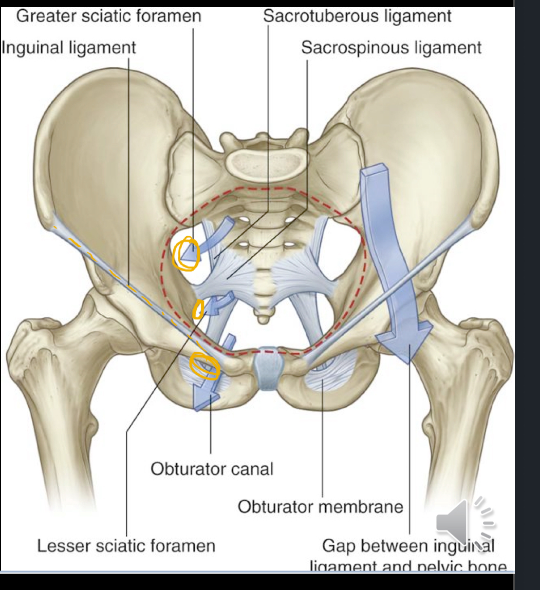
What is the obturator canal?
The obturator canal is a passageway in the pelvis that allows the obturator nerve, artery, and vein to exit the pelvis and enter the medial compartment of the thigh.

What is the greater sciatic foramen?
The greater sciatic foramen is an opening in the posterior pelvis that permits the passage of the sciatic nerve, along with other nerves and vessels, from the pelvis to the lower limb.
Piriformis muscle
Superior gluteal N, Sciatic N, Nerve to Obturator Internus, Nerve to Quadratus femoris
Gluteal vessels

What is the lesser sciatic foramen?
An opening in the pelvic girdle which allows for the pasage of the obturator internus.

The Inguinal Ligament
is a fibrous band that runs from the anterior superior iliac spine to the pubic tubercle, forming the floor of the inguinal canal.
Forms a roof like structure and allows the iliopsoas muscle, pectineus, femoral AVN, and lymphatics to pass deep to it.