BIOL 2052 - Brain development
1/16
There's no tags or description
Looks like no tags are added yet.
Name | Mastery | Learn | Test | Matching | Spaced | Call with Kai |
|---|
No analytics yet
Send a link to your students to track their progress
17 Terms
stages of brain development
neurogenesis
migration
differentiation
target innervation
synapse formation
neurogenesis
creation of neurons
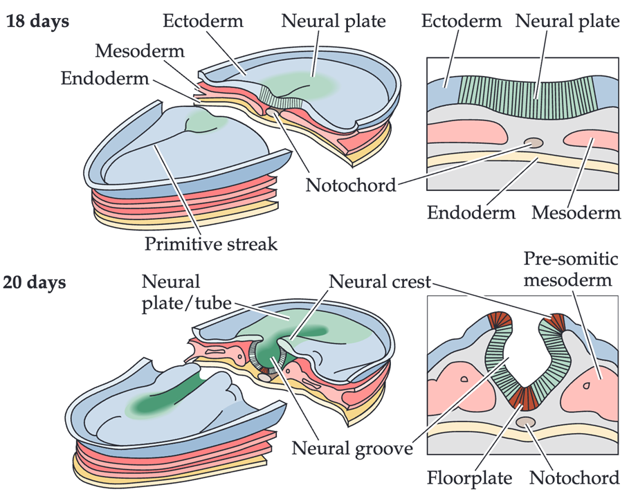
occurs during the first 18 days of development
a plate like structure formed which has 3 germ line layers
ENDODERM: gives rise to the lining of the digestive tract and organs
MESODERM: gives rise to the muscle, skeleton, circulatory system
ECTODERM: gives rise to the skin and brain —> from the neuroectoderm the neural plate is formed which gives rise to the whole nervous system
the notochord is formed at the midline
After formation of the neural plate it folds inwards and closes forming the neural tube
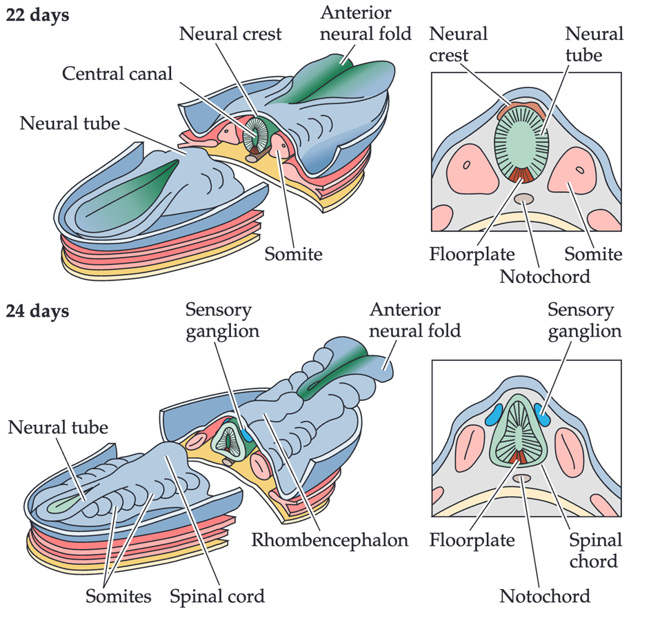
the neural tube at one end gives rise to the brain, whilst at the other end gives rise to the spinal chord
the lumen of the tube will become the ventricles which hold the CSF
where the tube closes is called the roofplate
the floorplate is found above the notochord
the notochord, floorplate and roofplate are transient structures which are important for instructing the nervous system
the neural crest will then separate from the neural tube and will give rise to the PNS
patterning in the neural tube
anterior and posterior regions along the length of the tube
dorsal and ventral along the cross section of the tube
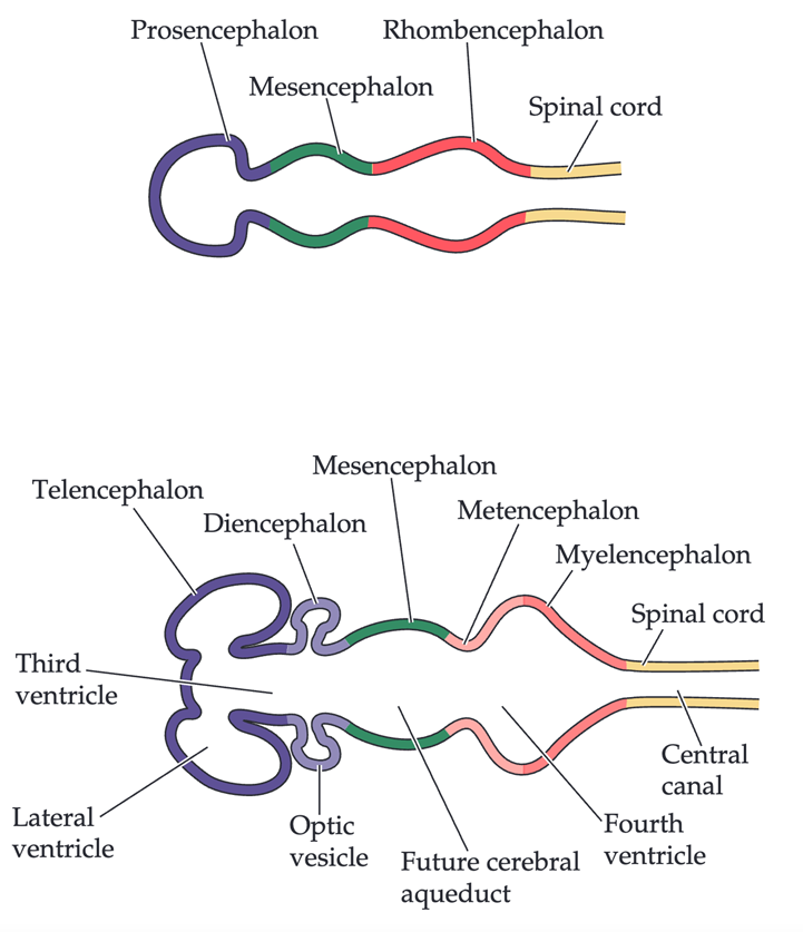
proliferation and segmentation determines the early spinal chord and forms 3 primary vesicles, the brainstem, midbrain and the forebrain
patterning and differentiation of cells is determined by morphogens
differentiation
when and where a neuron is born will determine its ultimate fate
differentiation is driven by morphogen gradient
morphogens bind to receptors to activate or repress transcription factors
gene expression profiles determine the cell identity
the response of the cell is determined by
availibility of ligand
Prescence of receptors
distance from secreting cell gradients
EXAMPLES:
cells in posterior secrete and produce less wnt and BMP compared to cells in the anterior
at the roofplate high expression of BMP, low Shh, whilst at the floorplate high Shh and low BMP
gene expression during development is so tightly controlled that receptor must be already expressed before the morphogen is secreted
hox gene
family of transcription factors
in human have A-D
hox helps to establish segmentation of anterior and posterior axis
how do we know cell fate can be induced
graft of tissue from pigmented to non amphibian embryo
secondary neural tube is developed with a mixed origin
transplanted cells instructed by genes but also by the cells arund them
migration
how neurons get to the correct place
cells use the scaffold of the ECM to migrate
neuroblasts migrate to the pial surface
they migrate from the marginal zone vertically and they differentiate into neurons
newer neuroblasts migrate past they older cousins to the cortex is built indie out
EXAMPLE: building the cortex
neuroepithelial progenitor cells in the neural tube are the neural precursor cells
the neural precursor cells form the ventricular zone
the radial glia connect the ventricular and pial surface and they divide slowly and symmetrically
the precursor cells divide asymmetrically in the ventricular zone (transit amplifying cells)
these generate new progenitors and post mitotic neuroblasts (they dont divide and give rise to neurons)
symmetric vs asymmetric division
RADIAL GLIA
dividing cells will give rise to identical cells
new cells provide more cells to expand that region
radial glia will undergo symmetric and asymmetric division but will form at least 1 ore radial glial cell with each division
NEUROBLASTS
dividing gives rise to different cell types which will have a different role and will migrate to a different area
origins of glia
generated from the neuroepithelium
glioblasts either
remain attached to the epithelium
become ependymal cells which produce CSF
move to the marginal layer and become astrocytes involved in the maintenance and repair o oligodendrocytes
interneurons
have an inhibitory function rather than being an excitable neuron
have a different origin —> born in the ganglionic eminences
migrate tangentially at the periphery
target innervation
neurons must polarize: reach in all directions and axons and dendrites are established
processes use cues and signals to help them navigate
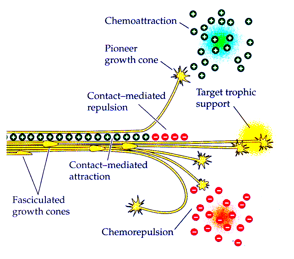
or a group of neurons will fasciculate - piggy back eachother and travel in a buncle with already developed neurons
how do axons find their target neurons
motor neurons have a clear target of where they must innervate
they have stops and checkpoints on the way to their destination and they use cues and signals to help them navigate called guidance signals
guidance signals can be both attractive and repulsive and can be short range or long range
guidance signals are interpreted by the growth cone which is a structure in the axon
if a guidance signal is chemo repulsive the growth cone will collapse and the process will not be able to grow further —> no longer see this as a target
TYES OF GUIDANCE SIGNALS
NON DIFFUSABLE
short range
substrate derived
in ECM or presented on the target cells
cadherins or ephrins
DIFFUSABLE
act as gradients
long range/short range
netrin and semaphorins
contact mediated repulasion
when fasciculation occurs the other axons will continue to produce chemo attractive signals until the growing axon needs to separate out
in which case chemo repulsive signals sent out and a new axon branches out and forms a growth cone
how are signals sensed
growth cone structures have filopodia and lamellipodia which have receptors on their surface which sense guidance cues
they have cytoskeletal proteins actin and microtubules which allow them to reach out once the direction has been determined
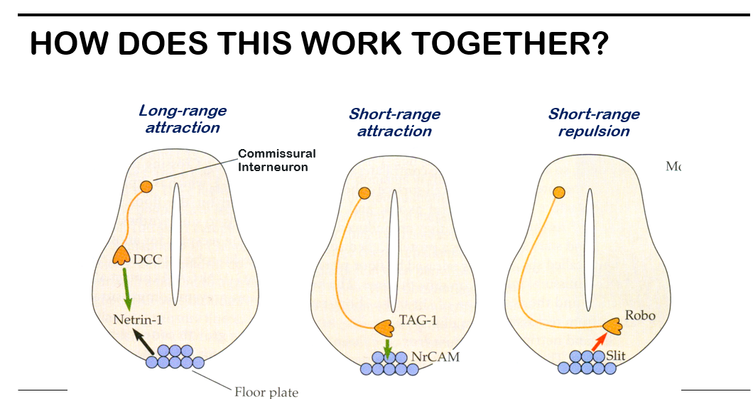
EXAMPLE: commissural neuron
DCC talks to netrin 1 in a long range attraction process
when the neuron gets close to the floorplate NrCAM attracts the neurons which is detected by TAG1 in a short range attraction
short range repulsion of the neuron slit is detected by Robo
why dont neurons cross?
motor neurons follow guidance signals and exit the cell to reach out to target muscle tissue
therefore, they travel outside the cell
also DSCAM acts as a chemo repulsive signal which prevents them from crossing over
synapse formation
mediated by synaptic cell adhesion molecules
molecules which help maintain the connection between synapses
neurexins (NXN) expessed in the presynaptic terminal which interact with the neuroligands in the postsynaptic terminal
several cell adhesion molecules will hold together the synapses
neuroligands on the postsynaptic terminal will recruit other entities which will detect the release of neurotransmitters so helps relieve the signal
abandoning synapses
some synapses kept, some abandoned
neurotrophins and electrical activity determine the final pattern of the contacts
not only are synapses abandoned when theyre not used but cells are abandoned too
axons which have more activity will become more stabilised as the binding of neurotransmitters and calcium signalling molecules cause more electrical activity therefore more stable
axons frequently used together will come together to form neural circuits