Special Areas of the Lower Limb
1/8
There's no tags or description
Looks like no tags are added yet.
Name | Mastery | Learn | Test | Matching | Spaced | Call with Kai |
|---|
No analytics yet
Send a link to your students to track their progress
9 Terms
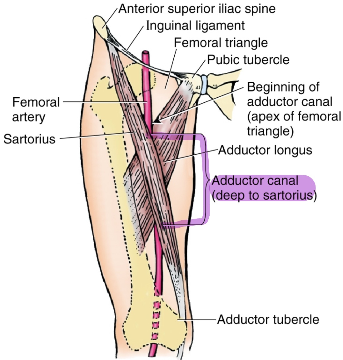
Femoral Triangle
Triangular space in the superoanterior third of the thigh; found at the junction b/w the trunk & the lower limb
Femoral artery pulse can be palpated here
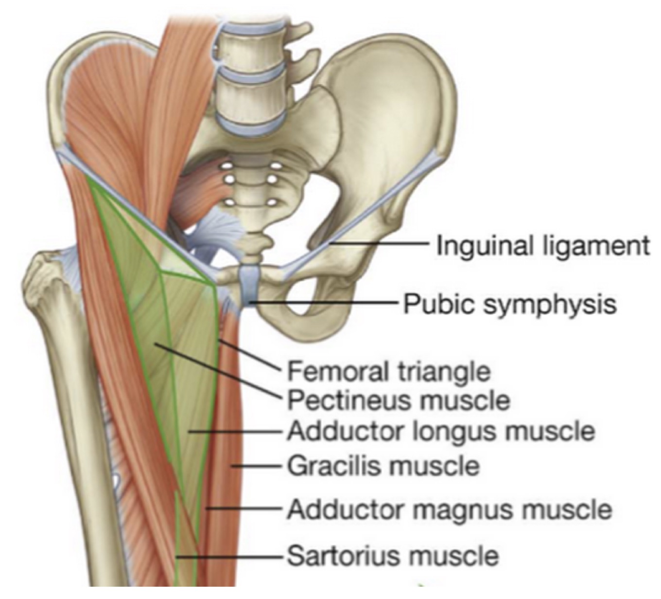
Borders & Floor of the Femoral Triangle
Borders:
1. Inguinal Ligament
2. Sartorius Muscle
3. Adductor Longus Muscles
Floor:
A. Iliopsoas Muscle
B. Pectineus Muscle
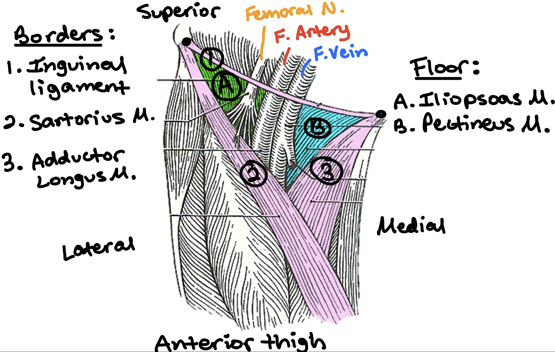
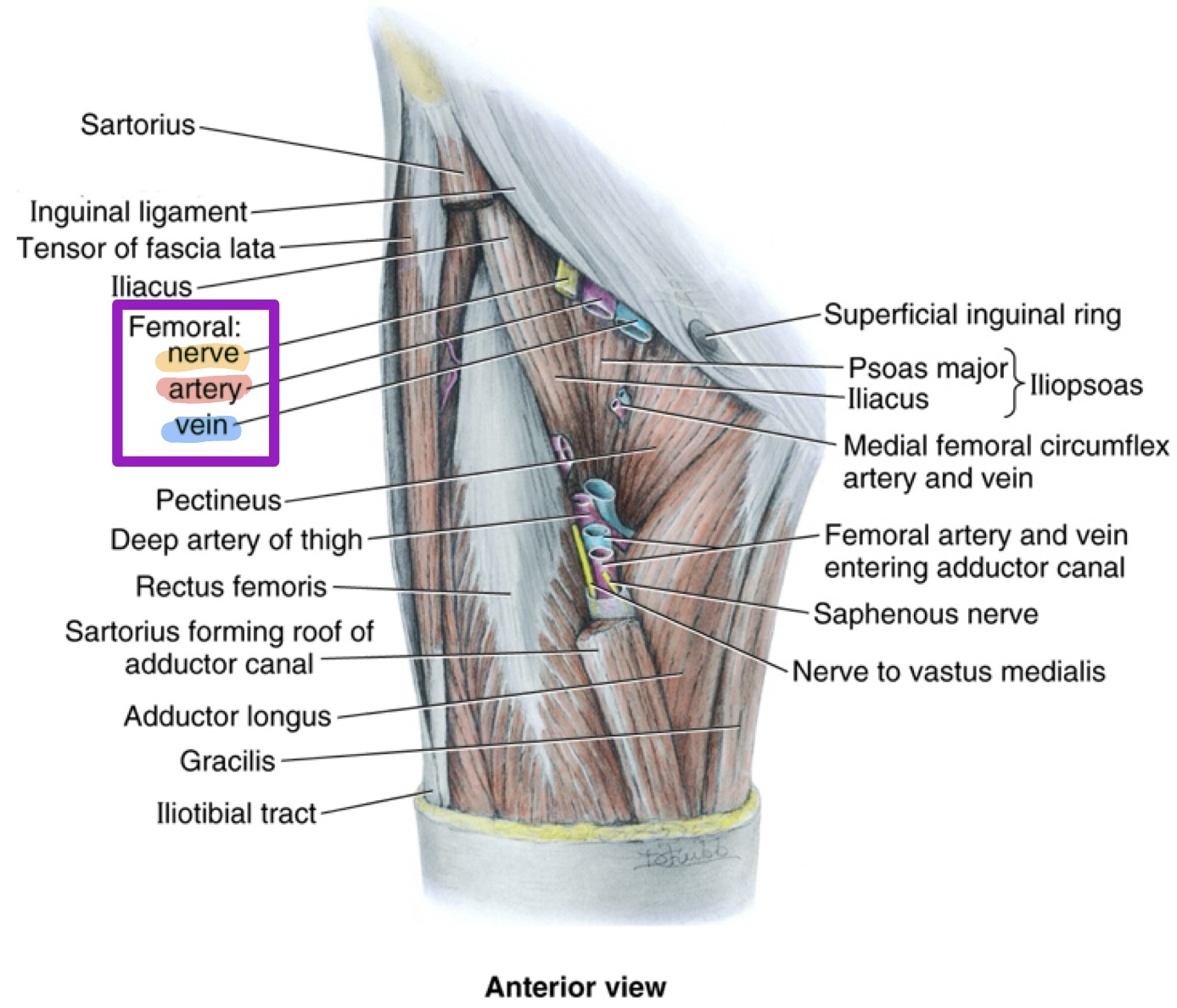
Contents of the Femoral Triangle
Content from medial → lateral:
Femoral vein
Femoral artery
Femoral nerve
V.A.N. drives out of femoral triangle (medial → lateral)
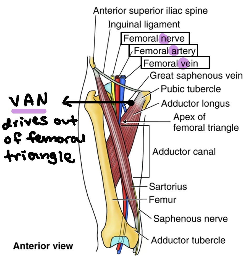
Adductor Canal
Compartment found b/w the quadriceps femoris muscles & the medial femoral muscles
Found deep to the satorius muscle
Contents of the Adductor Canal
Femoral artery
Femoral vein
Saphenous nerve (branch of the femoral nerve)
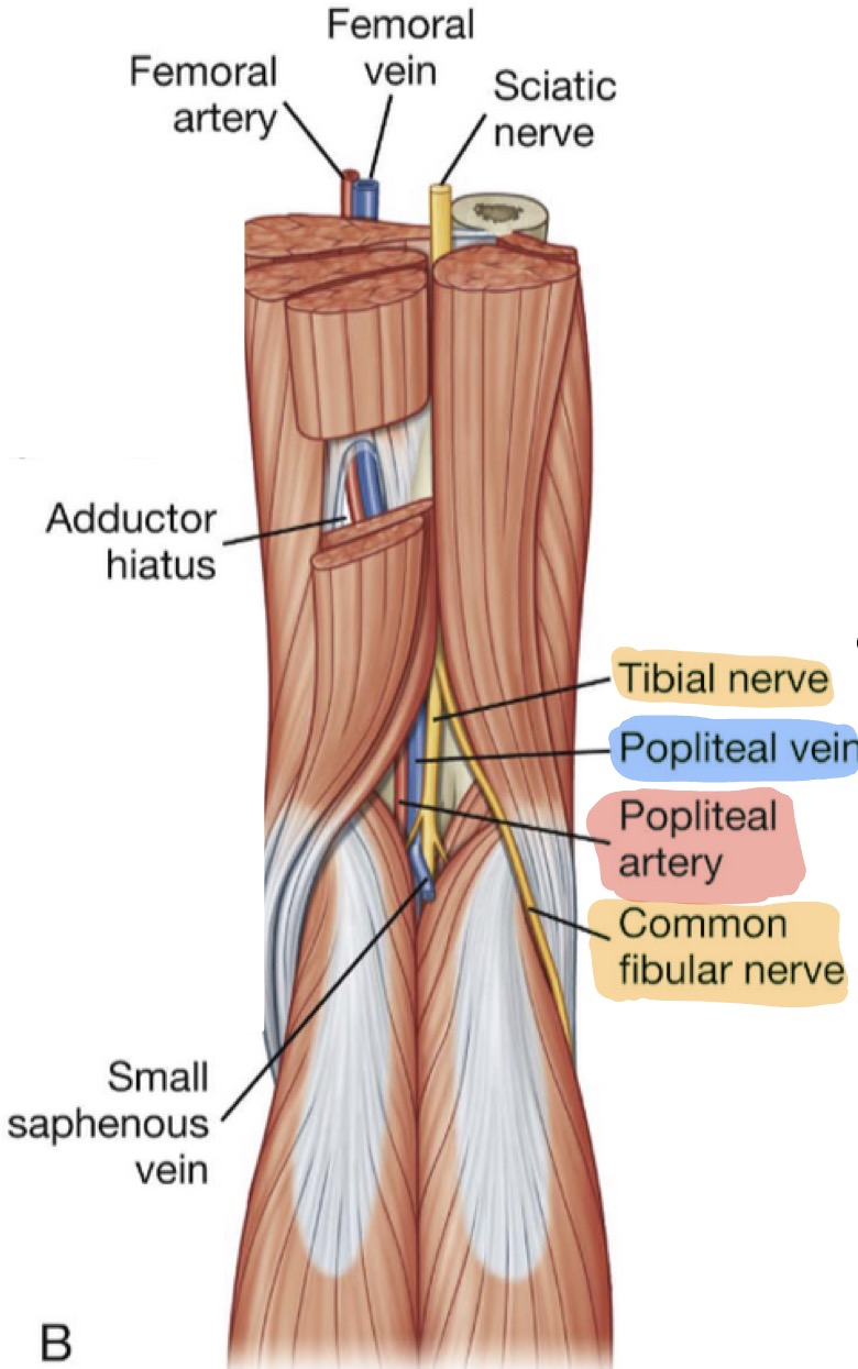
Popliteal Fossa
Diamond-shaped area in the back of the knee
Popliteal artery pulse is difficult to palpate due to depth
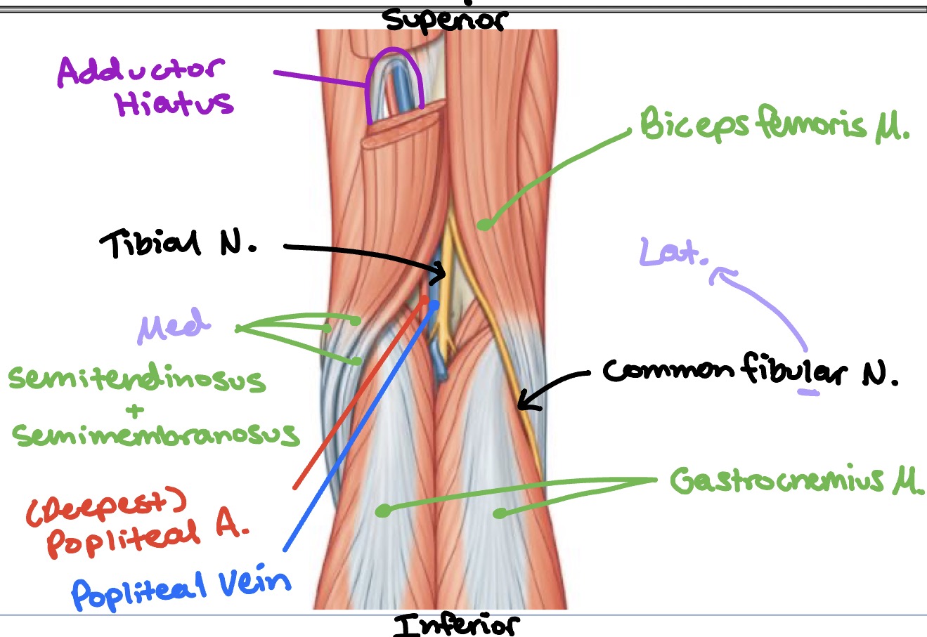
Content of the Popliteal Fossa
Content from deep → superficial:
Popliteal artery (deep)
Popliteal vein (deep)
Tibial nerve (superficial)
Common fibular nerve (superficial)
(Tibial nerve & common fibular nerve are same level superficial)
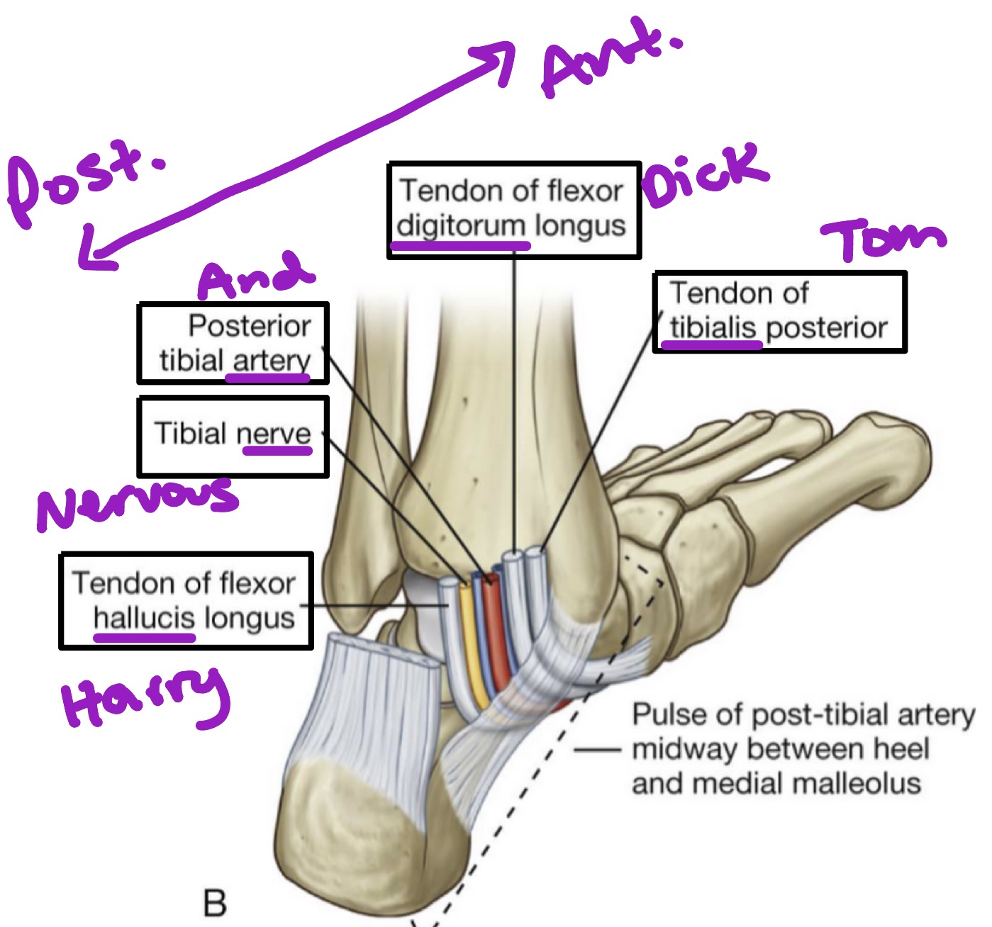
Tarsal Tunnel
Important relationship of structures located posterior to the medial malleolus
Bound by flexor retinaculum
Begins at the medical malleolus & ends at the calcaneus
Also known as “Tom, Dick, And Nervous Harry”
Contents of the Tarsal Tunnel
Contents from medial malleolus (anterior) to calcaneous (posterior):
Tendon of tibialis posterior muscle
Tendon of flexor digitorum longus muscle
Posterior tibial artery
Tibial nerve
Tendon of flexor hallucis longus muscle