Histo Lab-Cardiovascular Sysytem (Aughey)
1/36
There's no tags or description
Looks like no tags are added yet.
Name | Mastery | Learn | Test | Matching | Spaced |
|---|
No study sessions yet.
37 Terms
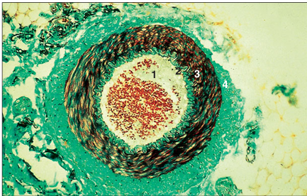
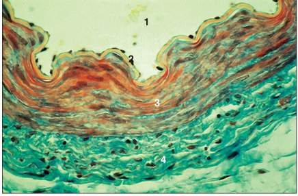
name of histologic slide:
Specie:
Parts
1.__ 2. __ 3.__ 4.__
Stain Used:
Artery (dog). (1) Lumen. (2) Tunica intima. (3) Tunica media. (4) Tunica adventitia. Masson’s trichrome.×62.5.
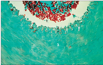
name of histologic slide:
Specie:
Parts
1.__ 2. __ 3.__
Stain Used:
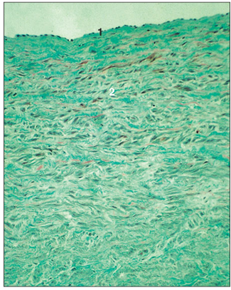
Artery (dog). (1) Lumen. (2) Tunica intima with the internal elastic lamina (arrowed). (3) Tunica media. Masson’s trichrome. ×125.
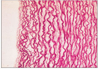
name of histologic slide:
Specie:
Stain Used:
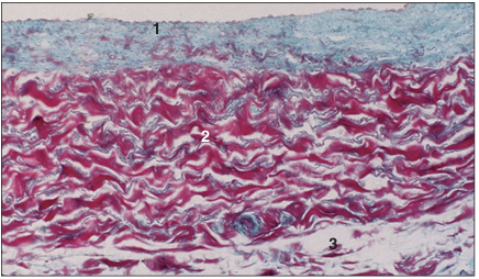
Aorta (horse). Elastic artery stained to illustrate the wavy elastic fibres. Weigert’s elastin. ×100.
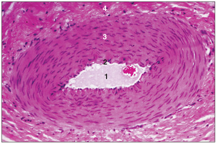
name of histologic slide:
Specie:
Parts
1.__ 2. __ 3.__ 4.__
Stain Used:
Muscular artery (sheep). (1) Lumen. (2) Tunica intima with the internal elastic lamina. (3) Tunica media. (4) Tunica adventitia. H& E. ×125
name of histologic slide:
Specie:
Parts
1.__ 2. __ 3.__ 4.__
Stain Used:
Muscular artery (sheep). (1) Lumen. (2) Tunica intima. (3) Tunica media. (4) Tunica adventitia. Masson’s trichrome.×250.
name of histologic slide:
Specie:
Parts
1.__ 2. __ 3.__
Stain Used:
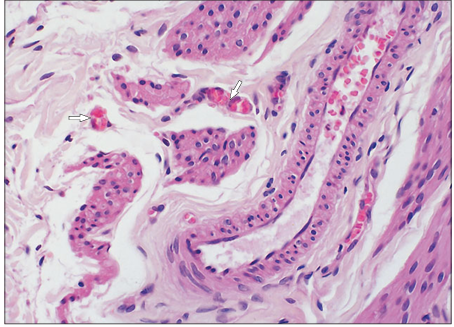
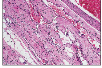
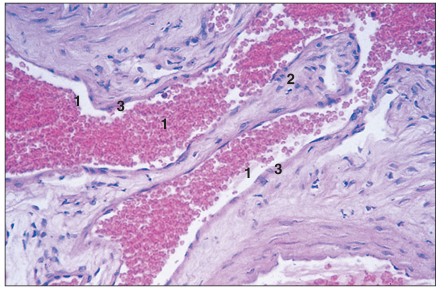
Artery and vein. Stomach (dog). (1) Small artery. (2) Small vein. (3) Lymphatic vessel. H & E. ×62.5.
name of histologic slide:
Specie:
Parts
1.__ 2. __ 3.__
Stain Used:
Arteriole, vein and a Iymphatic vessel in connective tissue. Tongue (ox). (1) Arteriole. (2) Vein. (3) Lymphatic vessel. H & E. ×62.5.
name of histologic slide:
Specie:
Parts
1.__ 2. __ 3.__
Stain Used:
Arterioles in the wall of a small artery (horse). (1) Arteriole. (2) Veins. (3) Artery. H & E. ×160.
Three types of Artery:
elastic,
muscular (distributing)
arteriole.
Structure of the arterial wall
tunica intima (inner lining layer),
tunica media (middle layer)
tunica adventitia (outer layer
tunica intima consists of
elongated flattened endothelial cells resting on loose areolar connective tissue.
tunica media of the elastic arteries has a
-high proportion of concentric lamellae of fenestrated elastic fibres interspersed with smooth muscle fibres (these allow the vessels to dilate)
-consist of only one layer of smooth muscle cells, and the luminal diameter is less than the thickness of the wall
composition of muscular (distributing) arteries
The elastic content is reduced and the smooth muscle increased.
Internal and external elastic lamina are present
arterioles
reduce the pressure of the blood and supply the capillary bed.
tunica adventitia is composed of
collagen and elastic fibres, and contains the vasa vasorum, the small nutrient arteries and veins in the walls of the larger blood vessels
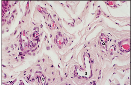
name of histologic slide:
Specie:
Stain Used:
Capillaries (sheep) in the connective tissue of the cervix (arrowed). Masson’s trichrome. ×125.
capillary
capillary is the smallest unit of the vascular system.
layers of capillary wall (two-layered)
a tunica intima of one or two squamous endothe lial cells resting on a basal lamina,
a fine tunica adventitia of collagen and elastic fibres.
pericytes:
undifferentiated cells believed capable of becoming fibroblasts or muscle cells
Venules
collect the blood from the capillaries.
Their lumina are wider than those of the arterioles.
The tunica intima (there is no tunica media and adventitia) in each venule consists of a continuous layer of endothelial cells and areolar connective tissue.
Pericytes are also present.
name of histologic slide:
Specie:
Parts
1.__ 2. __
Stain Used:
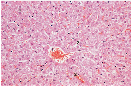
Liver (sheep). (1) Central vein. (2) Liver sinusoids. H & E. ×62.5.
name of histologic slide:
Specie:
Stain Used:
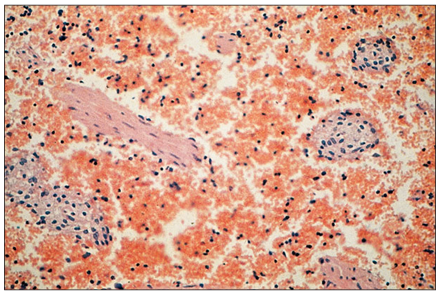
Spleen (horse). Sinusoids filled with erythrocytes. H & E. ×125.
name of histologic slide:
Specie:
Parts
1.__ 2. __
Stain Used:
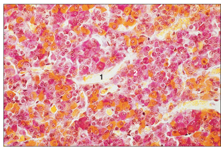
Pars distalis of the adenohypophysis (cat). (1) Sinusoids. (2) Cords of hypophyseal cells. Orange G. ×250.
Sinusoids
found in the liver, spleen, bone mar row and adenohypophysis.
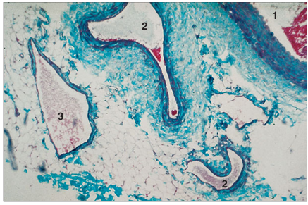
name of histologic slide:
Specie:
Stain Used:
Spermatic cord (horse). Venous plexus (arrowed). H& E. ×62.5.
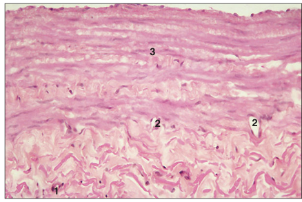
name of histologic slide:
Specie:
Parts
1.__ 2.__ 3.__
Stain Used:
Caudal vena cava (dog). (1) Tunica intima. (2) Tunica media. (3) Tunica adventitia with vasa vasorum (arrowed). Gomori’s trichrome.×125.
name of histologic slide:
Specie:
Parts
1.__ 2.__
Stain Used:
Caudal vena cava (horse). (1) Tunica intima. (2) Tunica media. Masson’s trichrome. ×125.
name of histologic slide:
Specie:
Parts
1.__ 2.__
Stain Used:
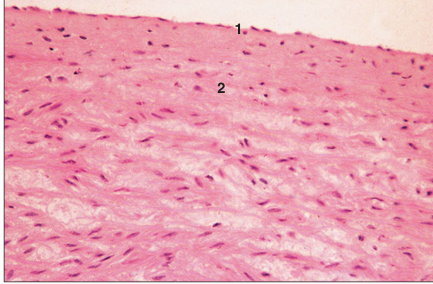
Cranial vena cava (horse). (1) Tunica intima. (2) Tunica media. H & E. ×125.
name of histologic slide:
Specie:
Parts
1.__ 2.__ 3.__ 4.__ 5.__
Stain Used:
Femoral vein with valves (cat). (1) Lumen. (2) Valve. (3) Tunica intima. (4) Tunica media. (5) Tunica adventitia. Masson’s trichrome. ×25
name of histologic slide:
Specie:
Parts
1.__ 2.__ 3.__
Stain Used:
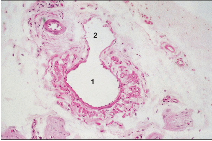
Valve in the brachial vein (cat). (1) Lumen filled with erythrocytes. (2) Valve. (3) Tunica intima. H & E. ×125.
Veins
lined by a continuous layer of endothelial cells and areolar connective tissue
name of histologic slide (clinical correlates):
Specie:
Parts
1.__ 2.__
Stain Used:
Arteriovenous anastomosis in loose connective tissue (cat). (1) Artery. (2) Vein. H & E. ×62.5.
Arteriovenous anastomoses
are special areas of the skin of the nose, lips and pads where the arteriole opens directly into a venule without going 6.19 through the capillary bed
This provides an alternative channel of blood supply and regulation of heat loss
name of histologic slide:
Specie:
Parts
1.__ 2.__ 3.__
Stain Used:
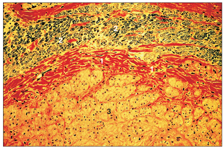
Cardiac muscle (ox). (1) Cardiac muscle. (2) Connective tissue of the fibrous skeleton. (3) Atrioventricular node. Masson’s trichrome. ×125.
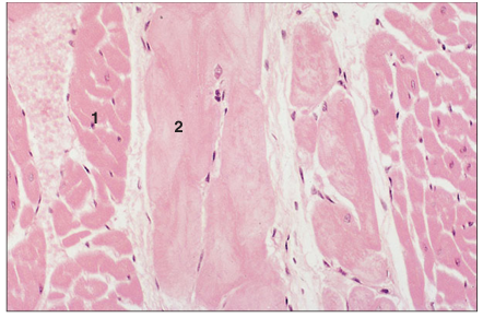
name of histologic slide:
Specie:
Parts
1.__ 2.__
Stain Used:
Cardiac muscle (horse). (1) Cardiac muscle fibres. (2) Conducting (Purkinje) fibres. H & E. ×125.
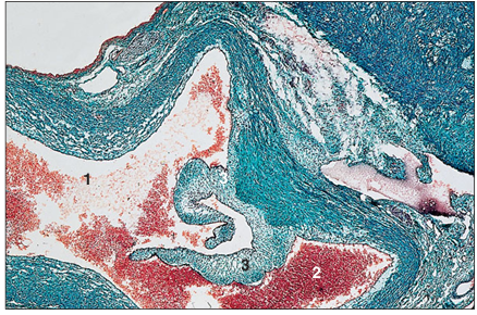
name of histologic slide:
Specie:
Parts
1.__ 2.__ 3.__
Stain Used:
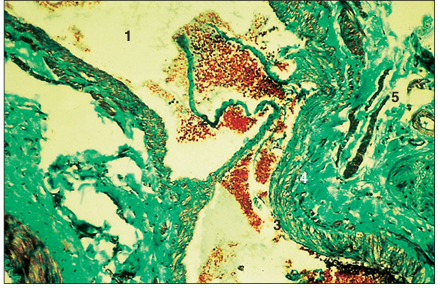
Heart (dog). (1) Lumen of the atrium. (2) Lumen of the pulmonary artery. (3) Valve cusps. Masson’s trichrome. ×62.5.
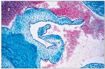
name of histologic slide :
Specie:
Parts
1.__ 2.__ 3.__
Stain Used:
Heart (dog). (1) Valve cusps. (2) Dense connective tissue part of the fibrous skeleton of the heart. Masson’s trichrome. ×62.5.