Duel-Energy X-ray Absorptiometry (DEXA) and Adaptix
1/26
There's no tags or description
Looks like no tags are added yet.
Name | Mastery | Learn | Test | Matching | Spaced | Call with Kai |
|---|
No study sessions yet.
27 Terms
What does DEXA stand for?
Duel-energy x-ray absorptiometry
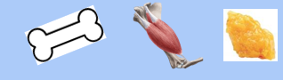
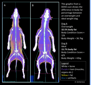
DEXA is a 2-dimentional technology which is used to measure ..
Bone mass
Lean mass (muscle)
Fatty mass (adipose tissue)
What is DEXA?
Originally developed for use in human patients
Precise method in determining body fat distribution
Not available at standard veterinary practices

How does it work?
Uses two low-dose energy x-rays
Although radiation exposure is minimal, some health and safety aspects still apply
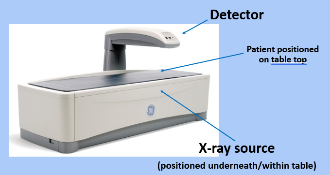
X-ray source positioned underneath the machine table supporting the patient and the director is encased in an arm above
DEXA uses two low-dose energy rays to distinguish the type and amount of tissue begin scanned. These two energies are used for ..
Separating mineral (bone) and soft-tissue components
One energy is absorbed mainly by soft tissue and the other by bone
There is a high energy photon and a low energy photon
Radiation exposure is much lower than standard radiography - depending on the area of the body being examined - at just 10% of normal everyday background radiation. Lead PPE (apron, thyroid guard) is not warranted, however ..
Dosimeters must be worn by the operator to monitor individual radiation exposure levels
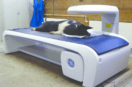
DEXA machine
Large, heavy machines and require a lot of floor space in practice
Expensive to purchase
Often why these machines aren’t used in general practice

During the scan
The arm presses over the body as a beam of x-rays are passed through the area being scanned
X-ray source and detector move together over the patient
X-ray photons of the 2 different energy levels are absorbed differently by bone, adipose and lean tissue
X-ray detector in arm measures the amount of x-rays passed through the body to produce a two-dimensional image and the scan results

The scan takes approximately ..
5-10 minutes to complete
The accuracy and precision of the scan has been determined with ..
Cadaver analysis of dogs and cats
DEXA - Patient preparation and support
Withhold food the evening before the scan - due to the administration of a sedative or GA
Sedation or GA is required to ensure the patient remains absolutely still during scan - movement blur may occur
Metal items must be removed - collars, harnesses, dog coats etc ..
Be await of metal implants as they are dense objects which will contribute towards density/mass - hip replacements, internal fixators etc ..
Check patient history → DEXA scan must not be performed within 10-14 days of a contrast study
DEXA relies on the assumption that lean mass is uniformly hydrate at 0.73ml water/g. This must be taken into consideration when performing the scan on certain patients, for example ..
Dehydration, renal disease, oncology patients etc ..
This is because the results may not be entirely accurate
Clients should be advised to remove the patients water in the morning of the scan rather than withholding water overnight
Patient positioning for DEXA
Dorsal, ventral and lateral positioning possible - as patient will be sedated or GA
Whichever position is used, sequential scans must be performed in the same position to prevent variable and to standardised practice
Scan limit borders marked on table top mattress - the patients must be positioned within the borders
Patient positioning depends on the area being scanned and the type of scanner. What else will influence the patient position?
The thickness of the tissue/area
How else may we use DEXA in veterinary patients?
Systemic disease - neoplasia cachexia
Orthopaedic disease
Meat/livestock industry
Pros of DEXA
Non-invasive and a quick procedure
Instant results
No primary complication
Precise method in determining body fat distribution
Can be used to monitor progress/deterioration
Cons of DEXA
Room/space required
Initial start up cost
Staff training required
Uses ionising radiation
Sedation/DA required
Unable to predict who will experience a fracture (but can be used to prevent and determine if treatment is required)
Same machine required for repeat scans due to variation in results (if using different machines)

Adaptix uses ..
Uses Digital Tomosynthesis to create a 3D x-ray
Originally designed for human breast mammography
What is Adaptic and how does it work?
X-ray head creates low-dose x-rays that perform a ‘sweeping’ motion over the patient to make x-ray slices then create a 3D image from the slices
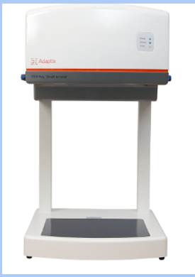
Is a mobile system and can be ‘table-top’ so easily stored and can be used outside of the veterinary practice
Can shown soft tissue and bone, like a 2D radiograph

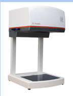
Adaptix machine
Patient to be positioned on the x-ray plate
Recumbency type isn’t a concern unless multiple x-rays are being taken, then the same recumbency must be used
X-ray head, internal beam moves - no outward movement
X-ray plate
During the Adaptix scan
The scan itself take about 30 seconds to complete
Changing the x-ray imaging slices from 2D to 3D is instantaneous within the computer programme
A 3D digital x-ray is produced that can be manipulated and ‘zoomed’ between the slices
Patient preparation for Adaptix
May need to be sedated/anaesthetised - very much depends on the image being taken
Otherwise, preparation is that of a normal x-ray
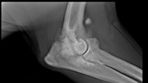
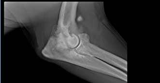
Clinical use of Adaptix - Orthopaedics
Excellent definition of bones and joints
Can be used in theatre during fracture repair
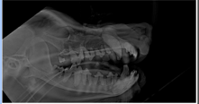
Clinical uses of Adaptix - Dentistry
Excellent definition of jaw and teeth
Patients can be put in 1 lateral recumbency and whole jaw x-rayed
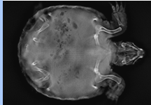
Clinical use of Adaptix - Exotics
Excellent definition of finer bones
Very good for small mammals and reptiles
Is easier to get ‘through’ Chelonia shell
Pros of Adaptix
Non-invasive and a quick procedure
Instant results
No primary complications
Mobile machine
Cons of Adaptix
Initial start up cost Staff training required
Uses ionising radiation
Sedation/GA sometimes required
is yet to be tested with contrast media
Not in many practices .. yet
Still patented so can only buy from one company