Chapter 3: The Cellular Level of Organization
Structure and Composition of the Cell Membrane
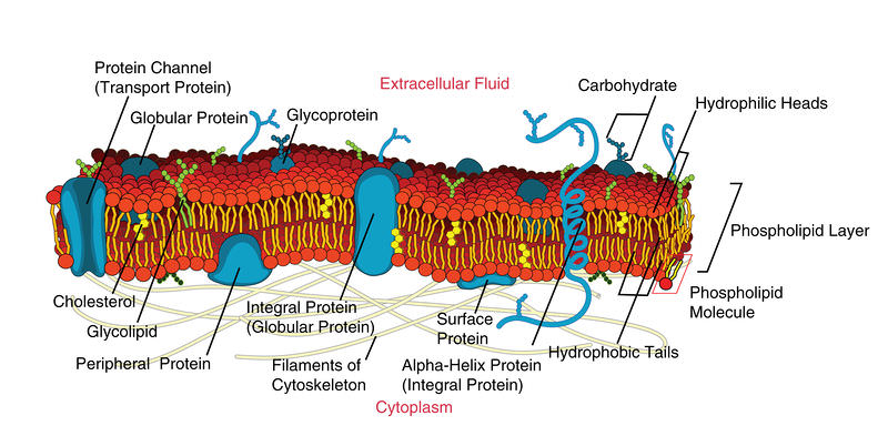
The cell membrane is an extremely pliable structure composed primarily of back-to-back phospholipids (a “bilayer”).
A single phospholipid molecule has a phosphate group on one end, called the “head,” and two side-by-side chains of fatty acids that make up the lipid tails.
- The phosphate group is negatively charged, making the head polar and hydrophilic—or “water loving.”
A hydrophilic molecule (or region of a molecule) is one that is attracted to water.
A hydrophobic molecule (or region of a molecule) repels and is repelled by water.
An amphipathic molecule is one that contains both a hydrophilic and a hydrophobic region.
Intracellular fluid (ICF) is the fluid interior of the cell.
Extracellular fluid (ECF) is the fluid environment outside the enclosure of the cell membrane.
Interstitial fluid (IF) is the term given to extracellular fluid not contained within blood vessels.
Membrane Proteins
- An integral protein is a protein that is embedded in the membrane.
- A channel protein is an example of an integral protein that selectively allows particular materials, such as certain ions, to pass into or out of the cell.
- A receptor is a type of recognition protein that can selectively bind a specific molecule outside the cell, and this binding induces a chemical reaction within the cell.
- A ligand is the specific molecule that binds to and activates a receptor.
- A glycoprotein is a protein that has carbohydrate molecules attached, which extend into the extracellular matrix.
- The glycocalyx is a fuzzy-appearing coating around the cell formed from glycoproteins and other carbohydrates attached to the cell membrane.
- Peripheral proteins are typically found on the inner or outer surface of the lipid bilayer but can also be attached to the internal or external surface of an integral protein.
Transport across the Cell Membrane
- A membrane that has selective permeability allows only substances meeting certain criteria to pass through it unaided.
- Passive transport is the movement of substances across the membrane without the expenditure of cellular energy.
- A concentration gradient is the difference in concentration of a substance across a space.
- Diffusion is the movement of particles from an area of higher concentration to an area of lower concentration.
- Facilitated diffusion is the diffusion process used for those substances that cannot cross the lipid bilayer due to their size and/or polarity
- Osmosis is the diffusion of water through a semipermeable membrane.
- A solution that has a higher concentration of solutes than another solution is said to be hypertonic, and water molecules tend to diffuse into a hypertonic solution.
- A solution that has a lower concentration of solutes than another solution is said to be hypotonic, and water molecules tend to diffuse out of a hypotonic solution.
- Active transport is the movement of substances across the membrane using energy from adenosine triphosphate (ATP).
- The sodium-potassium pump, which is also called Na+ /K+ ATPase, transports sodium out of a cell while moving potassium into the cell.
- An electrical gradient is a difference in electrical charge across a space.
- Endocytosis (bringing “into the cell”) is the process of a cell ingesting material by enveloping it in a portion of its cell membrane, and then pinching off that portion of membrane
- A vesicle is a membranous sac—a spherical and hollow organelle bounded by a lipid bilayer membrane.
- Phagocytosis (“cell eating”) is the endocytosis of large particles.
- Pinocytosis (“cell drinking”) brings fluid containing dissolved substances into a cell through membrane vesicles.
- Receptor-mediated endocytosis is endocytosis by a portion of the cell membrane that contains many receptors that are specific for a certain substance.
- Exocytosis (taking “out of the cell”) is the process of a cell exporting material using vesicular transport.
The Cytoplasm and Cellular Organelles
Cytosol, the jelly-like substance within the cell, provides the fluid medium necessary for biochemical reactions.
- Eukaryotic cells, including all animal cells, also contain various cellular organelles.
An organelle (“little organ”) is one of several different types of membrane-enclosed bodies in the cell, each performing a unique function.
The organelles and cytosol, taken together, compose the cell’s cytoplasm.
The nucleus is a cell’s central organelle, which contains the cell’s DNA.
Endoplasmic Reticulum
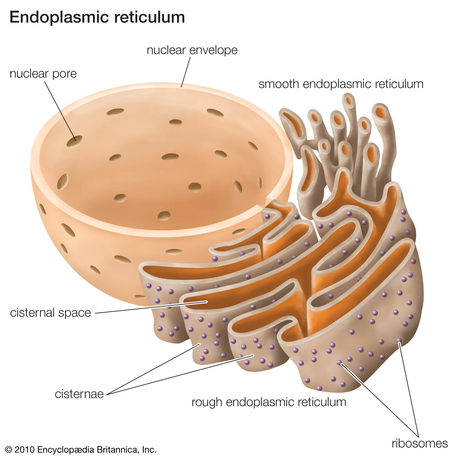
The endoplasmic reticulum (ER) is a system of channels that is continuous with the nuclear membrane (or “envelope”) covering the nucleus and composed of the same lipid bilayer material.
A ribosome is an organelle that serves as the site of protein synthesis.
The Golgi Apparatus: The Golgi apparatus is responsible for sorting, modifying, and shipping off the products that come from the rough ER, much like a post-office.
Lysosomes
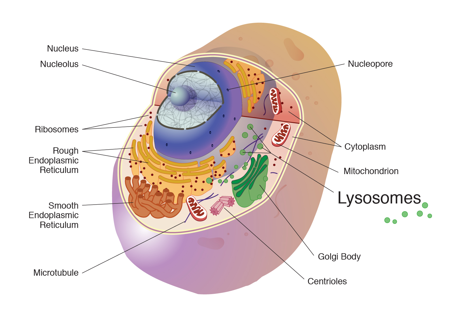
A lysosome is an organelle that contains enzymes that break down and digest unneeded cellular components, such as a damaged organelle.
Autophagy (“self-eating”) is the process of a cell digesting its own structures.
This “self-destruct” mechanism is called autolysis, and makes the process of cell death controlled (a mechanism called “apoptosis”).
Mitochondria
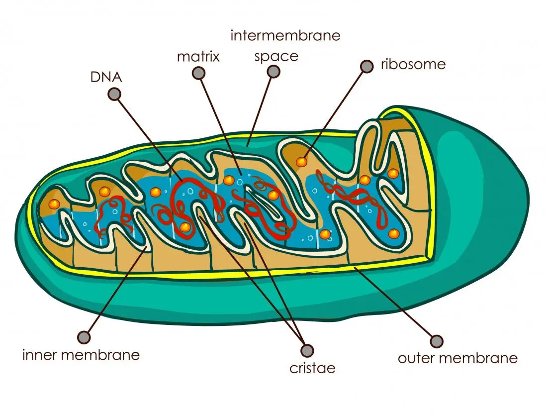
A mitochondrion (plural = mitochondria) is a membranous, bean-shaped organelle that is the “energy transformer” of the cell.
Mitochondria consist of an outer lipid bilayer membrane as well as an additional inner lipid bilayer membrane.
The inner membrane is highly folded into winding structures with a great deal of surface area, called cristae.
Peroxisomes
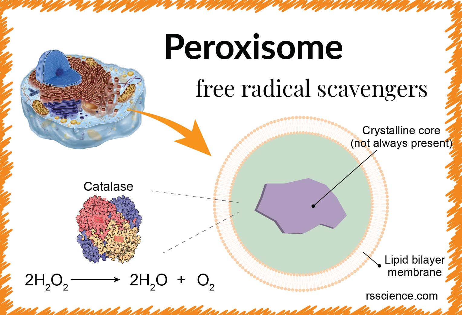
A peroxisome is a membrane-bound cellular organelle that contains mostly enzymes.
Peroxisomes perform a couple of different functions, including lipid metabolism and chemical detoxification.
Reactive oxygen species (ROS) such as peroxides and free radicals are the highly reactive products of many normal cellular processes, including the mitochondrial reactions that produce ATP and oxygen metabolism.
A mutation is a change in the nucleotide sequence in a gene within a cell’s DNA, potentially altering the protein coded by that gene.
The Cytoskeleton
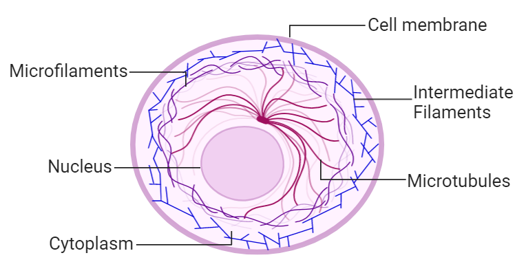
- The cytoskeleton is a group of fibrous proteins that provide structural support for cells, but this is only one of the functions of the cytoskeleton.
- The thickest of the three is the microtubule, a structural filament composed of subunits of a protein called tubulin.
- Cilia are found on many cells of the body, including the epithelial cells that line the airways of the respiratory system.
- A flagellum (plural = flagella) is an appendage larger than a cilium and specialized for cell locomotion.
- Centriole can serve as the cellular origin point for microtubules extending outward as cilia or flagella or can assist with the separation of DNA during cell division.
- The microfilament is a thinner type of cytoskeletal filament.
- An intermediate filament is a filament intermediate in thickness between the microtubules and microfilaments
Organization of the Nucleus and Its DNA
Like most other cellular organelles, the nucleus is surrounded by a membrane called the nuclear envelope.
There also can be a dark-staining mass often visible under a simple light microscope, called a nucleolus (plural = nucleoli).
Within the nucleus are threads of chromatin composed of DNA and associated proteins.
A nucleosome is a single, wrapped DNA-histone complex.
The chromosome is composed of DNA and proteins; it is the condensed form of chromatin. It is estimated that humans have almost 22,000 genes distributed on 46 chromosomes.
DNA Replication
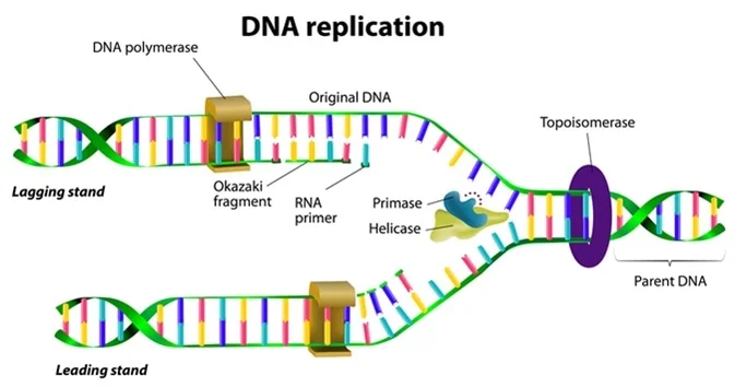
- DNA replication is the copying of DNA that occurs before cell division can take place.
- Stage 1: Initiation. The two complementary strands are separated, much like unzipping a zipper. Special enzymes, including helicase, untwist and separate the two strands of DNA.
- Stage 2: Elongation. Each strand becomes a template along which a new complementary strand is built. DNA polymerase brings in the correct bases to complement the template strand, synthesizing a new strand base by base. A DNA polymerase is an enzyme that adds free nucleotides to the end of a chain of DNA, making a new double strand. This growing strand continues to be built until it has fully complemented the template strand.
- Stage 3: Termination. Once the two original strands are bound to their own, finished, complementary strands, DNA replication is stopped and the two new identical DNA molecules are complete.
Protein Synthesis
- Just as the cell’s genome describes its full complement of DNA, a cell’s proteome is its full complement of proteins.
- A gene is a functional segment of DNA that provides the genetic information necessary to build a protein.
- Gene expression, which transforms the information coded in a gene to a final gene product, ultimately dictates the structure and function of a cell by determining which proteins are made.
- A triplet is a section of three DNA bases in a row that codes for a specific amino acid.
From DNA to RNA: Transcription
- The intermediate messenger is messenger RNA (mRNA), a single-stranded nucleic acid that carries a copy of the genetic code for a single gene out of the nucleus and into the cytoplasm where it is used to produce proteins.
- Gene expression begins with the process called transcription, which is the synthesis of a strand of mRNA that is complementary to the gene of interest.
- Stage 1: Initiation. A region at the beginning of the gene called a promoter—a particular sequence of nucleotides—triggers the start of transcription.
- Stage 2: Elongation. Transcription starts when RNA polymerase unwinds the DNA segment. One strand, referred to as the coding strand, becomes the template with the genes to be coded. The polymerase then aligns the correct nucleic acid (A,C, G, or U) with its complementary base on the coding strand of DNA. RNA polymerase is an enzyme that adds new nucleotides to a growing strand of RNA. This process builds a strand of mRNA.
- Stage 3: Termination. When the polymerase has reached the end of the gene, one of three specific triplets (UAA, UAG, or UGA) codes a “stop” signal, which triggers the enzymes to terminate transcription and release the mRNA transcript.
- Their function is still a mystery, but the process called splicing removes these non-coding regions from the pre-mRNA transcript.
- A spliceosome—a structure composed of various proteins and other molecules—attaches to the mRNA and “splices” or cuts out the non-coding regions.
- The removed segment of the transcript is called an intron.
- An exon is a segment of RNA that remains after splicing.
From RNA to Protein: Translation
- Translation is the process of synthesizing a chain of amino acids called a polypeptide.
- Ribosomal RNA (rRNA) is a type of RNA that, together with proteins, composes the structure of the ribosome.
- Transfer RNA (tRNA) is a type of RNA that ferries the appropriate corresponding amino acids to the ribosome, and attaches each new amino acid to the last, building the polypeptide chain one-by-one.
- This sequence of three bases on the tRNA molecule is called an anticodon.
Cell Growth and Division
- A somatic cell is a general term for a body cell, and all human cells, except for the cells that produce eggs and sperm (which are referred to as germ cells), are somatic cells.
- A homologous pair of chromosomes is the two copies of a single chromosome found in each somatic cell.
- The human is a diploid organism, having 23 homologous pairs of chromosomes in each of the somatic cells. The condition of having pairs of chromosomes is known as diploidy.
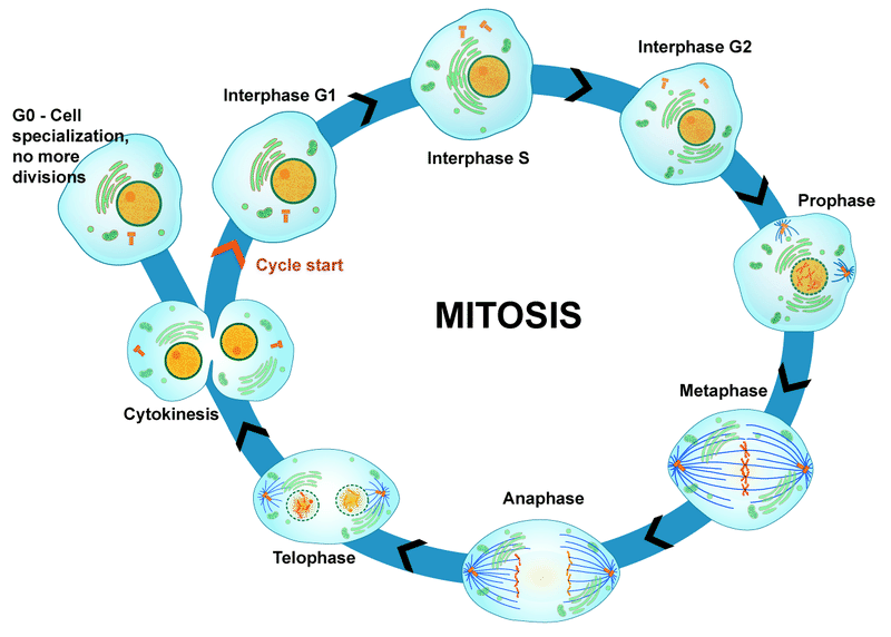
- The cell cycle is the sequence of events in the life of the cell from the moment it is created at the end of a previous cycle of cell division until it then divides itself, generating two new cells.
- Interphase is the period of the cell cycle during which the cell is not dividing. The majority of cells are in interphase most of the time.
- Mitosis is the division of genetic material, during which the cell nucleus breaks down and two new, fully functional, nuclei are formed.
- Cytokinesis divides the cytoplasm into two distinctive cells.
Interphase
- G1 phase (gap 1 phase) is the first gap, or growth phase in the cell cycle.
- The S phase (synthesis phase) is the period during which a cell replicates its DNA.
- The G2 phase is a second gap phase, during which the cell continues to grow and makes the necessary preparations for mitosis.
- The G0 phase is a resting phase of the cell cycle.
The Structure of Chromosomes
Each copy of the chromosome is referred to as a sister chromatid and is physically bound to the other copy.
The centromere is the structure that attaches one sister chromatid to another.
Mitosis and Cytokinesis
- The mitotic phase of the cell typically takes between 1 and 2 hours. During this phase, a cell undergoes two major processes.
- Prophase is the first phase of mitosis, during which the loosely packed chromatin coils and condenses into visible chromosomes.
- A centrosome is a pair of centrioles together.
- The mitotic spindle is the structure composed of the centrosomes and their emerging microtubules.
- The kinetochore is a protein structure on the centromere that is the point of attachment between the mitotic spindle and the sister chromatids.
- Metaphase is the second stage of mitosis.
- The metaphase plate is the name for the plane through the center of the spindle on which the sister chromatids are positioned.
- Anaphase is the third stage of mitosis.
- Anaphase takes place over a few minutes, when the pairs of sister chromatids are separated from one another, forming individual chromosomes once again.
- Telophase is the final stage of mitosis.
- Telophase is characterized by the formation of two new daughter nuclei at either end of the dividing cell.
- The cleavage furrow is a contractile band made up of microfilaments that forms around the midline of the cell during cytokinesis.
Mechanisms of Cell Cycle Control
A checkpoint is a point in the cell cycle at which the cycle can be signaled to move forward or stopped.
A cyclin is one of the primary classes of cell cycle control molecules.
A cyclindependent kinase (CDK) is one of a group of molecules that work together with cyclins to determine progression past cell checkpoints.
Stem Cells
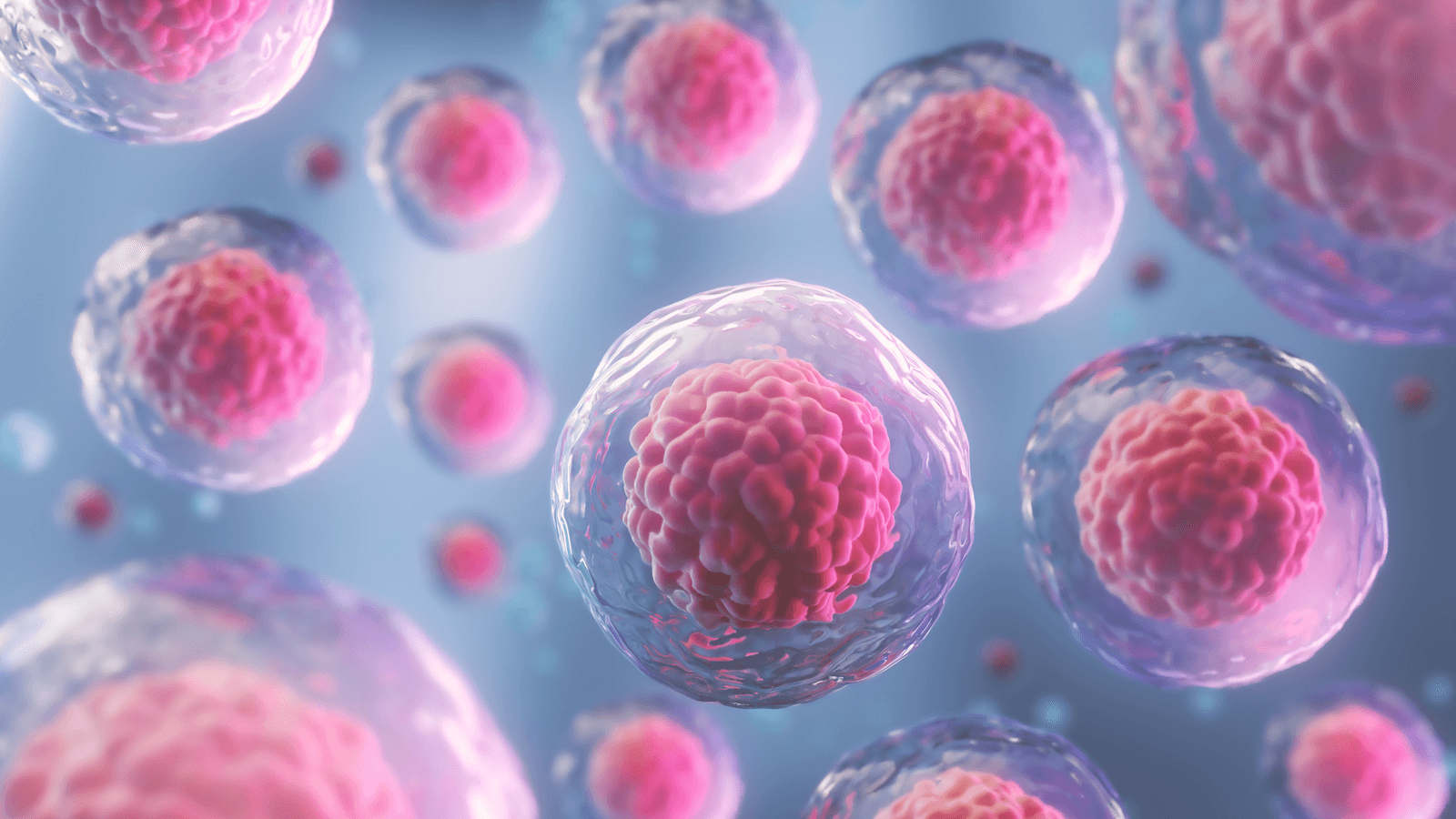
A stem cell is an unspecialized cell that can divide without limit as needed and can, under specific conditions, differentiate into specialized cells.
The first embryonic cells that arise from the division of the zygote are the ultimate stem cells; these stem cells are described as totipotent because they have the potential to differentiate into any of the cells needed to enable an organism to grow and develop.
A pluripotent stem cell is one that has the potential to differentiate into any type of human tissue but cannot support the full development of an organism.
A multipotent stem cell has the potential to differentiate into different types of cells within a given cell lineage or small number of lineages, such as a red blood cell or white blood cell.
An oligopotent stem cell is limited to becoming one of a few different cell types.
A unipotent cell is fully specialized and can only reproduce to generate more of its own specific cell type.
Differentiation: A transcription factor is one of a class of proteins that bind to specific genes on the DNA molecule and either promote or inhibit their transcription.