Nervous System
4.5(2)
Studied by 31 peopleCard Sorting
1/58
Earn XP
Description and Tags
Last updated 4:20 PM on 12/6/22
Name | Mastery | Learn | Test | Matching | Spaced | Call with Kai |
|---|
No analytics yet
Send a link to your students to track their progress
59 Terms
1
New cards
general functions of a neuron
1) respond to chemical and physical stimuli
2) conduct electrochemical impulses
3) release chemical regulators
4) enable perception of sensory stimuli, learning, memory, and control of muscles and glands
2) conduct electrochemical impulses
3) release chemical regulators
4) enable perception of sensory stimuli, learning, memory, and control of muscles and glands
2
New cards
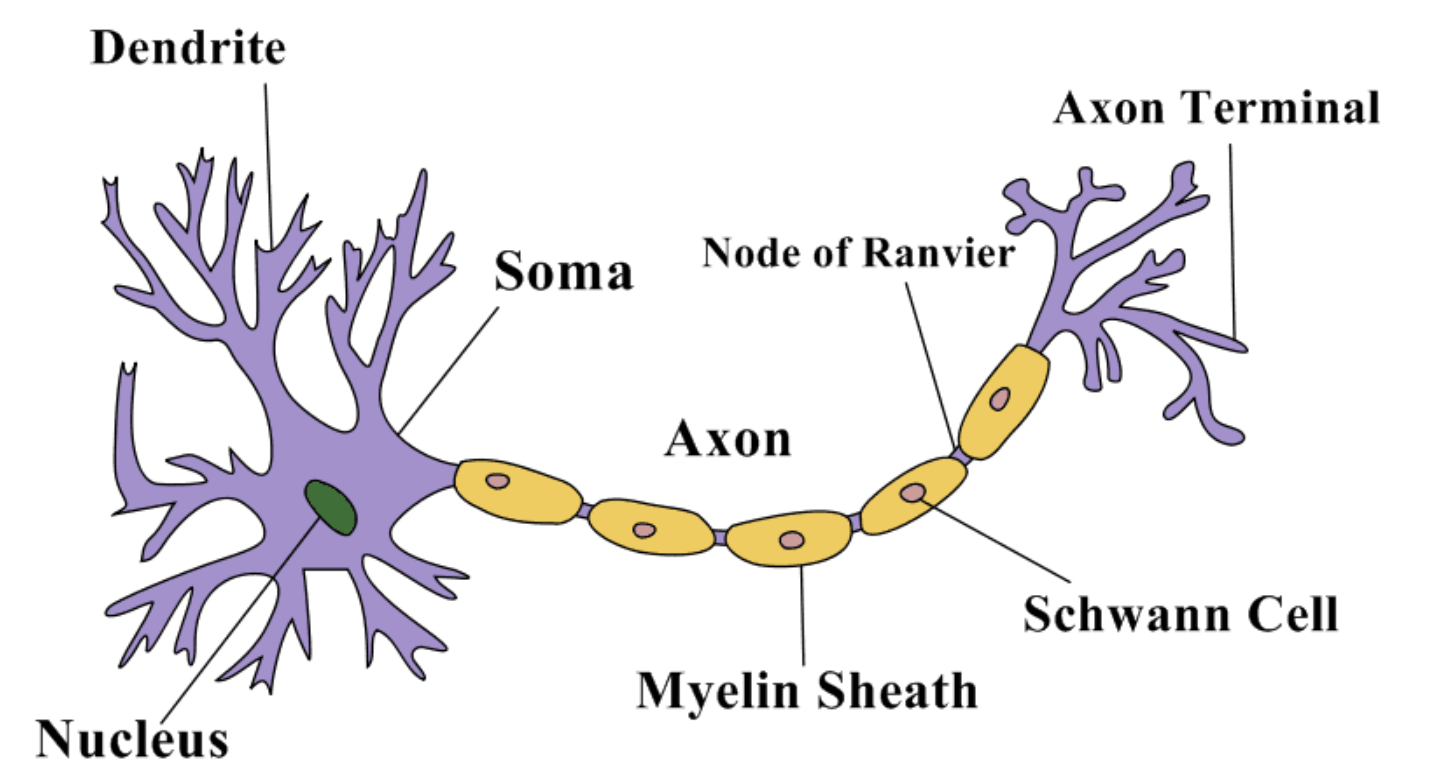
basic structure of a neuron
1) soma
2) dendrites
3) axon hillock
4) axon
5) axon terminals
2) dendrites
3) axon hillock
4) axon
5) axon terminals
3
New cards
axon
connected to cell body/soma by the axon hillock- where action potentials are generated at the initial segment of the axon
covered in myelin with empty spots called "nodes of ranvier"; insulate signal and allow for quicker propagation of signal
can form many branches called "axon collaterals"
vary in length from a few millimeters to a meter
covered in myelin with empty spots called "nodes of ranvier"; insulate signal and allow for quicker propagation of signal
can form many branches called "axon collaterals"
vary in length from a few millimeters to a meter
4
New cards
nerve
neurons+glia cells
5
New cards
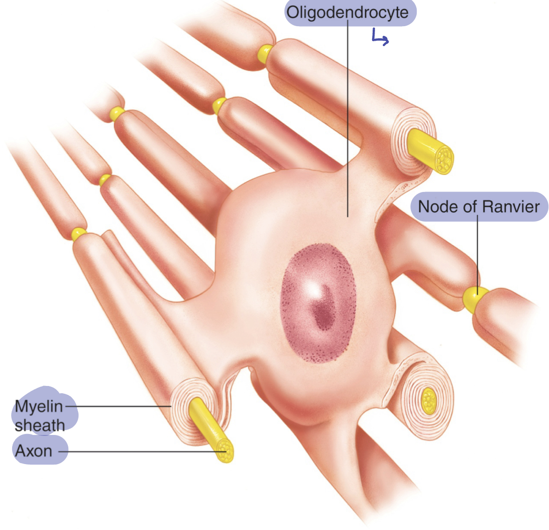
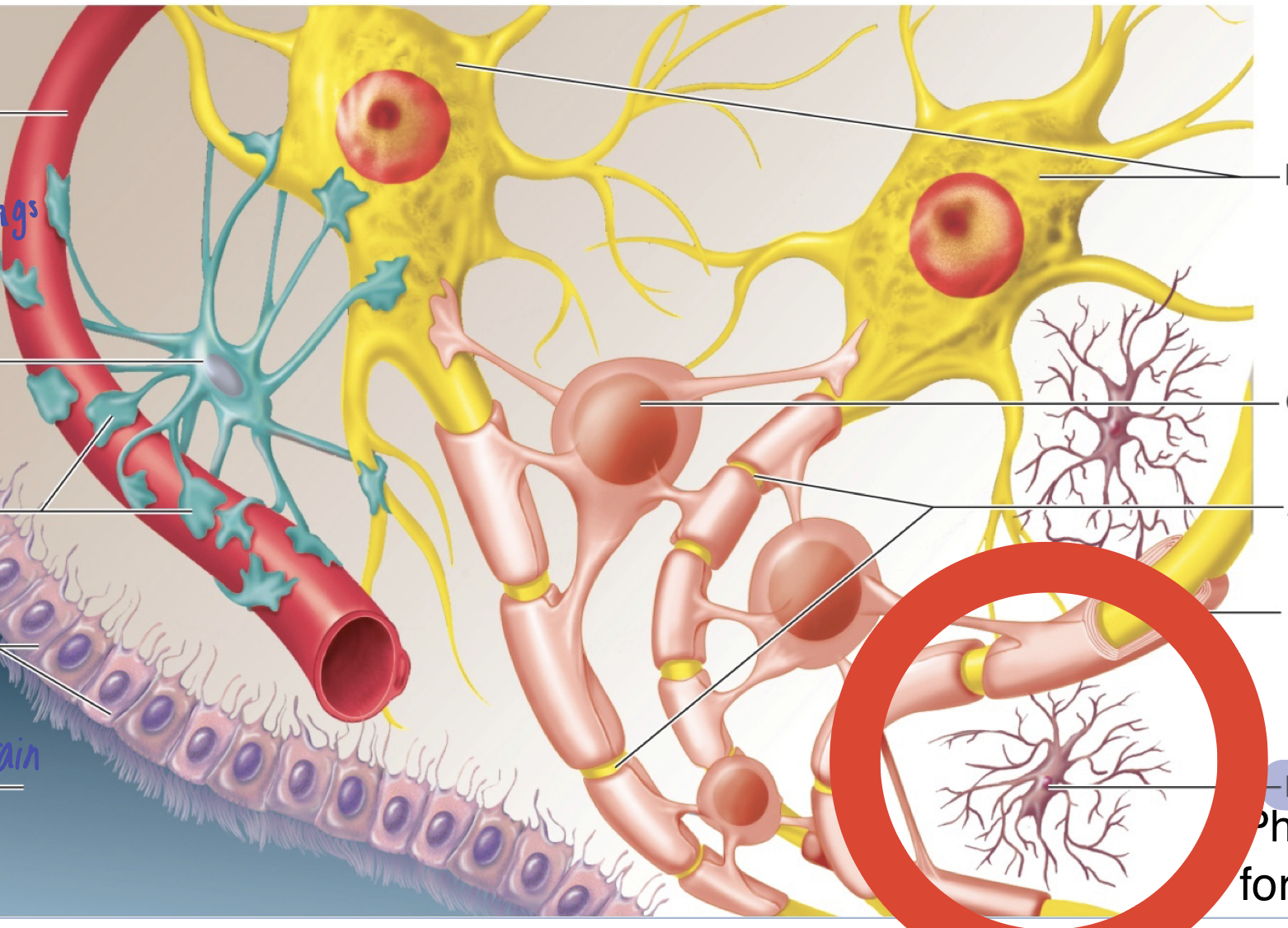
glia cells
supportive cells
oligodendrocytes and schwann cells
oligodendrocytes and schwann cells
6
New cards
oligodendrocyte
supply myelin in CNS; insulation
mostly membrane and proteins (and some cytoplasm) cover the axon
one wraps around many axons (1: many)
mostly membrane and proteins (and some cytoplasm) cover the axon
one wraps around many axons (1: many)
7
New cards
schwann cells
supply myelin in PNS
one wraps around axon (1:1)
one wraps around axon (1:1)
8
New cards
microglia
supportive cell
phagocytosis of foreign object/debris (similar to immune cell)
phagocytosis of foreign object/debris (similar to immune cell)
9
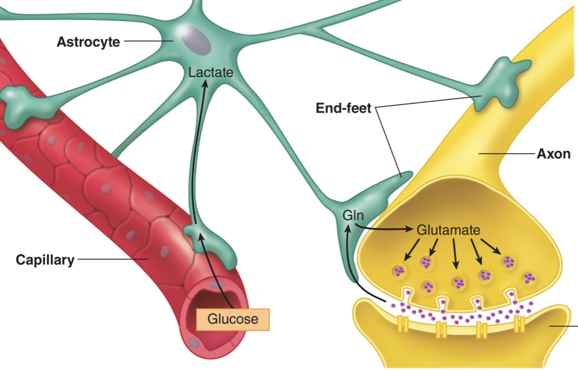
New cards
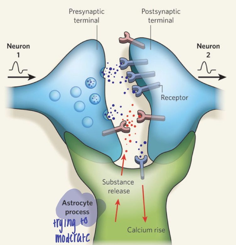
astrocyte
helps regulate external environment of neurons; regulate blood- brain barrier; keep certain thing out of the brain
end-feet cover capillary surfaces or are adjacent to synapses between two neurons
guide neuron growth, control chemical environment around neurons, and absorbs extra neurotransmitters from synapse (moderates interaction and monitor communication of neurons)
capillary cells in the brain are joined by tight junctions
10
New cards
information flow of a neuron
reception: dendrites
integration/summation: axon hillock
conduction: axon
transmission: axon terminals
integration/summation: axon hillock
conduction: axon
transmission: axon terminals
11
New cards
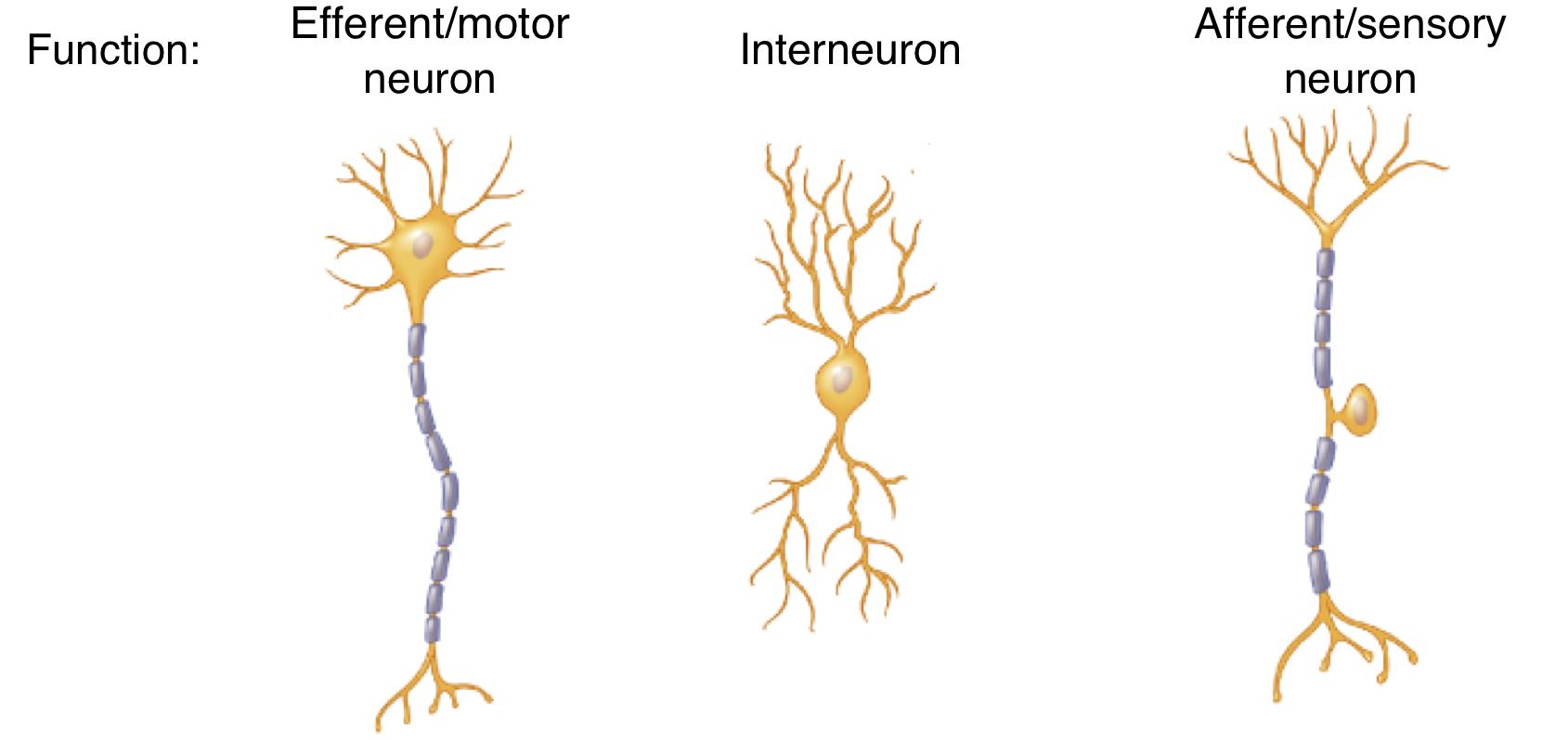
functional classification of neuron
1) efferent/motor neuron (signal "exits")
2) interneuron
3) afferent/sensory neuron (send signal inward)
2) interneuron
3) afferent/sensory neuron (send signal inward)
12
New cards
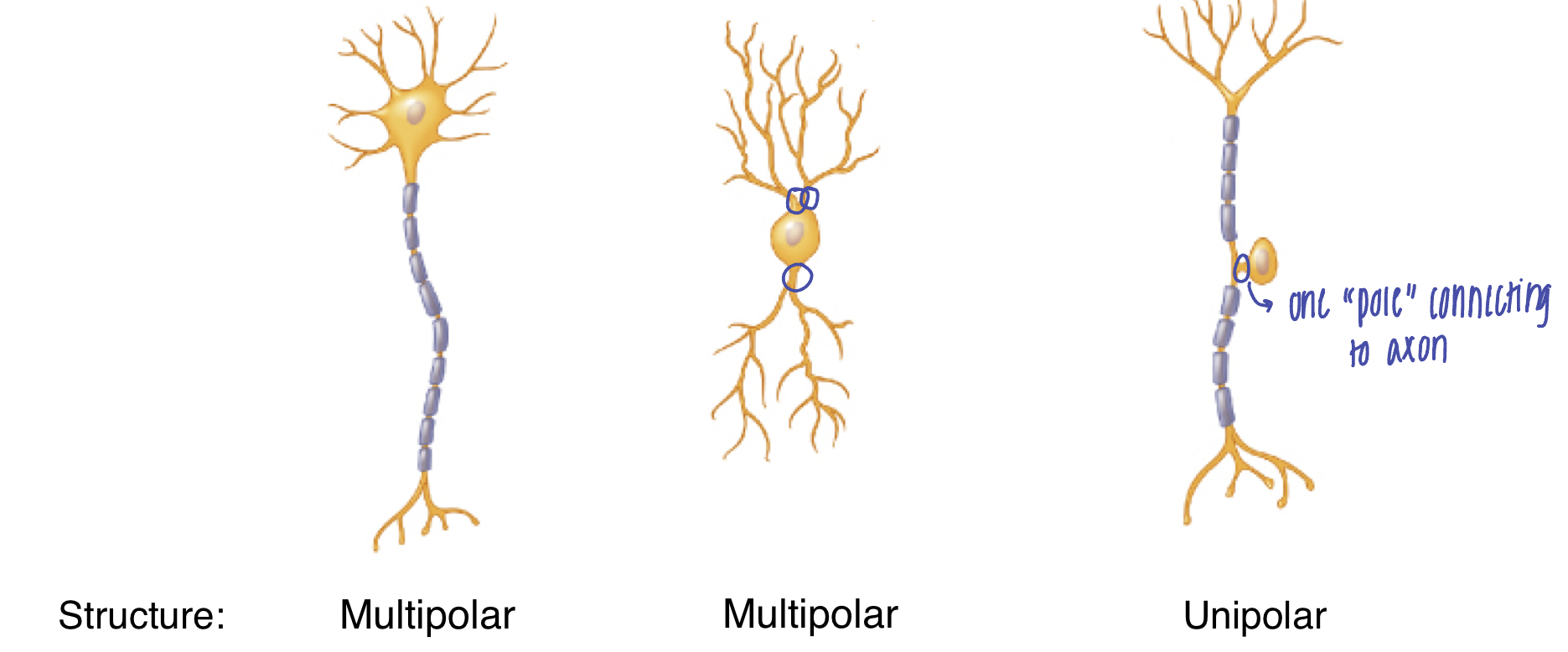
structural classification of neuron
unipolar or multipolar
based on number of "poles" connecting to the soma
based on number of "poles" connecting to the soma
13
New cards
membrane/resting potential
the difference in electrical potential (change in charge) between the interior and exterior of the cell
requires an electrochemical gradient
mostly negative inside the cell- greater amount of K+ inside the cell; greater amount of Na+ outside the cell
cell has a negative membrane potential (polarized): -70mV
requires an electrochemical gradient
mostly negative inside the cell- greater amount of K+ inside the cell; greater amount of Na+ outside the cell
cell has a negative membrane potential (polarized): -70mV
14
New cards
electrochemical gradient
chemical and electrical gradients
chemical gradient: difference in solute concentration across a membrane
electrical gradient: difference in charge across a membrane
chemical gradient: difference in solute concentration across a membrane
electrical gradient: difference in charge across a membrane
15
New cards
why is membrane potential of neurons important?
potential energy, when activated, can be used to send signals; ions that shift, stimulate
16
New cards
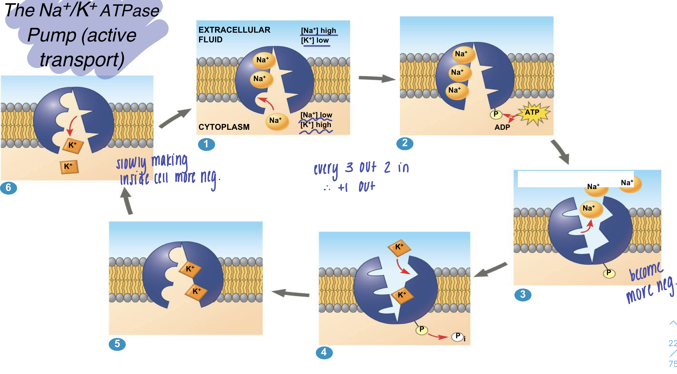
the Na+/K+/ATP pump (active transport)
1) Na+ binds to the sodium potassium pump. the affinity for Na+ is high when the protein has this shape
2) Na+ binding stimulates phosphorylation by ATP
3) phosphorylation leads to a change in protein shape, reducing its affinity for Na+, which is released outside the cell; becomes more negative inside the cell
4) the new shape has a high affinity fro K+, which binds on the extracellular side and triggers release of the phosphate group
5) loss of the phosphate group restores the protein's shape, which has a lower affinity for K+
6) K+ is released; affinity fro Na+ is high again, and the cycle repeats
17
New cards
leak channel proteins
allow ions to "leak" back in to keep charge balance
slower rate than being pumped in/out by sodium-potassium pump
slower rate than being pumped in/out by sodium-potassium pump
18
New cards
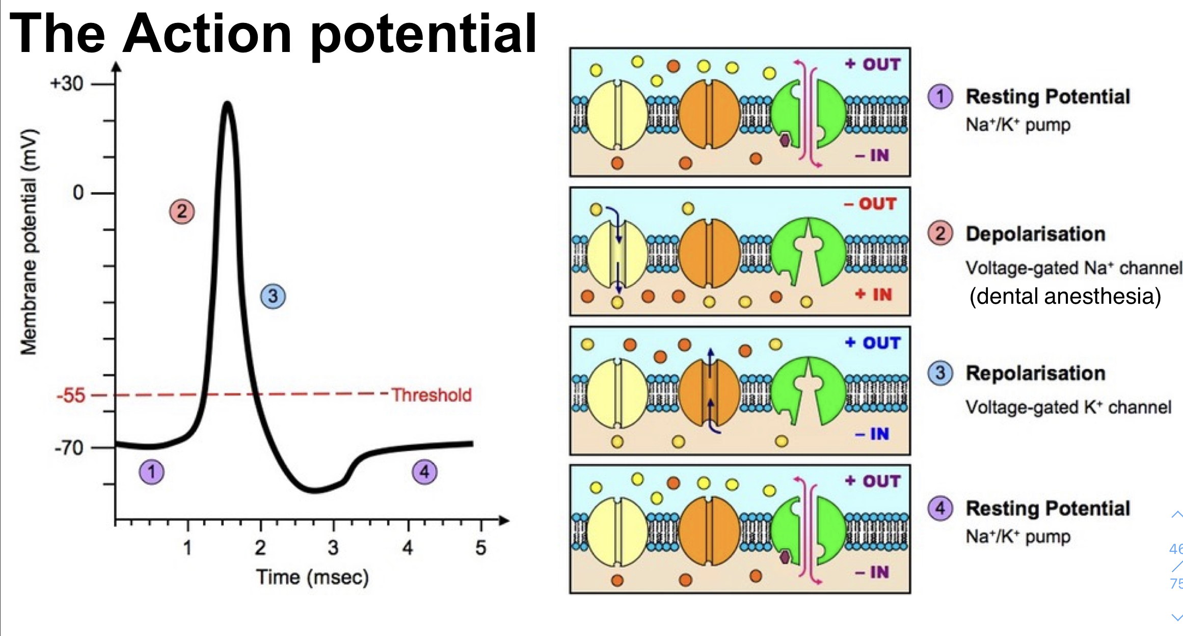
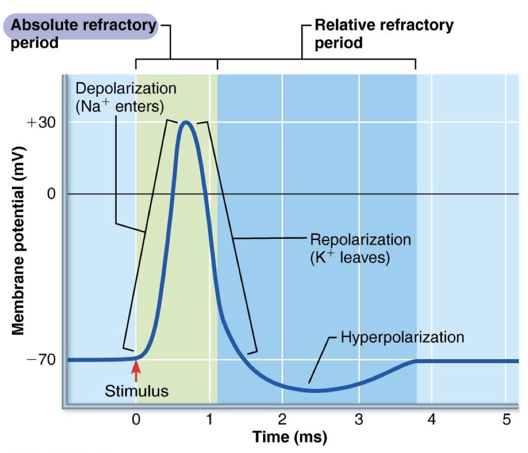
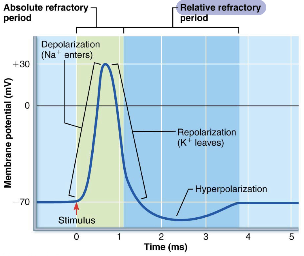
action potential
large and rapid changes in the membrane potential made possible by voltage gated Na+ and K+ channels
allows for rapid conduction down the axon
allows for rapid conduction down the axon
19
New cards
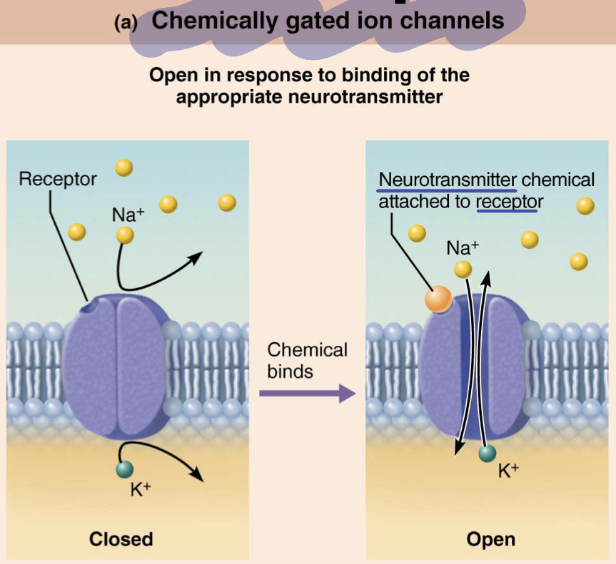
graded potentials- initiation
activation of post-synaptic receptors allow for + or - charges to flow into the cell which changes the membrane potential
20
New cards
chemically gated ion channels
open in response to binding of appropriate neurotransmitter
21
New cards
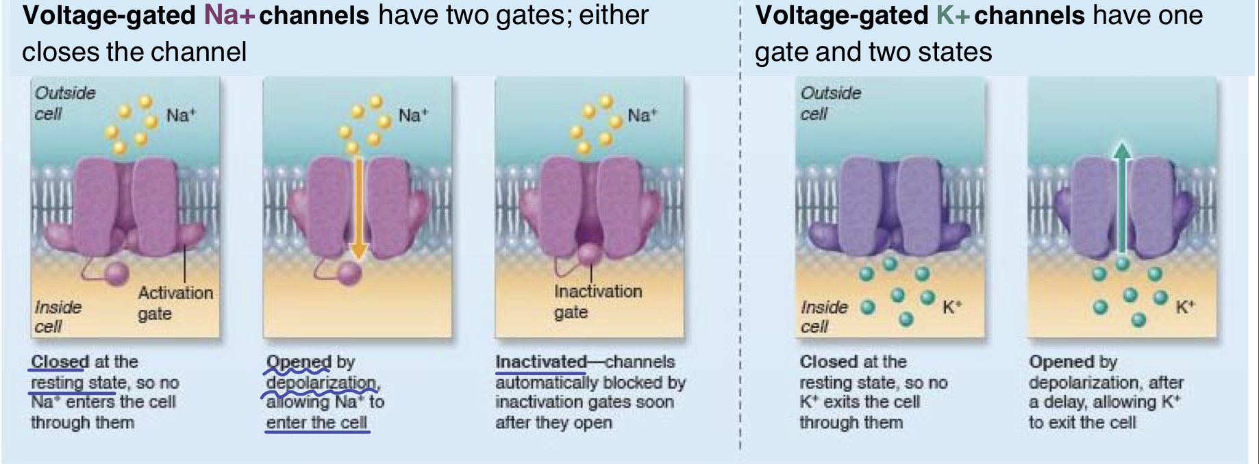
voltage gated channels
change in voltage allows channel to open, allowing for ions to rush through, down the concentration gradient
22
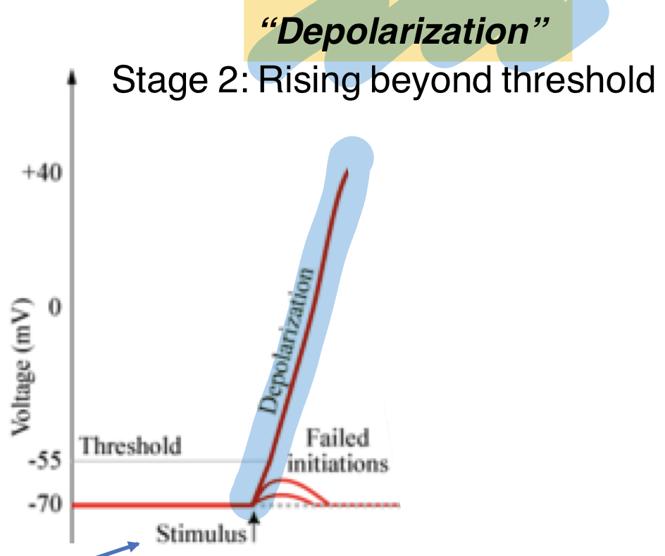
New cards
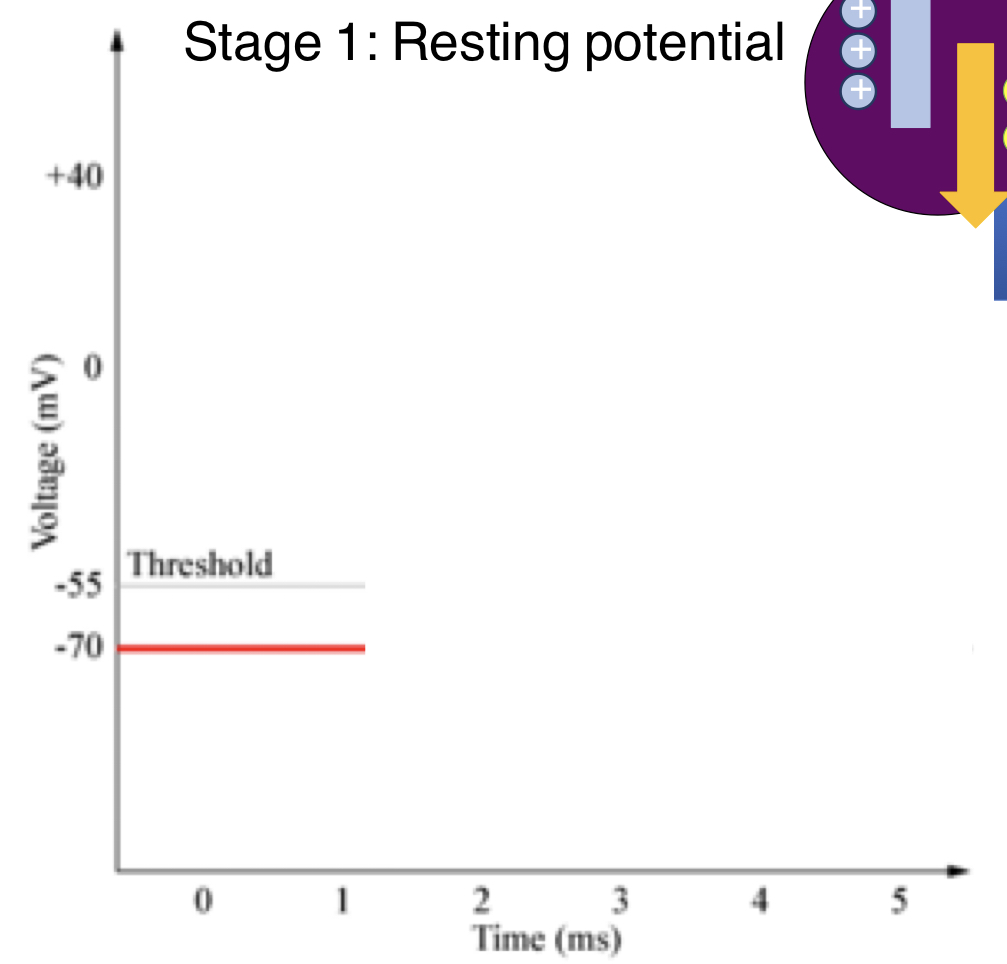
stage 1 of action potential
resting/membrane potential
-70mV
channels involved:
leak channels for K+ and Na+
Na+/K+/ATPase
-70mV
channels involved:
leak channels for K+ and Na+
Na+/K+/ATPase
23
New cards
stage 2 of action potential
depolarization; rising beyond threshold
channels involved:
1) leak channels K+ and Na+
2) Na+/K+/ATPase
3) voltage gated Na channel (VGNaC): open immediately and close quickly; Na+ rush in quickly, making the inside of the cell more positive
channels involved:
1) leak channels K+ and Na+
2) Na+/K+/ATPase
3) voltage gated Na channel (VGNaC): open immediately and close quickly; Na+ rush in quickly, making the inside of the cell more positive
24
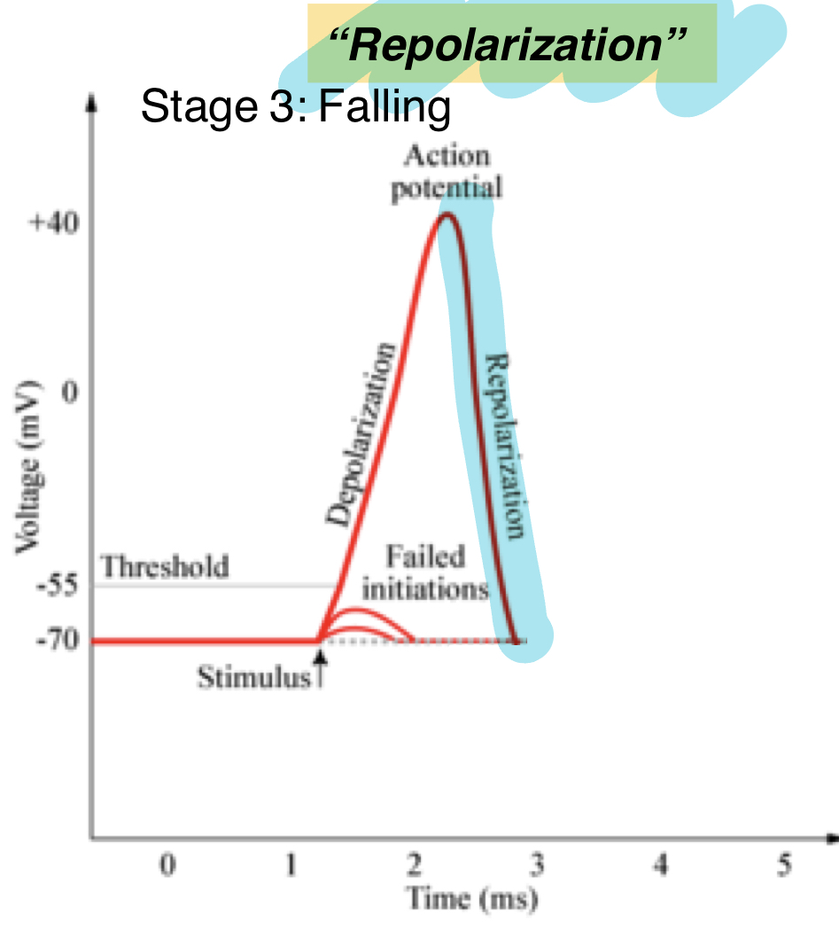
New cards
stage 3 of action potential
repolarization; falling
channels involved:
1) leak channels K+ and Na+
2) Na+/K+/ATPase
3) VGKC: K+ rush out of cell to bring cell back to more negative; open after delay and takes more time to close
channels involved:
1) leak channels K+ and Na+
2) Na+/K+/ATPase
3) VGKC: K+ rush out of cell to bring cell back to more negative; open after delay and takes more time to close
25
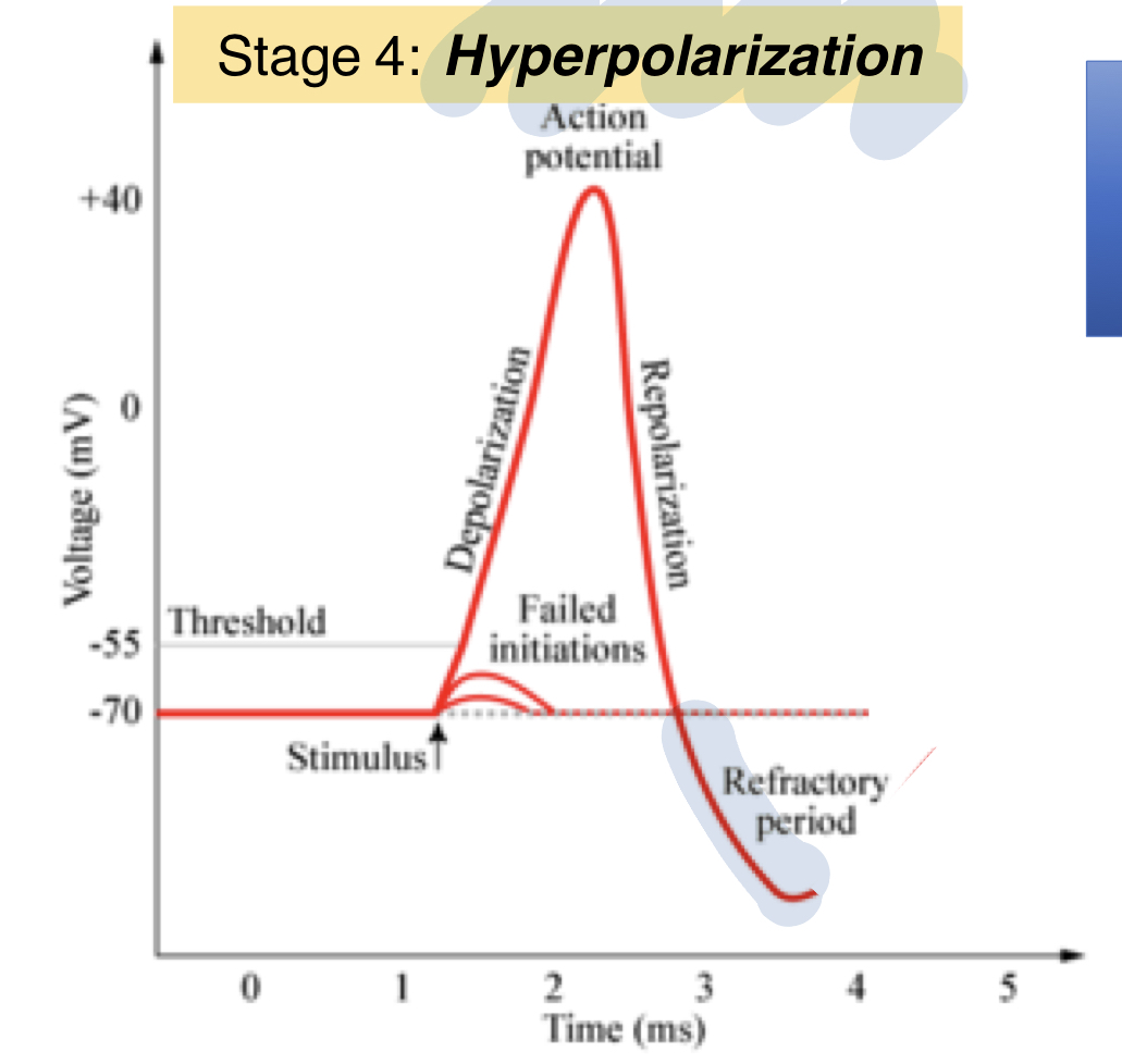
New cards
stage 4 of action potential
hyperpolarization
part of refractory period
cannot fire at this point
channels involved:
1) leak channels K+ and Na+
2) Na+/K+/ATPase
3) VGKC: remain open, close slowly
part of refractory period
cannot fire at this point
channels involved:
1) leak channels K+ and Na+
2) Na+/K+/ATPase
3) VGKC: remain open, close slowly
26
New cards
stage 5 of action potential
recovery; goes back up to resting state/membrane potential
channels involved:
1) leak channels K+ and Na+
2) Na+/K+/ATPase
channels involved:
1) leak channels K+ and Na+
2) Na+/K+/ATPase
27
New cards
as the inside of the cell becomes more positive, is this depolarization or hyperpolarization?
depolarization
28
New cards
the more _____ the inside of the cell, the stronger the electrical attraction for Na+ ions to enter the cell
negative
29
New cards
Na+ and K+ are present on both sides of the cell. which of these factor into the chemical driving force fro Na+ to enter the cell?
Na+; bc the chemical concentration gradient
30
New cards
why does it take a stronger stimulus to re-fire the neuron during the relative refractory period?
the inside of the cell is much more negative/farther from the threshold
K+ is flooding out (losing positives) of the cell quickly, therefore making the inside of the cell more negative quickly; need to let more positives into the cell, but would also be trying to get Na+ ions to go against its concentration gradient
K+ is flooding out (losing positives) of the cell quickly, therefore making the inside of the cell more negative quickly; need to let more positives into the cell, but would also be trying to get Na+ ions to go against its concentration gradient
31
New cards
absolute refractory
a depolarized membrane can't depolarize again once it's just fired; can't fire again
occurs during depolarization and most of repolarization
occurs during depolarization and most of repolarization
32
New cards
relative refractory period
once the VGNaC reset, you could depolarize again IF the signal is strong enough to reopen the Na+ channels and overcome the K+ mvmt
occurs during some of repolarization, hyperpolarization, and refractory
occurs during some of repolarization, hyperpolarization, and refractory
33
New cards
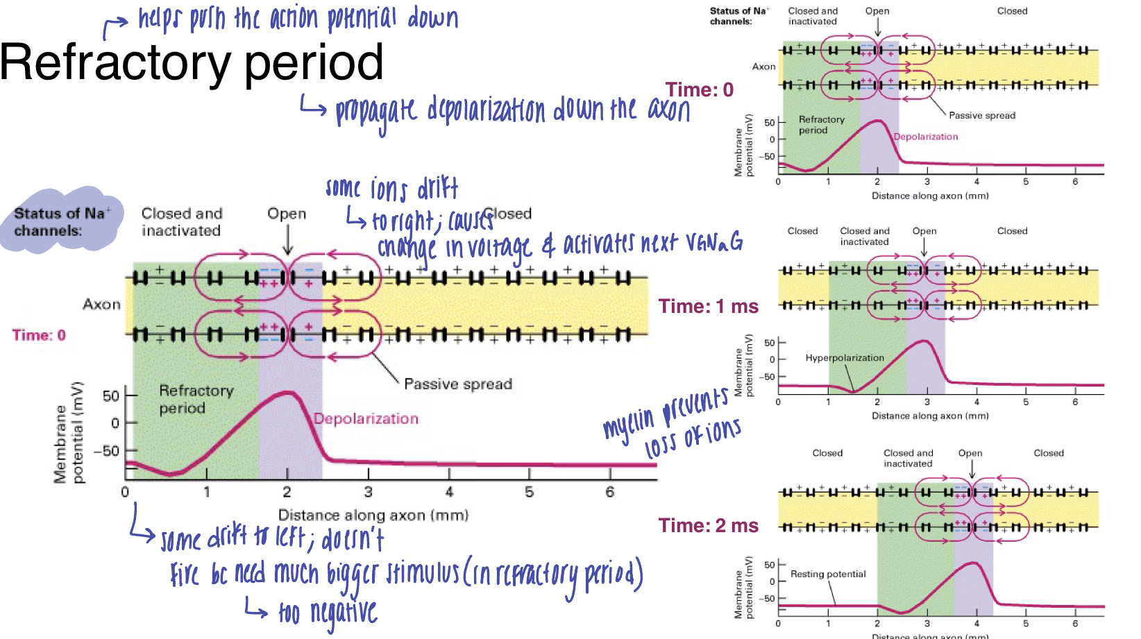
function of the refractory period
propagate depolarization down the axon; help push the action potential down
when Na floods into the axon, some ions drift to the right (toward more negative), which causes a change in voltage and activates the next VGNaC
some ions drift to the left, but doesnt fire bc need a much bigger stimulus (too negative in the refractory period)
when Na floods into the axon, some ions drift to the right (toward more negative), which causes a change in voltage and activates the next VGNaC
some ions drift to the left, but doesnt fire bc need a much bigger stimulus (too negative in the refractory period)
34
New cards
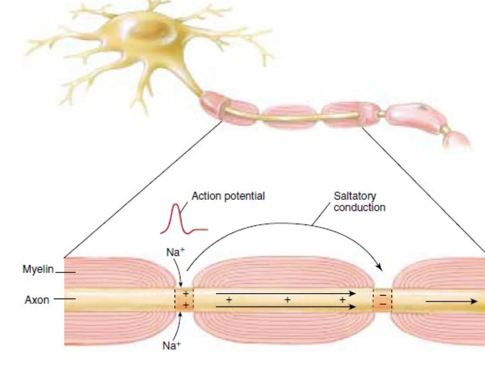
optimize speed of action potential conduction
low resistance (larger axon diameter): with a wider diameter, there is less resistance and pressure, therefore allowing quick flow
insulation: myelin sheath insulates axon
insulation: myelin sheath insulates axon
35
New cards
saltatory conduction
signal "jumps" down the axon
36
New cards
nodes of ranvier
gaps in myelin on the axon; allow signal to "jump"
37
New cards
mechanism of nodes of ranvier
1) when an action potential comes down the axon and reaches a node/gap, it causes an influx of Na at the node
2) Na rushes into the axon at the node, creating an electrical force that pushes on the ions already inside the axon
3) the signal reaches the next node and creates another action potential, therefore refreshing the signal
2) Na rushes into the axon at the node, creating an electrical force that pushes on the ions already inside the axon
3) the signal reaches the next node and creates another action potential, therefore refreshing the signal
38
New cards
synapse
1) axon terminus (presynaptic cell)
2) synaptic cleft
3) post synaptic cell
2) synaptic cleft
3) post synaptic cell
39
New cards
neurotransmitter release (transmission)
1) action potential (change in voltage) arrives at the axon terminal
2) Voltage-gated calcium channels open; calcium enters the axon terminal
3) calcium entry causes synaptic vesicles to release neurotransmitter by exocytosis
4) neurotransmitters diffuse across the synaptic cleft and bind to specific receptors on the postsynaptic membrane
5) binding of neurotransmitters opens ion channels in postsynaptic cell
40
New cards
neurotransmitter elimination
1) diffusion; some get absorbed by astrocytes and other diffuse out in to the body
2) enzyme degradation
3) re-uptake: repackage left over NT
ex. SSRI= re-uptake inhibitor ( results in more NT spending more time in synaptic cleft; increases time and ability to bind to receptors)
2) enzyme degradation
3) re-uptake: repackage left over NT
ex. SSRI= re-uptake inhibitor ( results in more NT spending more time in synaptic cleft; increases time and ability to bind to receptors)
41
New cards
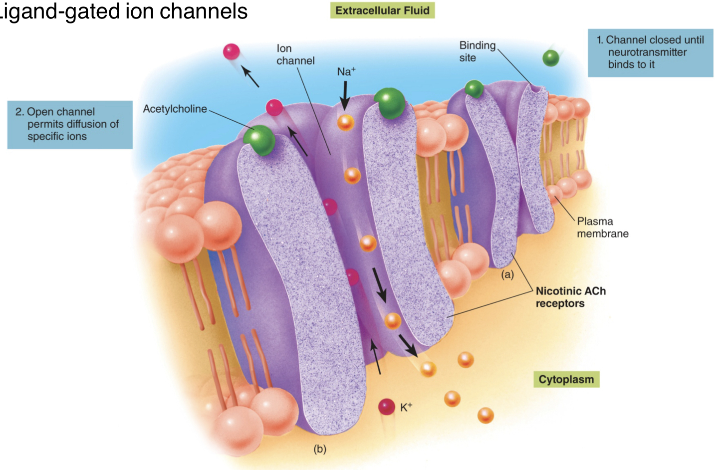
ionotropic receptors (ligand-gated ion channels)
direct-acting NT (similar to primary messenger)
1) channel closed until NT binds to it
2) open channel allows diffusion of specific ions
1) channel closed until NT binds to it
2) open channel allows diffusion of specific ions
42
New cards
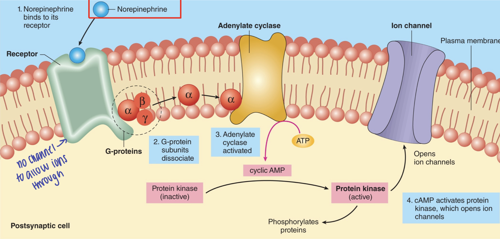
G- protein coupled receptors
indirect-acting NT (similar to second messenger)
1) NT binds to its receptor
2) G-protein subunits dissociate
3) adenylate cyclase activated
4) cAMP activates protein kinase, which opens ion channels
1) NT binds to its receptor
2) G-protein subunits dissociate
3) adenylate cyclase activated
4) cAMP activates protein kinase, which opens ion channels
43
New cards
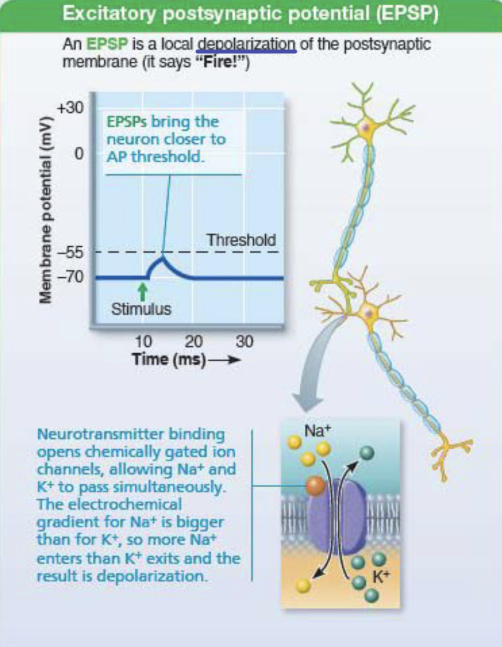
excitatory postsynaptic potential (EPSP)
local depolarization of the postsynaptic membrane; brings the neuron closer to action potential threshold
one won't be enough to fire
one won't be enough to fire
44
New cards
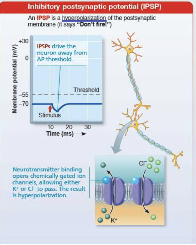
inhibitory postsynaptic potential (IPSP)
hyperpolarization of the postsynaptic membrane
drive neuron away from action potential threshold
drive neuron away from action potential threshold
45
New cards
what must occur for the postsynaptic neuron to reach the threshold to fire?
EPSPs must outweigh IPSPs
46
New cards
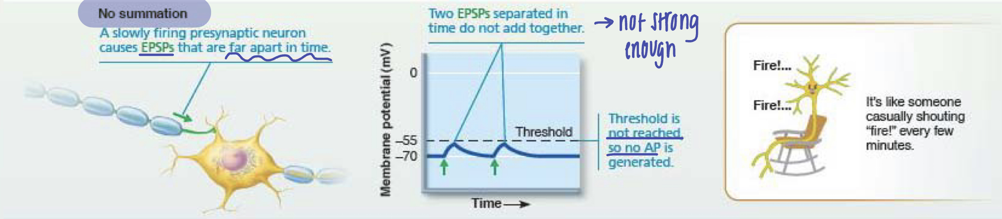
no summation
presynaptic neuron causes EPSPs that are too far apart or too weak --> they do not add together and is not strong enough --> threshold is not reached, so no action potential is generated
signals summated --> too weak/far --> no fire
signals summated --> too weak/far --> no fire
47
New cards
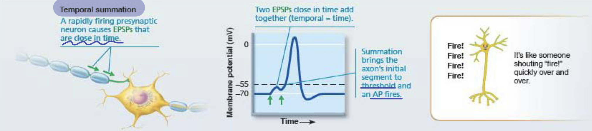
temporal summation
a rapidly firing presynaptic neuron causes EPSPs that are close in time --> summation brings axon to threshold --> action potential fires
multiple rapid signals--> summation --> reaches threshold --> neuron fires
multiple rapid signals--> summation --> reaches threshold --> neuron fires
48
New cards
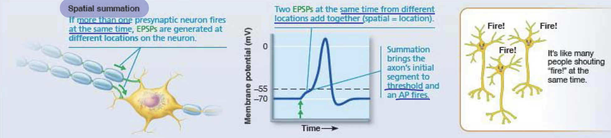
spatial summation
more than one presynaptic neuron fires at the same time --> EPSPs are generated at different locations of the neuron --> add together/summation brings axon to threshold --> neuron fires
49
New cards
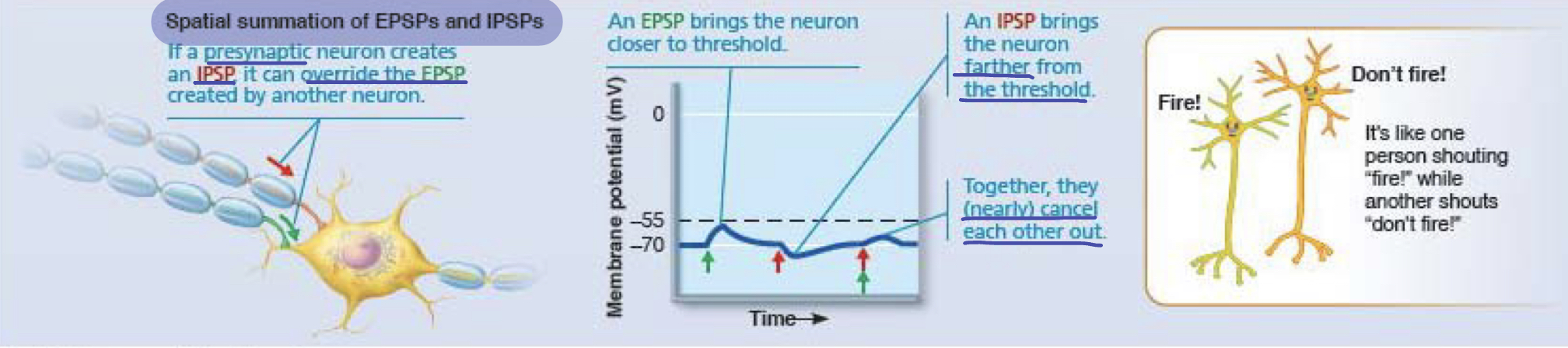
spatial summation of EPSPs and IPSPs
presynaptic neuron creates an IPSP it can override the EPSP created by another neuron --> EPSP brings neuron closer to threshold, IPSP brings the neuron farther from threshold --> together, (nearly) cancel each other out
EPSPs and IPSPs compete; if EPSPs win, neuron fires
EPSPs and IPSPs compete; if EPSPs win, neuron fires
50
New cards
serial neural processing
whole system works in a predictable, all or nothing manner
reflexes: stimulus always gives the same response (ex. touch something hot --> hand pulls back)
reflexes: stimulus always gives the same response (ex. touch something hot --> hand pulls back)
51
New cards
parallel neural processing
input is segregated into many pathways; different parts of the nervous system can respond differently to same info
helps brain put parts together to understand the whole
ex. smelling a pickle may invoke a memory, remind you of a dislike, remind you to buy some, all at once
helps brain put parts together to understand the whole
ex. smelling a pickle may invoke a memory, remind you of a dislike, remind you to buy some, all at once
52
New cards
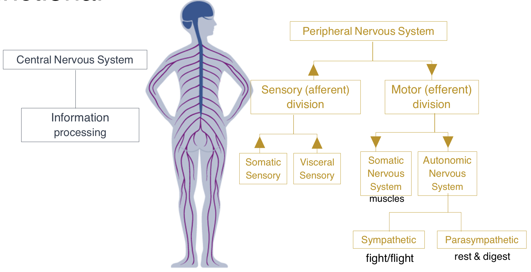
general organization: structural
CNS: brain and spinal cord (higher functions)
PNS: spinal nerves and cranial nerves (basic functions)
PNS: spinal nerves and cranial nerves (basic functions)
53
New cards
general organization: functional
CNS: information processing
PNS: sensory (afferent) division and motor (efferent) division
sensory: somatic sensory (sense outside the body; touch, temp, sharp) and visceral sensory (sense inside the body; organs; urge to pee, eat, etc)
motor: somatic nervous system (muscles; info going out)
autonomic nervous system --> sympathetic (fight or flight) and parasympathetic (rest and digest)
PNS: sensory (afferent) division and motor (efferent) division
sensory: somatic sensory (sense outside the body; touch, temp, sharp) and visceral sensory (sense inside the body; organs; urge to pee, eat, etc)
motor: somatic nervous system (muscles; info going out)
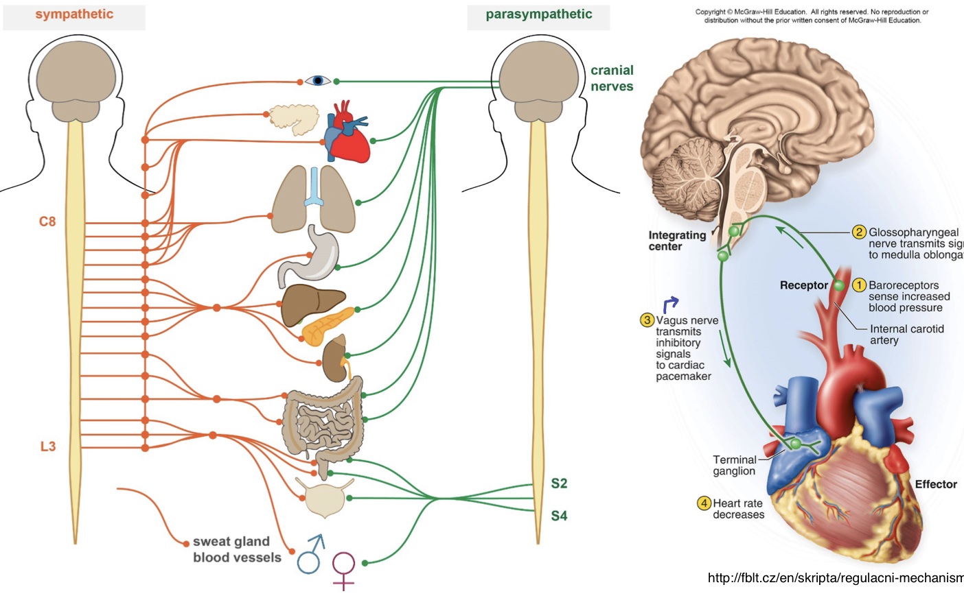
autonomic nervous system --> sympathetic (fight or flight) and parasympathetic (rest and digest)
54
New cards
what do the sympathetic and parasympathetic systems control?
glands, cardiac muscle, and smooth muscle
55
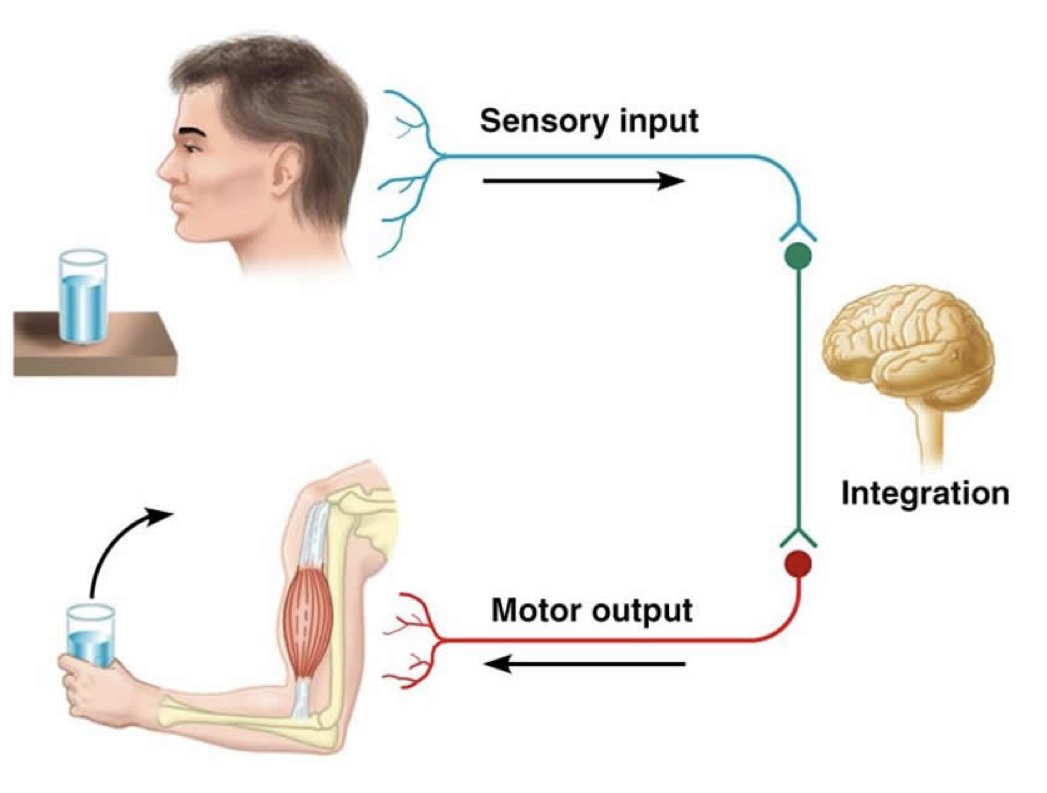
New cards
reflex arc
the neural path of a reflex
the sensory pathway: somatosensations --> CNS
the motor pathway: CNS --> neuromuscular junction
the sensory pathway: somatosensations --> CNS
the motor pathway: CNS --> neuromuscular junction
56
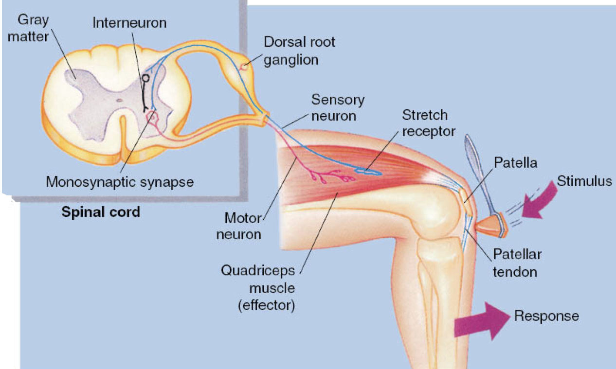
New cards
patellar reflexe
1) physically stretching the patellar tendon stretches the quads; activates the sensory neuron
2) nerve impulse goes to motor neuron to quad, telling it to contract, causing the knee to jerk
keeps us upright as knees buckle
2) nerve impulse goes to motor neuron to quad, telling it to contract, causing the knee to jerk
keeps us upright as knees buckle
57
New cards
somatosensations
REWATCH LECTURE
58
New cards
upper motor neurons
efferent pathways primarily relaying info from the cerebrum to the brainstem or spinal cord
synapse with interneurons
if damaged, partial recovery possible
synapse with interneurons
if damaged, partial recovery possible
59
New cards
lower motor neurons
neurons having direct influence on muscles
cell bodies originate in gray matter of spinal cord, but their axons extend into the PNS
if destroyed, permanent paralysis
cell bodies originate in gray matter of spinal cord, but their axons extend into the PNS
if destroyed, permanent paralysis