ISD4 Pulpotomy Demonstration
Pulpotomy
Removal of carious infected/affected coronal pulp
Maintenance of healthy radicular pulp
Can be difficult to differentiate and selectively remove diseased tissue
Aim: to maintain pulp vitality (until exfoliation)
Medication is applied to the exposed radicular pulp stumps to enhance healing and recovery of the remaining tissue (form dentine bridge)
Success rate 70-90% with MTA or Biodentine
Contraindications
Resorption
Furaction perforation
Insufficient root structure
Unrestorable tooth
Periradicular pathology
Immunocompromised child
The child unable to accept LA and a rubber dam
Equipment:
High-speed diamond bur to access the occlusal caries
Anaesthetise patient with LA
A rubber dam would be placed
In order to access the full roof of the pulp chamber, it is likely that the occlusal access will be fairly extensive
Do not undermine the cusps where it isn’t necessary
→ revise pulp morphology
High-speed diamond round bur to access deeper caries and the roof and the full width of the pulp chamber
In a pulpotomy, we are expecting that the coronal pulp may be infected or affected by a carious bacterial process.
We are expecting the radicular pulp to be healthy.
Now use an endo Z bur
You will expect the orifices to each root canal to appear where morphology indicates they are.
At this stage, you can access the pulp with an excavator.
A sterile cotton wool pellet soaked in saline can be inserted into the cavity prepared.
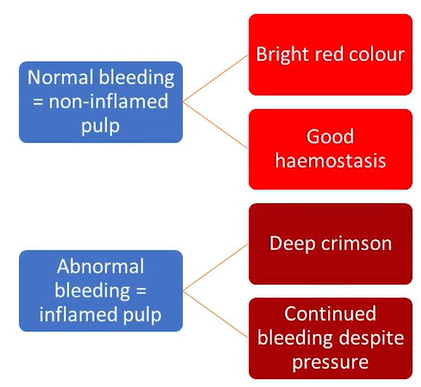
The aim is to achieve haemostasis, with gentle pressure, over 3-5mins with this cotton wool in situ.
If the pulp is still haemorrhagic bright crimson in colour then it is likely it is still infected
Remove more pulp
Or decide that a pulpotomy procedure is no longer indicated
Direct Evaluation of Pulp Stumps
Insert a dry cotton wool pellet to ensure the cavity itself has moisture control
Pulpotomy: Medication Choices
MTA: creates a bridge of dentine, biocompatible, excellent sealing, high pH, bone morphogenic proteins and growth factors act through their osteogenic potential in pulp repair. Outperforms previous material choices.
Biodentine: tricalcium silicate, biocompatible and bioactive (induces hard tissue generations, dentine bridge formation), excellent sealing and handling properties, high pH (antimicrobial). May be more readily available in primary care than MTA.
Pulpotomy: MTA vs Biodentine
Both have abilities to induce reparative dentine through the modulation of transforming growth factor-b1 secretion by pulp cells
Both have similar pulp-dentine complex responses to vital pulp therapy
Similar clinical and radiographic success rates for pulpotomy
Biodentine has reduced setting time and porosity, enhanced compressive and push-out bond strength, and increased microhardness
Therefore - similar but Biodentine has better handling properties
Put Biodentine in an amalgam packer and place it onto the pulp stumps.
Gently pack into pump stumps to ensure an optimal biocompatible seal.
Following pulpotomy, an optimal coronal seal can be achieved with a PMC
Pulpotomy Follow Up
Radicular pulp should remain asymptomatic without adverse clinical signs or symptoms
There should be no postop radiographic evidence of external root resorption
Internal root resorption may be self-limiting and stable (monitor)
If resorption causes perforation/loss of supportive bone and/or clinical signs of infection and inflammation → extract.