12. Generating T cells

0.0(0)
studied byStudied by 0 people
0.0(0)
full-widthCall Kai
learnLearn
examPractice Test
spaced repetitionSpaced Repetition
heart puzzleMatch
flashcardsFlashcards
GameKnowt Play
Card Sorting

1/45

encourage image

There's no tags or description

Looks like no tags are added yet.

Study Analytics
Name
Mastery
Learn
Test
Matching
Spaced

No study sessions yet.

46 Terms

1
New cards

How are unactivated (“naïve”) B and T cells generated against any possible antigen?

Through VDJ recombination, which creates immense diversity of BCRs and TCRs, allowing recognition of virtually any antigen before activation.

<p>Through <strong>VDJ recombination</strong>, which creates immense <strong>diversity of BCRs and TCRs</strong>, allowing recognition of virtually any antigen before activation.</p>
2
New cards

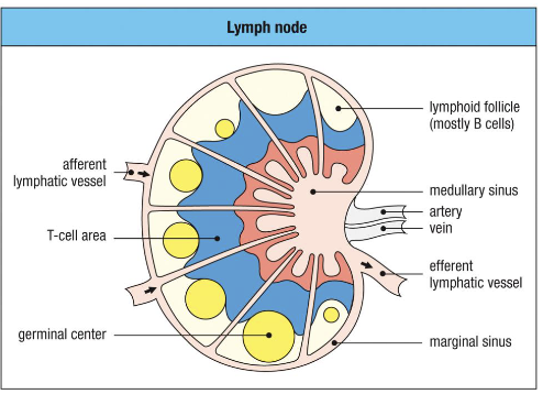
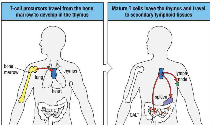
Where do naïve T cells come from before they’re activated in lymph nodes?

  • Origin: Precursors from bone marrow → develop in thymus.

  • Before activation: Enter lymph nodes and wait for dendritic cell presentation.

  • After activation: Mature → move to peripheral tissues to perform effector functions.

<ul><li><p><strong>Origin</strong>: Precursors from bone marrow → develop in thymus.</p></li><li><p><strong>Before activation:</strong> Enter lymph nodes and wait for dendritic cell presentation.</p></li><li><p><strong>After activation:</strong> Mature → move to peripheral tissues to perform effector functions.</p></li></ul><p></p>
3
New cards

Outline the developmental pathway of T cells.

  1. Hematopoietic stem cells → T cell precursors (bone marrow)

  2. Precursors travel to the thymus (near the heart)

  3. Develop into naïve T cells in the thymus

  4. Naïve T cells exit → secondary lymphoid tissues
    “T” stands for “thymus.”

<ol><li><p>Hematopoietic stem cells → T cell precursors (bone marrow)</p></li><li><p>Precursors travel to the <strong>thymus </strong>(near the heart)</p></li><li><p>Develop into <strong>naïve T cells</strong> in the thymus</p></li><li><p>Naïve T cells exit → <strong>secondary lymphoid tissues</strong><br><em>“T” stands for “thymus.”</em></p></li></ol><p></p>
4
New cards

What key processes occur in the thymus during T cell development?

  1. VDJ recombination forms a functional TCR

  2. Cells differentiate into αβ or γδ T cells

  3. Cells further specialize into CD4+ helper or CD8+ cytotoxic T cells

5
New cards

What risk comes with VDJ recombination?

It can generate TCRs or BCRs that recognize self-antigens, causing autoimmune diseases (e.g., type 1 diabetes, lupus).

6
New cards

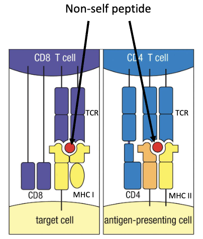
What four traits make a functional, non-self-reactive T cell?

  1. Functional TCR to detect and respond to antigens

  2. Can bind MHC molecules

  3. Does not bind self-proteins presented on MHC

  4. Is either CD4+ or CD8+

<ol><li><p>Functional <strong>TCR </strong>to detect and respond to antigens</p></li><li><p>Can bind <strong>MHC molecules</strong></p></li><li><p><strong>Does not</strong> bind self-proteins presented on MHC</p></li><li><p>Is either <strong>CD4+</strong> or <strong>CD8+</strong></p></li></ol><p></p>
7
New cards

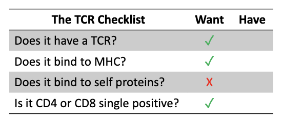
What is the goal of thymic T cell development?

To pass all 4 checkpoints:
Has TCR
Binds MHC
Doesn’t bind to self-proteins
Becomes single positive (CD4 or CD8)

<p>To pass all 4 checkpoints:<br><span>✅</span> Has TCR<br><span>✅</span> Binds MHC<br><span data-name="check_mark_button" data-type="emoji">✅</span> Doesn’t bind to self-proteins<br><span>✅</span> Becomes single positive (CD4 or CD8)</p>
8
New cards

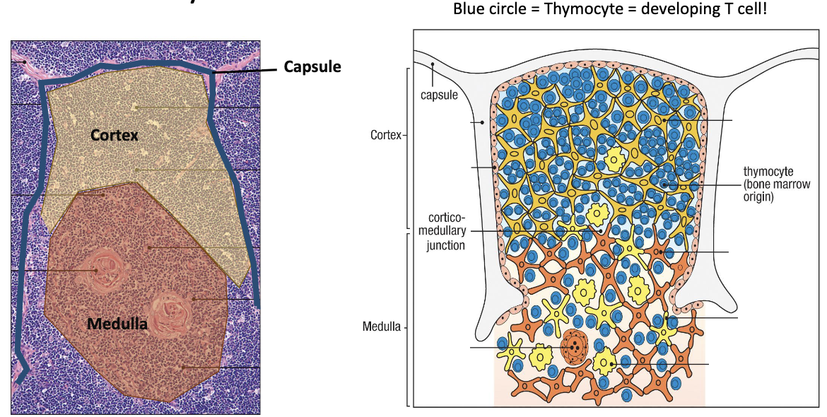
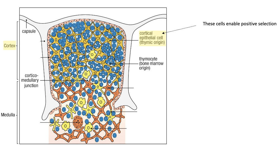
What are the main thymic regions?

  • Capsule (outer covering)

  • Cortex (outer dense region – early development)

    • Blue circles: Developing T cells = thymocytes.

  • Medulla (inner region – later stages)

<ul><li><p><strong>Capsule</strong> (outer covering)</p></li><li><p><strong>Cortex</strong> (outer dense region – early development)</p><ul><li><p>Blue circles: Developing T cells = thymocytes.</p></li></ul></li><li><p><strong>Medulla</strong> (inner region – later stages)</p></li></ul><img src="https://knowt-user-attachments.s3.amazonaws.com/75605104-5203-4534-91ad-bc35d1713ff6.png" data-width="100%" data-align="center"><p></p>
9
New cards

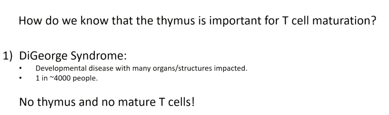
How does DiGeorge syndrome show the importance of the thymus for T-cell maturation?

  • Caused by deletion on chromosome 22.

  • Missing gene → thymus does not develop.

  • Without thymus → no mature T cellssevere immunodeficiency.

<ul><li><p>Caused by <strong>deletion on chromosome 22</strong>.</p></li><li><p>Missing gene → <strong>thymus does not develop</strong>.</p></li><li><p>Without thymus → <strong>no mature T cells</strong> → <strong>severe immunodeficiency</strong>.</p></li></ul><p></p>
10
New cards

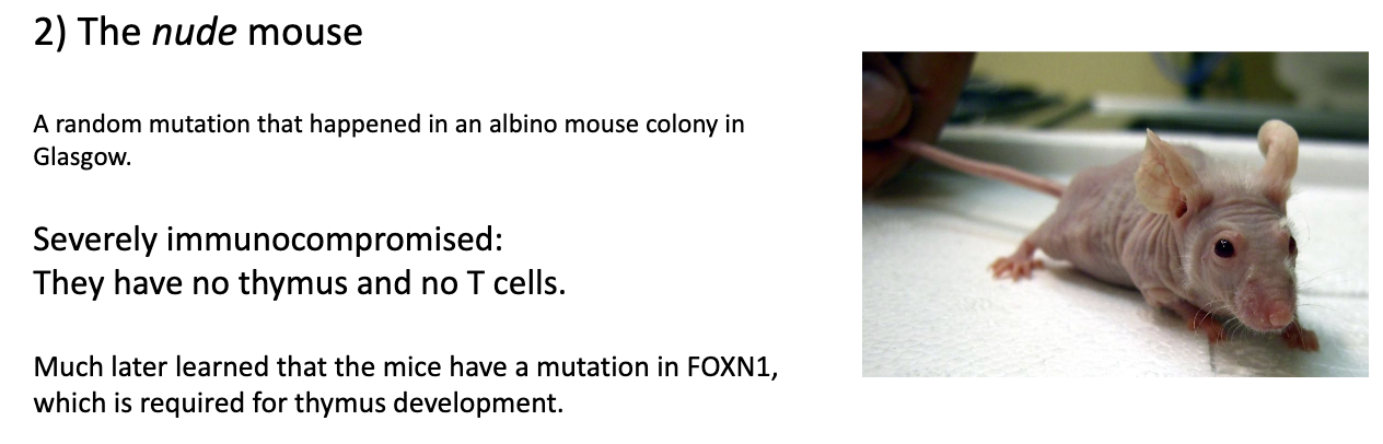
How do nude mice demonstrate the role of the thymus in T-cell maturation?

  • Nude mouse: spontaneous mutant discovered in albino mice → bred into own strain.

  • Key feature: no thymusno mature T cells.

  • Mutation: in FOXN1 gene, essential for thymus development.

<ul><li><p><strong>Nude mouse:</strong> spontaneous mutant discovered in albino mice → bred into own strain.</p></li><li><p><strong>Key feature:</strong> <strong>no thymus</strong> → <strong>no mature T cells</strong>.</p></li><li><p><strong>Mutation:</strong> in <strong>FOXN1 gene</strong>, essential for thymus development.</p></li></ul><p></p>
11
New cards

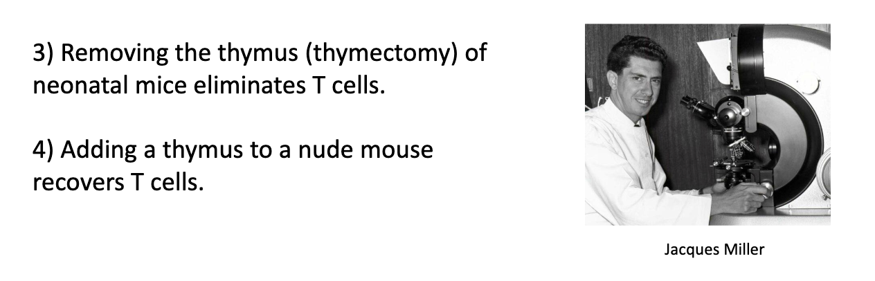
How did Jacques Miller demonstrate the thymus’s role in T-cell development?

  • Thymectomy in neonatal mice: no T cells develop.

  • Thymus transplant into nude mice: T cells are restored.

<ul><li><p><strong>Thymectomy in neonatal mice:</strong> no T cells develop.</p></li><li><p><strong>Thymus transplant into nude mice:</strong> T cells are restored.</p></li></ul><p></p>
12
New cards

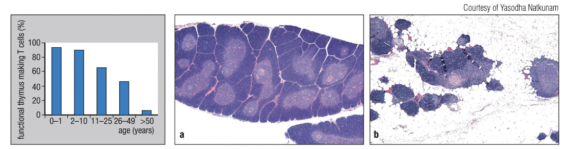
How does the thymus change with age?

  • It shrinks and fills with fat (thymic involution), so fewer new T cells are made.

  • Removing the thymus in adults doesn’t affect existing T cells.

<ul><li><p>It <strong>shrinks and fills with fat </strong>(thymic involution), so <strong>fewer new T cells</strong> are made.</p></li><li><p>Removing the thymus in adults doesn’t affect existing T cells.</p></li></ul><p></p>
13
New cards

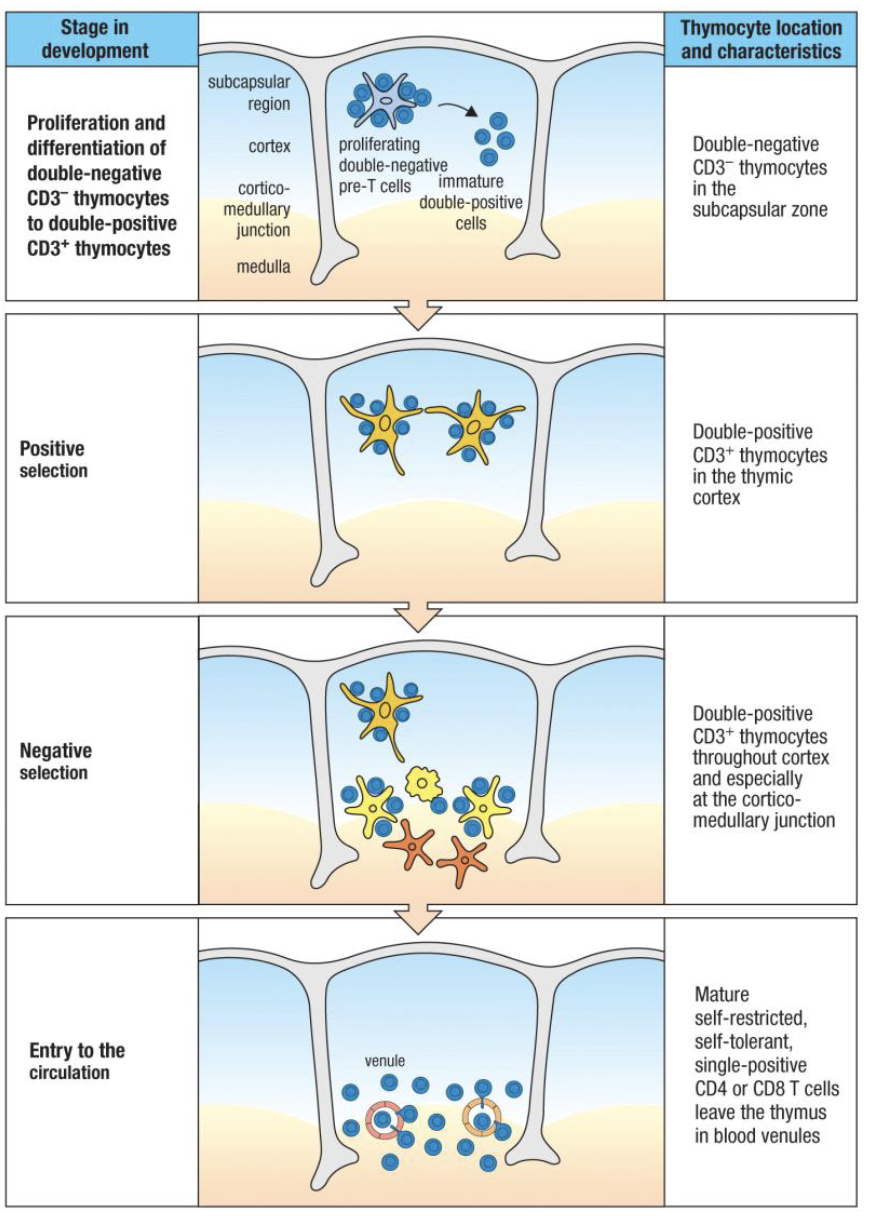
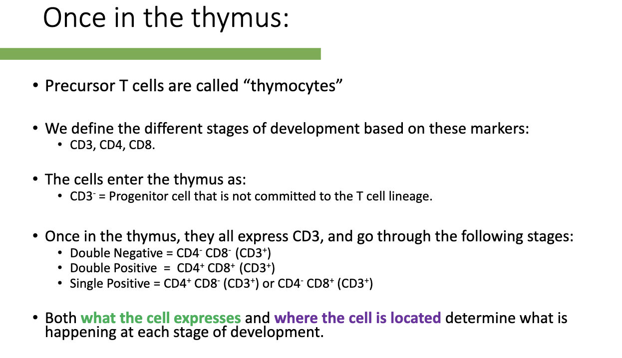
What are the stages of T-cell development in the thymus and how are they defined?

  • Precursor T cells in thymus: called thymocytes.

  • Markers used to define stages: CD3, CD4, CD8.

  • Entry stage: CD3- (uncommitted progenitor).

  • Development stages (all CD3+):

    1. Double Negative (DN): CD4- CD8-

    2. Double Positive (DP): CD4+ CD8+

    3. Single Positive (SP): CD4+ CD8- or CD4- CD8+

  • Function depends on: what markers the cell expresses and its location in the thymus.

<ul><li><p><strong>Precursor T cells in thymus:</strong> called thymocytes.</p></li><li><p><strong>Markers used to define stages:</strong> CD3, CD4, CD8.</p></li><li><p><strong>Entry stage:</strong> CD3- (uncommitted progenitor).</p></li><li><p><strong>Development stages (all CD3+):</strong></p><ol><li><p>Double Negative (DN): CD4- CD8-</p></li><li><p>Double Positive (DP): CD4+ CD8+</p></li><li><p>Single Positive (SP): CD4+ CD8- or CD4- CD8+</p></li></ol></li><li><p><strong>Function depends on:</strong> what markers the cell expresses and its location in the thymus.</p></li></ul><p></p>
14
New cards

How do thymocyte stages match the TCR “checklist”?

Development follows:

  1. Make TCR

  2. Test MHC binding

  3. Test self-reactivity

  4. Become CD4 or CD8 single positive

15
New cards

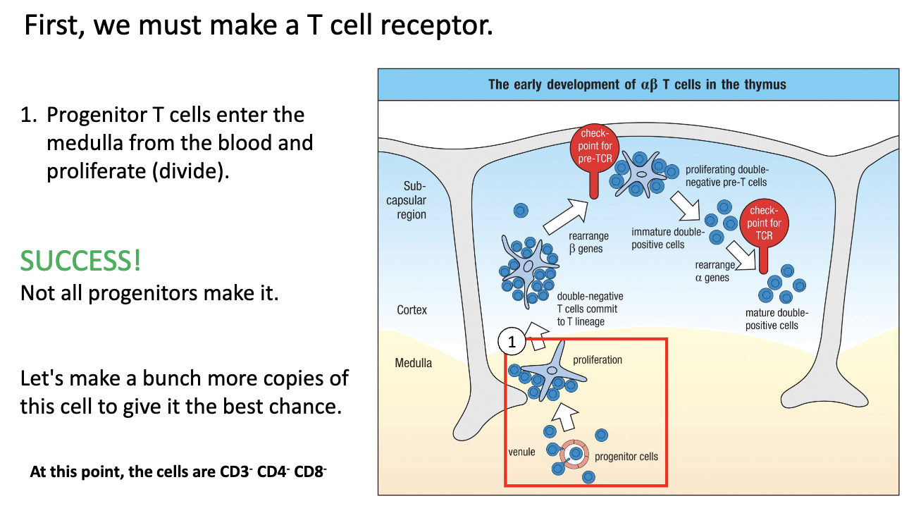
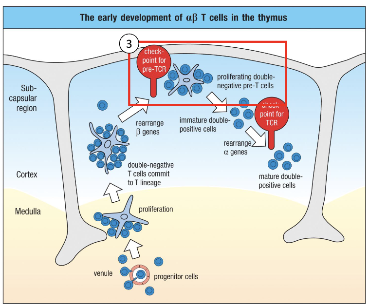
What happens when progenitor T cells first enter the thymus?

  • Enter thymus via venules into medulla/cortex.

  • Proliferate to make multiple copies (“save states”) of each progenitor.

  • Not all cells succeed; only some clones will continue T-cell development.

  • This expansion allows multiple chances for TCR rearrangement and successful T-cell maturation. 

<ul><li><p>Enter thymus <strong>via venules</strong> into <strong>medulla/cortex</strong>.</p></li><li><p><strong>Proliferate</strong> to make multiple copies (“save states”) of each progenitor.</p></li><li><p>Not all cells succeed; only some clones will continue T-cell development.</p></li><li><p>This <strong>expansion allows multiple chances</strong> for TCR rearrangement and successful T-cell maturation.&nbsp;</p></li></ul><p></p>
16
New cards

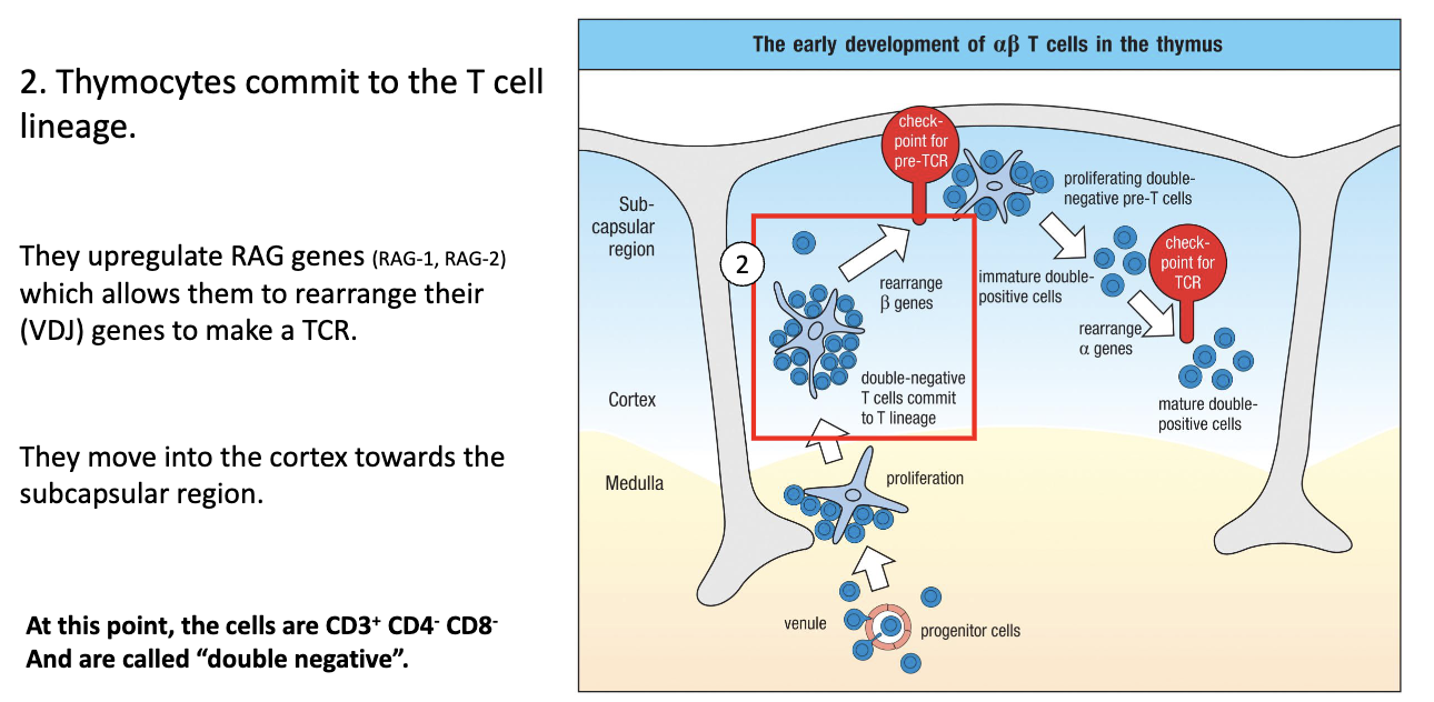
What happens to T cells in the thymic cortex during early development?

  • Move up into cortexcommit to T-cell lineage.

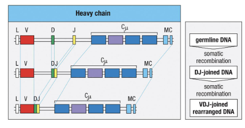
  • Turn on CD3 and begin TCR gene rearrangement.

  • RAG (RAG-1, RAG-2) genes are expressed → mediate VDJ recombination.

  • DJ joins first, then V joins DJ; specific timing for beta, gamma, delta, alpha chains.

  • At this stage: Double Negative (CD4- CD8-), CD3+, starting TCR rearrangement.

<ul><li><p>Move <strong>up into cortex</strong> → <strong>commit to T-cell lineage</strong>.</p></li><li><p><strong>Turn on CD3</strong> and begin <strong>TCR gene rearrangement</strong>.</p></li><li><p><strong>RAG</strong> (RAG-1, RAG-2) <strong>genes</strong> are expressed → mediate <strong>VDJ recombination</strong>.</p></li><li><p><strong>DJ joins first</strong>, then <strong>V joins DJ</strong>; specific timing for <strong>beta, gamma, delta, alpha chains</strong>.</p></li><li><p>At this stage: <strong>Double Negative (CD4- CD8-)</strong>, <strong>CD3+</strong>, starting TCR rearrangement.</p></li></ul><p></p>
17
New cards

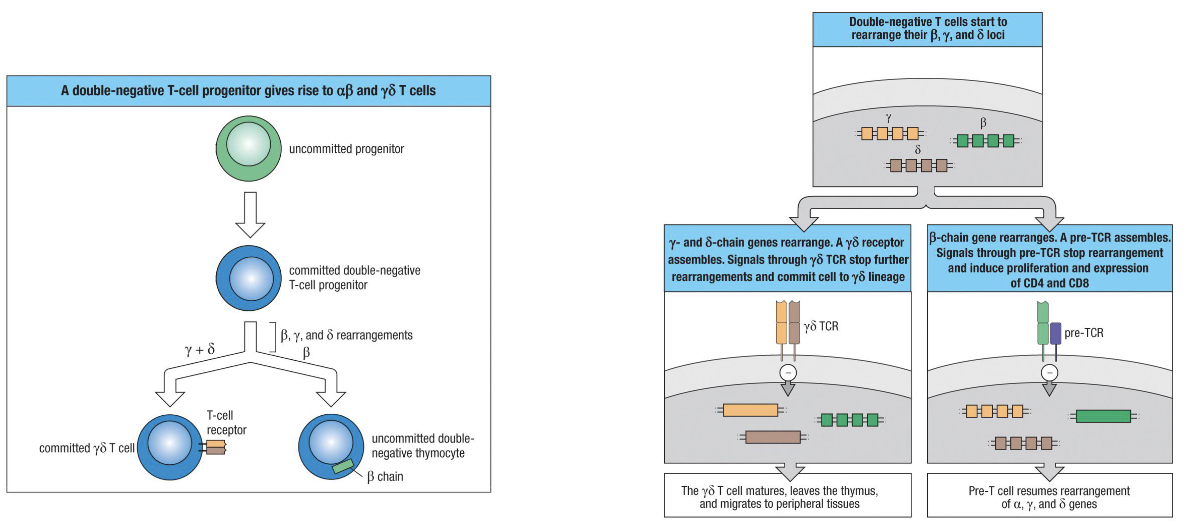
When and how is the αβ vs γδ decision made?

  • Committed progenitor expresses RAG genes → starts VDJ recombination at gamma, delta, and beta loci (NOT alpha).

  • Whichever locus rearranges successfully first determines cell type:

    • γδ TCR first → cell becomes γδ T cell, stops rearranging, leaves thymus mature.

    • Beta chain first (most common) → pairs with surrogate alpha chain, signals successful TCR → becomes αβ T cell.

  • TCR signaling required for survival: cells failing to make a functional receptor undergo apoptosis.

<ul><li><p>Committed progenitor expresses <strong>RAG genes</strong> → starts VDJ recombination at gamma, delta, and beta loci (<strong><u>NOT</u></strong> alpha).</p></li><li><p>Whichever locus rearranges successfully <strong>first</strong> determines cell type:</p><ul><li><p><strong>γδ TCR first</strong> → cell becomes γδ T cell, stops rearranging, leaves thymus mature.</p></li><li><p><strong>Beta chain first (most common) </strong>→ pairs with surrogate alpha chain, signals successful TCR → becomes αβ T cell.</p></li></ul></li><li><p>TCR signaling required for survival: cells failing to make a functional receptor undergo apoptosis.</p></li></ul><p></p>
18
New cards

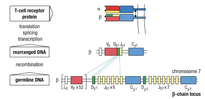
What does “rearranging” TCR genes mean?

VDJ recombination—random joining of gene segments to create unique receptors (same as BCRs).

<p><strong>VDJ recombination</strong>—random joining of gene segments to create unique receptors (same as BCRs).</p>
19
New cards

What are the key facts about αβ vs γδ T cells?

  • 95% of T cells: αβ TCR → majority, main focus in immunology.

  • γδ T cells:

    • Rare, mostly at mucosal sites, like intestines

    • Do not express CD4/CD8

    • Low diversity (low number of V genes)

    • Bind non-MHC targets (e.g., stress proteins)

    • Function more like innate immune cells

20
New cards

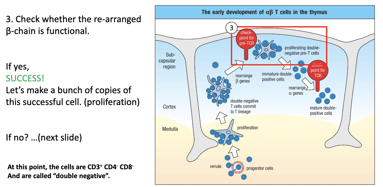
What is the next step after T cell lineage commitment?

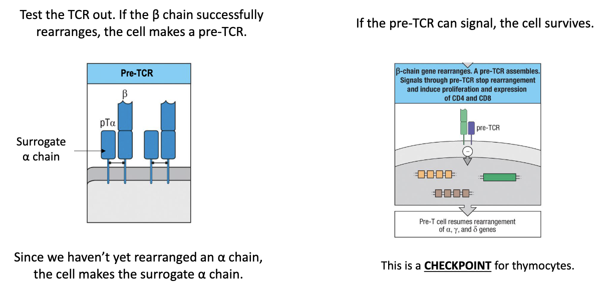
  • Test whether the rearranged β chain forms a functional pre-TCR.

  • Still CD3+ CD4– CD8–, i.e. double negative.

<ul><li><p>Test whether the <strong>rearranged β chain</strong> forms a <strong>functional pre-TCR</strong>.</p></li><li><p>Still <strong>CD3+ CD4– CD8–</strong>, i.e. <strong>double negative</strong>.</p></li></ul><p></p>
21
New cards

How is the β chain tested to ensure there is a functional TCR?

  • Surrogate alpha chain pairs with newly rearranged beta chain (before real alpha chain exists).

  • If beta + surrogate alpha forms a functional receptor → signals the cell → cell survives.

  • This is a checkpoint: ensures only cells with functional TCR survive.

  • Successful cells proliferate to make multiple copies (“safe states”) for further development.

<ul><li><p><strong>Surrogate alpha chain</strong> pairs with newly rearranged <strong>beta chain</strong> (before real alpha chain exists).</p></li></ul><ul><li><p>If <strong>beta + surrogate alpha</strong> forms a functional receptor → <strong>signals the cell</strong> → cell survives.</p></li><li><p>This is a <strong>checkpoint</strong>: ensures only cells with functional TCR <strong>survive</strong>.</p></li><li><p>Successful cells <strong>proliferate</strong> to make multiple copies (“safe states”) for further development.</p></li></ul><p></p>
22
New cards

What happens if β chain rearrangement works?

The cell proliferates, expanding successful clones (still double negative).

<p>The cell <strong>proliferates</strong>, expanding successful clones (still double negative).</p>
23
New cards

What happens if β chain rearrangement fails?

  • Non-productive rearrangement: cell did not make a functional beta chain.

  • Retry mechanism: cell tries other V + D + J combinations with alternative beta chain constant regions.

  • 2 attempts: cell has several chances to make a functional beta chain (2 alleles: 1 from each parent but each allele can try multiple VDJ combinations).

  • Outcome: if successful → cell survives and proliferates; if not → cell eventually dies.

<ul><li><p><strong>Non-productive rearrangement:</strong> cell did not make a functional beta chain.</p></li><li><p><strong>Retry mechanism:</strong> cell tries other V + D + J combinations with alternative beta chain constant regions.</p></li><li><p><strong>2 attempts:</strong> cell has several chances to make a functional beta chain (2 alleles: 1 from each parent but each allele can try multiple VDJ combinations).</p></li><li><p><strong>Outcome:</strong> if successful → <strong>cell survives and proliferates</strong>; if not → cell eventually dies.</p></li></ul><p></p>
24
New cards

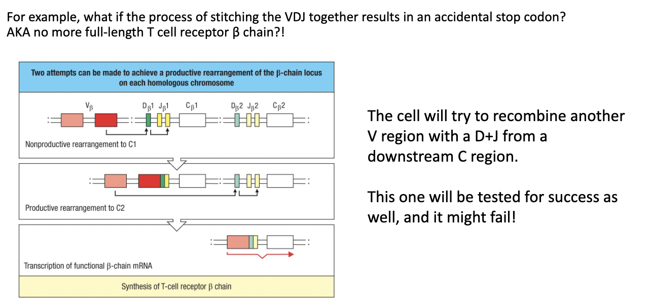
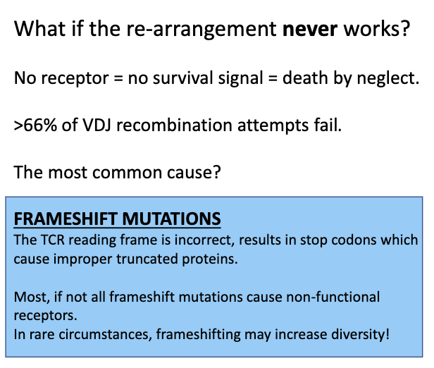
What if the β chain re-arrangement never works?

  • Randomness in VDJ recombination → may produce frame shifts or non-productive sequences.

  • No functional TCRno survival signal → cell dies (“death by neglect”).

  • Failure rate: ~66% of VDJ rearrangements fail.

  • Importance of proliferation: multiple “safe state” copies increase chances that at least one cell succeeds.

<ul><li><p><strong>Randomness in VDJ recombination</strong> → may produce <strong>frame shifts</strong> or non-productive sequences.</p></li><li><p><strong>No functional TCR</strong> → <strong>no survival signal</strong> → cell <strong>dies</strong> (“death by neglect”).</p></li><li><p><strong>Failure rate:</strong> ~66% of VDJ rearrangements fail.</p></li><li><p><strong>Importance of proliferation:</strong> multiple “safe state” copies increase chances that at least one cell succeeds.</p></li></ul><p></p>
25
New cards

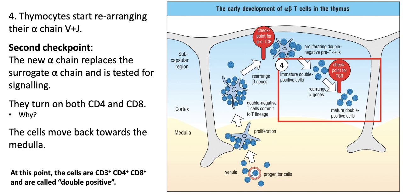
What happens when T cells rearrange their alpha chain, and why do they become double positive?

  • Alpha chain rearrangement: V + J segments (no D).

  • Surrogate alpha chain is replaced by functional alpha chain on the surface.

  • Functional alpha-beta TCR signals → cell survives.

  • CD4 and CD8 are turned on → cell becomes Double Positive (DP).

  • Reason for DP expression: needed to test the alpha-beta TCR with MHC I and II during positive selection.

  • Location: cells move up to cortex, then start moving back down toward medulla.

<ul><li><p><strong>Alpha chain rearrangement:</strong> V + J segments (no D).</p></li><li><p><strong>Surrogate alpha chain</strong> is replaced by <strong>functional alpha chain</strong> on the surface.</p></li><li><p><strong>Functional alpha-beta TCR signals</strong> → cell survives.</p></li><li><p><strong>CD4 and CD8 are turned on</strong> → cell becomes <strong>Double Positive (DP)</strong>.</p></li><li><p><strong>Reason for DP expression:</strong> needed to test the alpha-beta TCR with <strong>MHC I and II</strong> during positive selection.</p></li><li><p><strong>Location:</strong> cells move <strong>up to cortex</strong>, then start moving <strong>back down toward medulla</strong>.</p></li></ul><p></p>
26
New cards

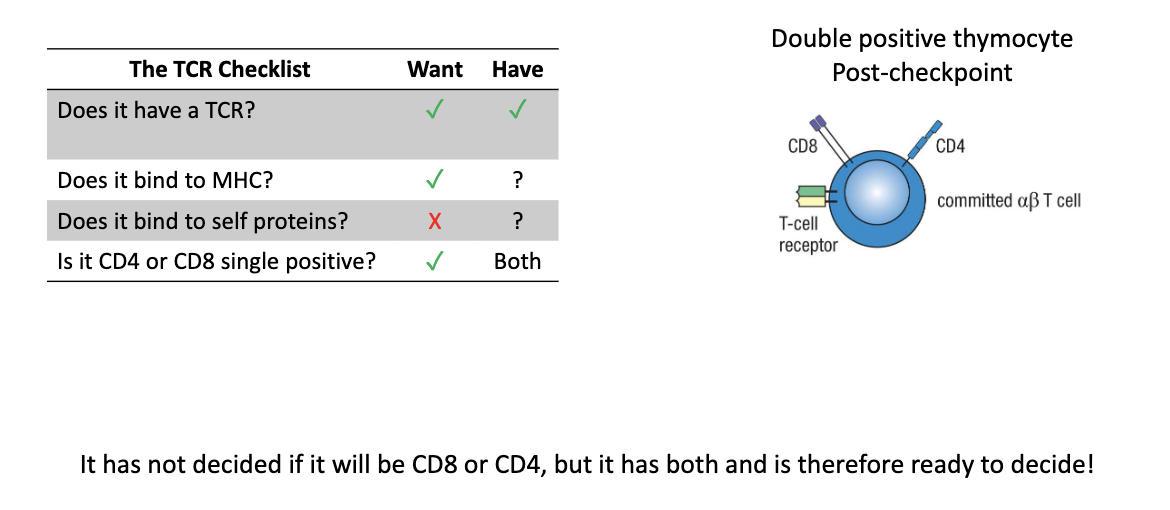
What is true of double positive (DP) thymocytes?

  • Have functional TCR

  • Express both CD4 and CD8

  • Next tests: MHC binding and self-reactivity

<ul><li><p>Have <strong>functional TCR</strong></p></li><li><p>Express <strong>both CD4 and CD8</strong></p></li><li><p>Next tests: <strong>MHC binding</strong> and <strong>self-reactivity</strong></p></li></ul><p></p>
27
New cards

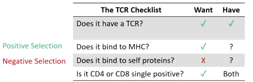
What two processes refine the TCR repertoire?

  1. Positive selection – keep T cells that bind MHC

  2. Negative selection – remove self-reactive T cells

<ol><li><p><strong>Positive selection</strong> – keep T cells that bind MHC</p></li><li><p><strong>Negative selection</strong> – remove self-reactive T cells</p></li></ol><p></p>
28
New cards

Where does positive selection occur?

In the cortex of the thymus, mediated by cortical epithelial cells (not immune cells).

<p>In the <strong>cortex</strong> of the thymus, mediated by <strong>cortical epithelial cells</strong> (not immune cells).</p>
29
New cards

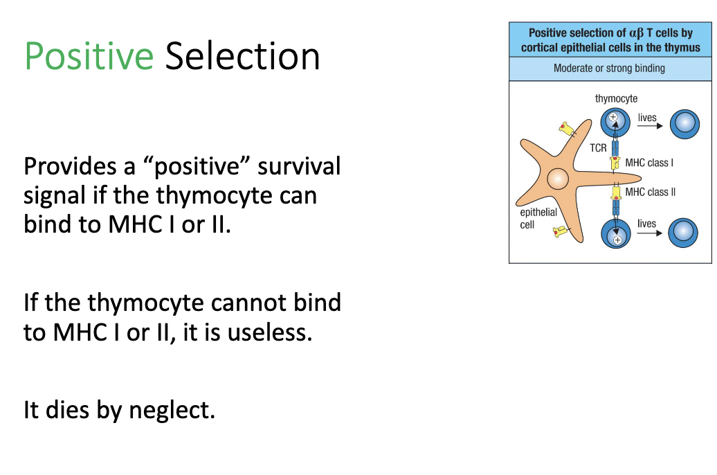
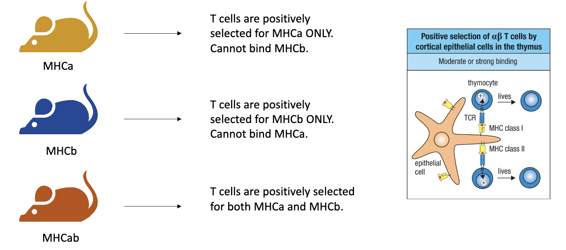
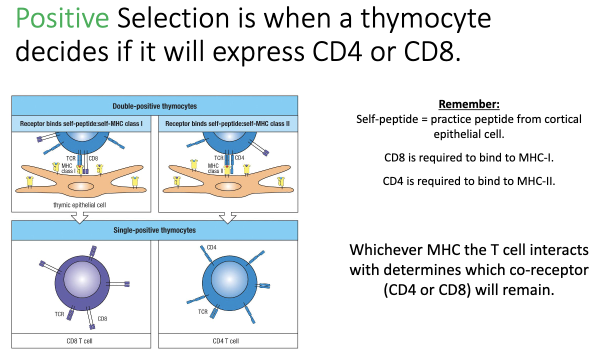
What happens during positive selection in the thymus?

  • Thymocyte tests TCR binding to MHC I or II.

  • Moderate to strong binding: receives positive survival signal → cell lives. 

  • No binding: fails positive selection → dies by neglect.

<ul><li><p><strong>Thymocyte tests TCR binding</strong> to <strong>MHC I or II</strong>.</p></li><li><p><strong>Moderate to strong binding:</strong> receives positive&nbsp;survival signal → cell lives.&nbsp;</p></li><li><p><strong>No binding:</strong> fails positive selection → <strong>dies by neglect</strong>.</p></li></ul><p></p>
30
New cards

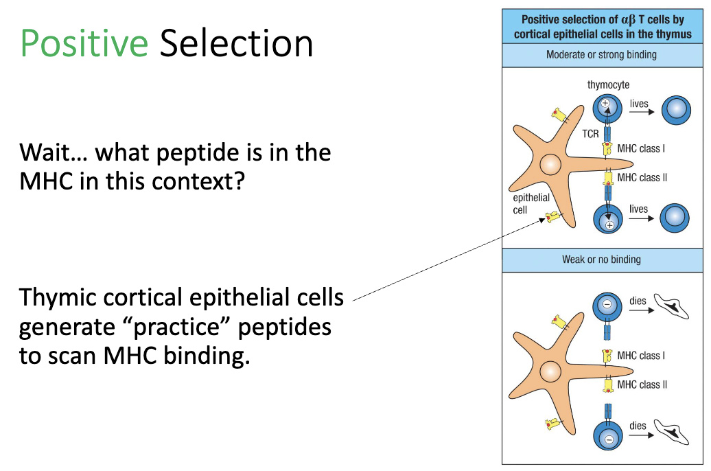
What peptides are presented during positive selection?

  • Thymic cortical epithelial cells load “practice peptides” on MHC

  • Purpose: T cells are testing MHC binding, not specific peptide recognition.

<ul><li><p>Thymic cortical epithelial cells load <strong>“practice peptides”</strong> on MHC</p></li></ul><ul><li><p><strong>Purpose:</strong> T cells are <strong>testing MHC binding</strong>, not specific peptide recognition.</p></li></ul><p></p>
31
New cards

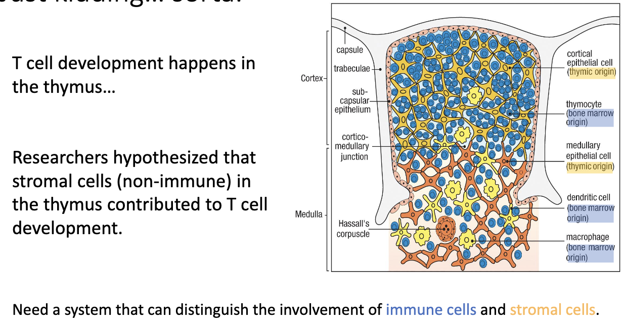
How was it discovered that T-cell development depends on thymic stromal cells?

  • Researchers (e.g., Jacques Miller) questioned why T-cell development must occur in the thymus.

  • Observations:

    • Mice with a thymus (even transplanted elsewhere) → still made T cells.

    • Mice without a thymus → no T cells.

  • Hypothesis: Some thymic stromal cells (non-immune cells) are essential for T-cell development.

  • To test this, they used chimeric mice (mix of different cell types) to distinguish whether immune cells or stromal cells were responsible.

<ul><li><p>Researchers (e.g., <strong>Jacques Miller</strong>) questioned <strong>why T-cell development must occur in the thymus</strong>.</p></li><li><p>Observations:</p><ul><li><p>Mice <strong>with a thymus (even transplanted elsewhere)</strong> → still made T cells.</p></li><li><p>Mice <strong>without a thymus</strong> → no T cells.</p></li></ul></li><li><p>Hypothesis: Some <strong>thymic stromal cells (non-immune cells)</strong> are essential for T-cell development.</p></li><li><p>To test this, they used <strong>chimeric mice</strong> (mix of different cell types) to distinguish whether <strong>immune cells</strong> or <strong>stromal cells</strong> were responsible.</p></li></ul><p></p>
32
New cards

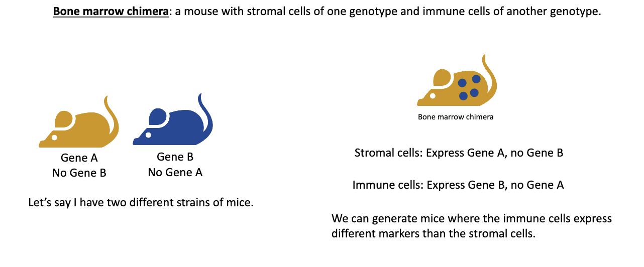
What is a bone marrow chimera and how is it made?

  • A bone marrow chimera is a mouse whose immune system comes from a different donor mouse’s bone marrow.

  • Steps:

    1. Two mouse strains are used — one has gene A (no gene B) and the other has gene B (no gene A).

    2. The recipient mouse is irradiated, which destroys its bone marrow progenitor cells, wiping out its immune system.

    3. Bone marrow from the donor mouse is transplanted into the irradiated mouse.

  • The new immune cells develop from the donor bone marrow, while the stromal (structural) cells remain from the original host.

<ul><li><p>A <strong>bone marrow chimera</strong> is a mouse whose <strong>immune system</strong> comes from a <strong>different donor mouse’s bone marrow</strong>.</p></li><li><p>Steps: </p><ol><li><p>Two mouse strains are used — one has <strong>gene A (no gene B)</strong> and the other has <strong>gene B (no gene A)</strong>.</p></li><li><p>The recipient mouse is <strong>irradiated</strong>, which <strong>destroys its bone marrow progenitor cells</strong>, wiping out its immune system.</p></li><li><p><strong>Bone marrow from the donor mouse</strong> is transplanted into the irradiated mouse.</p></li></ol></li></ul><ul><li><p>The <strong>new immune cells</strong> develop from the donor bone marrow, while the <strong>stromal (structural) cells</strong> remain from the original host.</p></li></ul><p></p>
33
New cards

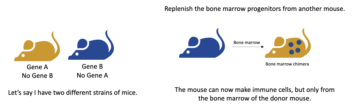
Example of bone marrow transplantation to create a bone marrow chimera.

  • Example: If a gene A mouse receives bone marrow from a gene B mouse →

    • Stromal cells (non-immune) = gene A

    • Immune cells = gene B

  • This setup lets scientists test whether immune functions depend on immune cells or stromal cells, making it a powerful tool in immunology.

<ul><li><p>Example: If a <strong>gene A</strong> mouse receives bone marrow from a <strong>gene B</strong> mouse →</p><ul><li><p><strong>Stromal cells</strong> (non-immune) = gene A</p></li><li><p><strong>Immune cells</strong> = gene B</p></li></ul></li></ul><ul><li><p>This setup lets scientists test <strong>whether immune functions depend on immune cells or stromal cells</strong>, making it a powerful tool in immunology.</p></li></ul><p></p>
34
New cards

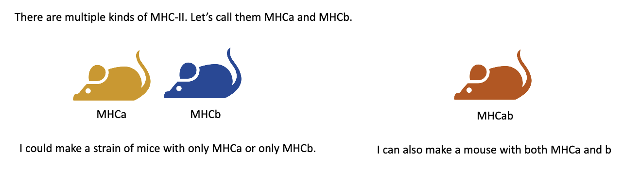
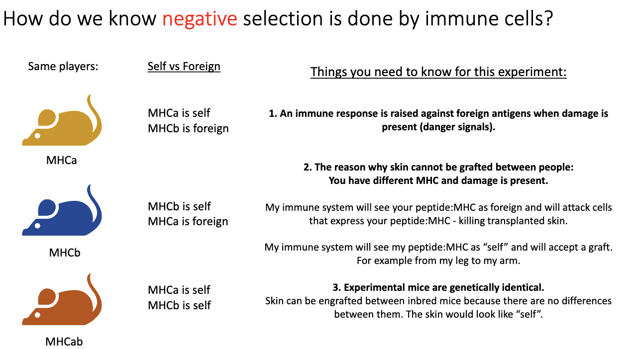
How did researchers use MHC variants in bone marrow chimera experiments to study T-cell development?

  • There are different types of MHC class II molecules (e.g., MHCA and MHCB).

  • Researchers used two mouse strains:

    • One mouse had MHCA only.

    • The other had MHCB only.

  • By crossing the two strains, they produced:

    • Homozygous mice → only MHCA or only MHCB.

    • Heterozygous mice → both MHCA and MHCB.

  • These mice were then used in bone marrow chimera setups to test whether T-cell selection depends on MHC type expressed by stromal (thymic) cells or by bone marrow–derived immune cells.

<ul><li><p>There are <strong>different types of MHC class II molecules</strong> (e.g., MHCA and MHCB).</p></li><li><p>Researchers used <strong>two mouse strains</strong>:</p><ul><li><p>One mouse had <strong>MHCA only</strong>.</p></li><li><p>The other had <strong>MHCB only</strong>.</p></li></ul></li><li><p>By <strong>crossing the two strains</strong>, they produced:</p><ul><li><p><strong>Homozygous mice</strong> → only MHCA or only MHCB.</p></li><li><p><strong>Heterozygous mice</strong> → both MHCA and MHCB.</p></li></ul></li><li><p>These mice were then used in <strong>bone marrow chimera setups</strong> to test whether <strong>T-cell selection</strong> depends on MHC type expressed by <strong>stromal (thymic)</strong> cells or by <strong>bone marrow–derived immune</strong> cells.</p></li></ul><p></p>
35
New cards

How does positive selection differ in MHCA, MHCB, and heterozygous (MHCA+MHCB) mice?

  • MHCA-only mice: T cells are positively selected for MHCA only → their TCRs can bind MHCA but not MHCB.

  • MHCB-only mice: T cells are positively selected for MHCB only → their TCRs can bind MHCB but not MHCA.

  • Heterozygous (MHCA+MHCB) mice: T cells can be selected for either MHCA or MHCB, producing a mix of TCR specificities for both MHC types.

But they didn’t know that at the time…

<ul><li><p><strong>MHCA-only mice:</strong> T cells are positively selected for MHCA only → their TCRs can bind MHCA but not MHCB.</p></li><li><p><strong>MHCB-only mice:</strong> T cells are positively selected for MHCB only → their TCRs can bind MHCB but not MHCA.</p></li><li><p><strong>Heterozygous (MHCA+MHCB) mice: </strong>T cells can be selected for either MHCA or MHCB, producing a mix of TCR specificities for both MHC types.</p></li></ul><p></p><p>But they didn’t know that at the time…</p><p></p>
36
New cards

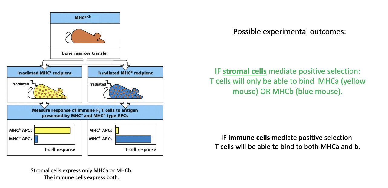
What did the bone marrow chimera experiment reveal about which cells mediate positive selection of T cells?

  • Setup:

    • Stromal cells express either MHCA or MHCB

    • Immune cells express both MHCA and MHCB

  • Predictions:

    • If stromal cells drive positive selection → T cells should only recognize either MHCA or MHCB type of the stromal cells.

    • If immune cells drive it → T cells should recognize both MHC types.

  • Result:

    • In MHCA-only mice → T cells recognized only MHCA.

    • In MHCB-only mice → T cells recognized only MHCB.

  • Conclusion: Positive selection is mediated by thymic stromal (epithelial) cells, not immune cells.

<ul><li><p><strong>Setup:</strong></p><ul><li><p>Stromal cells express <strong><u>either</u></strong> MHCA or MHCB</p></li><li><p>Immune cells express <strong><u>both</u></strong> MHCA and MHCB</p></li></ul></li><li><p><strong>Predictions:</strong></p><ul><li><p>If <strong>stromal cells</strong> drive positive selection → T cells should only recognize either MHCA or MHCB type of the stromal cells.</p></li><li><p>If <strong>immune cells</strong> drive it → T cells should recognize <strong>both</strong> MHC types.</p></li></ul></li><li><p><strong>Result:</strong></p><ul><li><p>In MHCA-only mice → T cells recognized only <strong>MHCA</strong>.</p></li><li><p>In MHCB-only mice → T cells recognized only <strong>MHCB</strong>.</p></li></ul></li><li><p><strong>Conclusion:</strong> Positive selection is mediated by thymic stromal (epithelial) cells, not immune cells.</p></li></ul><p></p>
37
New cards

How does positive selection determine CD4 vs CD8 expression in thymocytes?

  • During positive selection, thymocytes test their TCR binding to MHC molecules:

    • Binds MHC I → keeps CD8

    • Binds MHC II → keeps CD4

    • Cannot bind either → dies

  • Outcome: single positive (SP) thymocyte expressing either CD4 or CD8.

  • At this stage: TCR is functional and binds MHC, but self-reactivity has not yet been tested.

<ul><li><p>During <strong>positive selection</strong>, thymocytes test their <strong>TCR binding</strong> to MHC molecules:</p><ul><li><p><strong>Binds MHC I → keeps CD8</strong></p></li><li><p><strong>Binds MHC II → keeps CD4</strong></p></li><li><p><strong>Cannot bind either → dies</strong></p></li></ul></li><li><p>Outcome: <strong>single positive (SP) thymocyte</strong> expressing either <strong>CD4 or CD8</strong>.</p></li><li><p>At this stage: TCR is functional and binds MHC, but <strong>self-reactivity has not yet been tested</strong>.</p></li></ul><p></p>
38
New cards

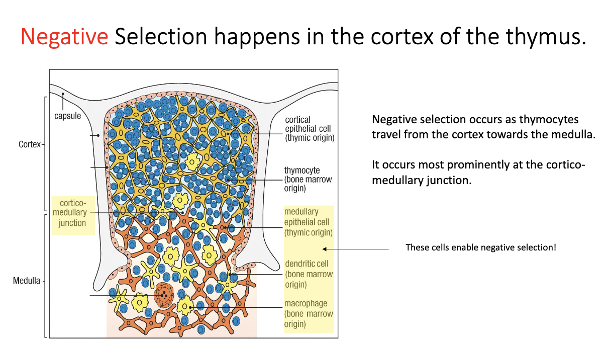
Where does negative selection occur in the thymus and what are the key cells involved?

  • Location: at the cortex–medulla junction of the thymus.

  • Key cells involved: medullary epithelial cells, dendritic cells, macrophages.

<ul><li><p><strong>Location:</strong> at the <strong>cortex–medulla junction</strong> of the thymus.</p></li><li><p><strong>Key cells involved:</strong> medullary epithelial cells, dendritic cells, macrophages.</p></li></ul><p></p>
39
New cards

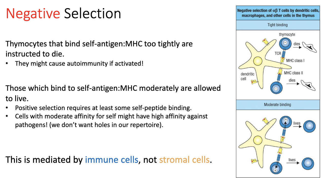
How does negative selection work, and what determines T cell survival or death?

  • Process:

    • T cells encounter self-peptides on MHC.

    • Strong binding → cell receives a death signal → eliminated.

    • Moderate binding → cell survives → allowed to continue (needed for proper positive selection).

    • Weak/no binding → cell would not have survived positive selection anyway.

  • Reason for threshold:

    • Ensures T cells recognize self-MHC but do not strongly react to self-peptides.

    • Maintains self-tolerance while avoiding gaps that pathogens could exploit by mimicking self.

  • Mediators: immune cells, not stromal cells.

<ul><li><p><strong>Process:</strong></p><ul><li><p>T cells encounter self-peptides on MHC.</p></li><li><p><strong>Strong binding</strong> → cell receives a <strong>death signal</strong> → eliminated.</p></li><li><p><strong>Moderate binding</strong> → cell survives → allowed to continue (needed for proper positive selection).</p></li><li><p><strong>Weak/no binding</strong> → cell would not have survived positive selection anyway.</p></li></ul></li><li><p><strong>Reason for threshold:</strong></p><ul><li><p>Ensures T cells <strong>recognize self-MHC</strong> but do not strongly react to self-peptides.</p></li><li><p>Maintains <strong>self-tolerance</strong> while avoiding gaps that pathogens could exploit by mimicking self.</p></li></ul></li><li><p><strong>Mediators:</strong> immune cells, not stromal cells.</p></li></ul><p></p>
40
New cards

How do we know that negative selection is mediated by immune cells, and what role does MHC play in skin graft experiments?

  • Negative selection: removes T cells that strongly react to self-peptides.

  • Evidence from skin grafts:

    • Genetically identical mice (same MHC, e.g., MHCA → MHCA): skin grafts are accepted because self-reactive T cells were removed by negative selection.

    • Genetically different mice (different MHC, e.g., MHCA → MHCB): skin grafts are rejected because T cells recognize the foreign MHC.

<ul><li><p><strong>Negative selection:</strong> removes T cells that strongly react to self-peptides.</p></li><li><p><strong>Evidence from skin grafts:</strong></p><ul><li><p><strong>Genetically identical mice (same MHC, e.g., MHCA → MHCA):</strong> skin grafts are <strong>accepted</strong> because self-reactive T cells were removed by negative selection.</p></li><li><p><strong>Genetically different mice (different MHC, e.g., MHCA → MHCB):</strong> skin grafts are <strong>rejected</strong> because T cells recognize the foreign MHC.</p></li></ul></li></ul><p></p>
41
New cards

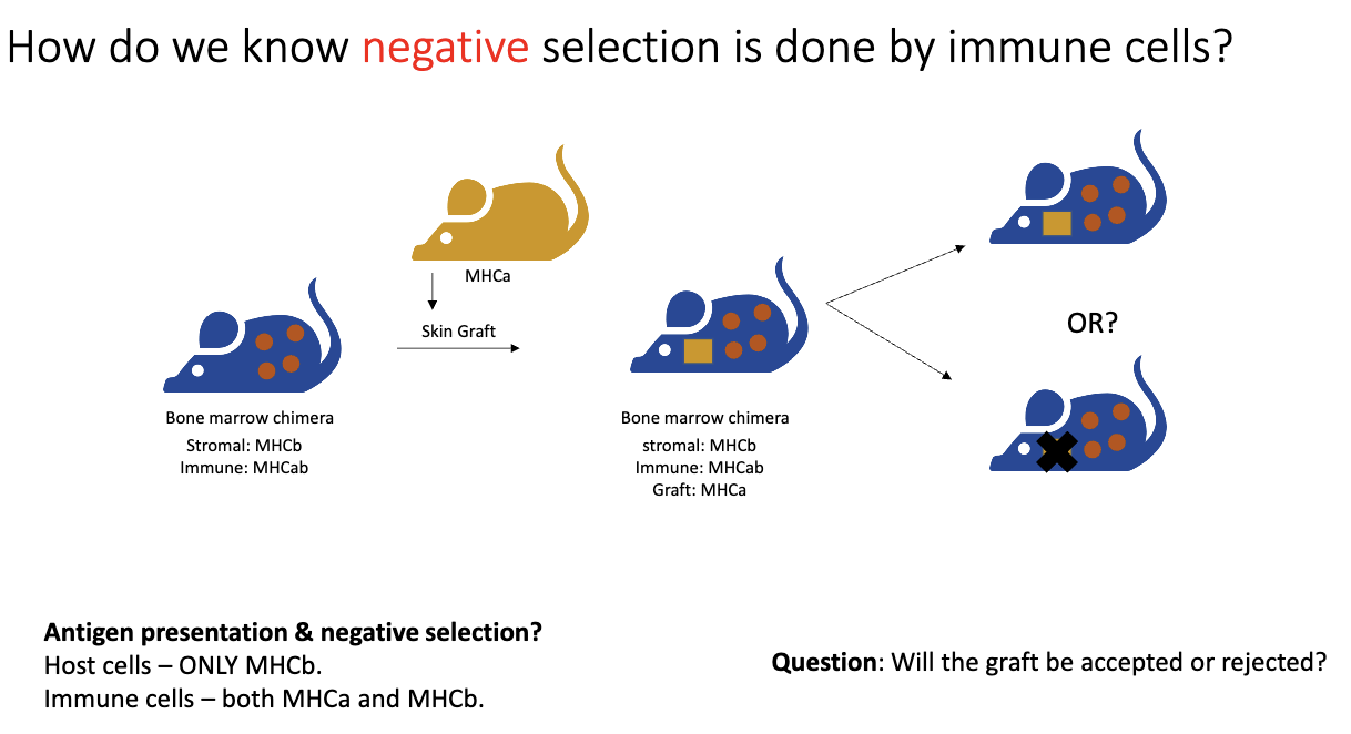
How did the bone marrow chimera experiment test whether negative selection is mediated by immune cells?

  • Setup:

    • Recipient mouse stromal cells: MHCB only.

    • Donor bone marrow (immune cells): MHCAB.

  • Test:

    • Skin graft from MHCA mouse (yellow) placed on recipient (blue, MHCB stromal cells).

  • Logic:

    • Normally, MHCA graft → rejected by T cells.

    • If negative selection is mediated by immune cells, T cells should be tolerized to MHCA because the immune cells express it.

    • Outcome shows whether immune cells can induce tolerance versus stromal cells.

<ul><li><p><strong>Setup:</strong></p><ul><li><p><strong>Recipient mouse stromal cells:</strong> MHCB only.</p></li><li><p><strong>Donor bone marrow (immune cells):</strong> MHCAB.</p></li></ul></li><li><p><strong>Test:</strong></p><ul><li><p>Skin graft from MHCA mouse (yellow) placed on recipient (blue, MHCB stromal cells).</p></li></ul></li><li><p><strong>Logic:</strong></p><ul><li><p>Normally, MHCA graft → <strong>rejected</strong> by T cells.</p></li><li><p>If <strong>negative selection is mediated by immune cells</strong>, T cells should be tolerized to MHCA because the immune cells express it.</p></li><li><p>Outcome shows <strong>whether immune cells can induce tolerance</strong> versus stromal cells.</p></li></ul></li></ul><p></p>
42
New cards

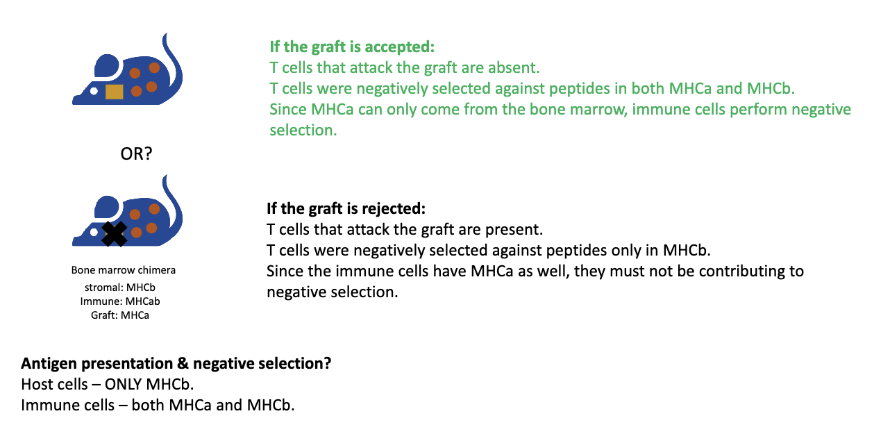
What were the results of bone marrow negative selection experiment?

  • Skin graft from MHCA mouse tested:

    • Outcome: Graft accepted → T cells recognizing MHCA removed → negative selection done by immune cells (not stromal cells).

    • If graft rejected → immune cells not mediating negative selection.

  • Result: Graft was accepted!

  • Key points:

    • Positive selection is mediated by stromal/epithelial cells.

    • Negative selection primarily by dendritic cells/macrophages (immune cells).

<ul><li><p>Skin graft from MHCA mouse tested:</p><ul><li><p><strong>Outcome:</strong> Graft accepted → T cells recognizing MHCA removed → negative selection done by <strong>immune cells</strong> (not stromal cells).</p></li><li><p>If graft rejected → immune cells not mediating negative selection.</p></li></ul></li><li><p><strong>Result: </strong>Graft was <strong>accepted</strong>!</p></li><li><p><strong>Key points:</strong></p><ul><li><p>Positive selection is mediated by stromal/epithelial cells.</p></li><li><p>Negative selection primarily by dendritic cells/macrophages (immune cells).</p></li></ul></li></ul><p></p>
43
New cards

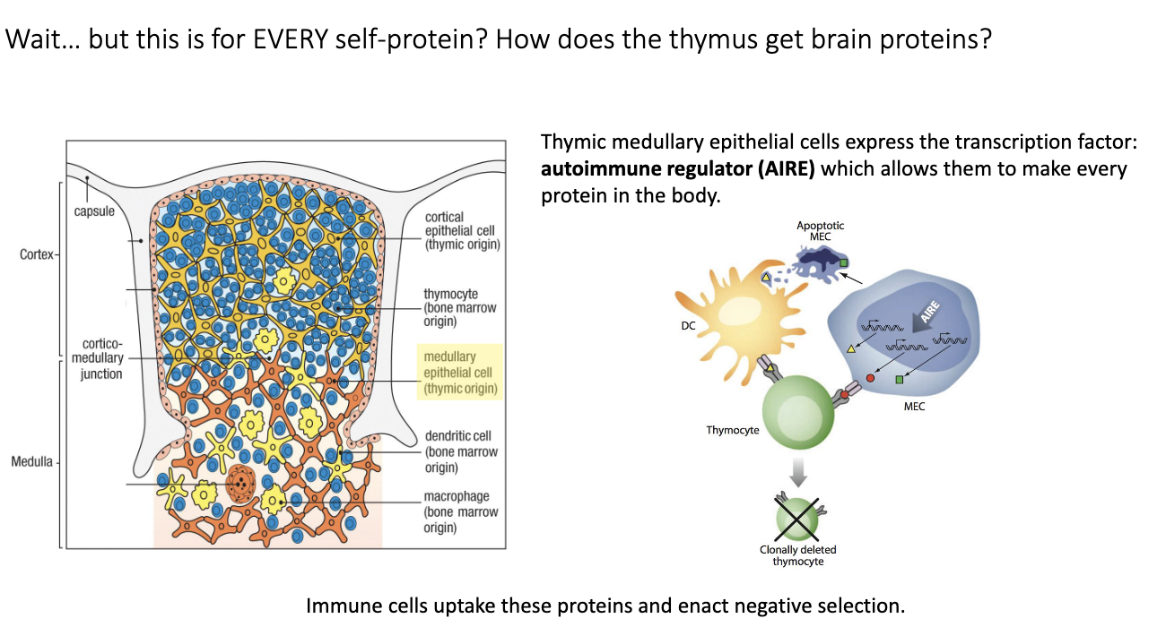
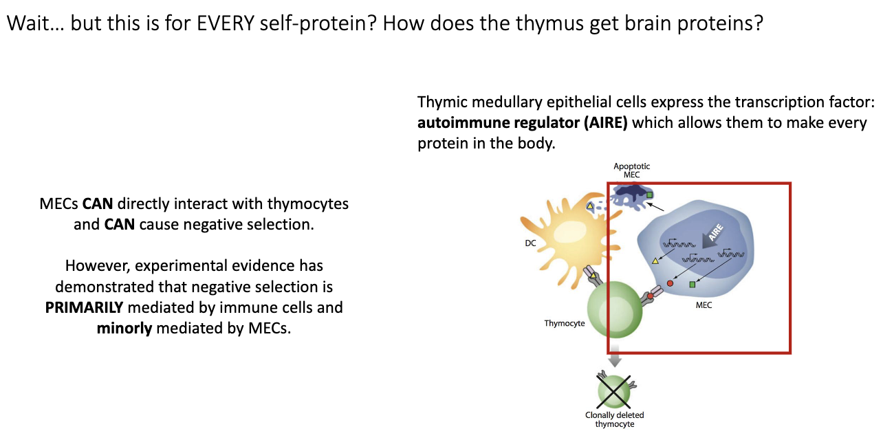
How do medullary thymic epithelial cells and the AIRE transcription factor contribute to negative selection of T cells?

  • Problem: Thymocytes need to be negatively selected against tissue-specific proteins (e.g., brain peptides), but thymus is not that tissue.

  • AIRE (Autoimmune Regulator):

    • Transcription factor in medullary thymic epithelial cells (mTECs).

    • Drives expression of many tissue-specific genes in thymus.

    • AIRE knockout → autoimmunity.

  • Mechanism:

    • mTECs produce self-proteins → dendritic cells uptake them → present on MHC → T cells that bind too strongly are deleted.

    • mTECs can also directly present to thymocytes, but mainly dendritic cells mediate negative selection.

  • Outcome:

    • Clonal deletion of self-reactive T cells.

    • Prevents autoimmunity while allowing functional T cells to survive.

<ul><li><p><strong>Problem:</strong> Thymocytes need to be negatively selected against tissue-specific proteins (e.g., brain peptides), but thymus is not that tissue.</p></li></ul><ul><li><p><strong>AIRE (Autoimmune Regulator):</strong></p><ul><li><p>Transcription factor in <strong>medullary thymic epithelial cells (mTECs)</strong>.</p></li><li><p>Drives expression of many tissue-specific genes in thymus.</p></li><li><p>AIRE knockout → autoimmunity.</p></li></ul></li><li><p><strong>Mechanism:</strong></p><ul><li><p>mTECs produce self-proteins → dendritic cells uptake them → present on MHC → T cells that bind too strongly are deleted.</p></li><li><p>mTECs can also directly present to thymocytes, but <strong>mainly dendritic cells mediate negative selection</strong>.</p></li></ul></li><li><p><strong>Outcome:</strong></p><ul><li><p><strong>Clonal deletion</strong> of self-reactive T cells.</p></li><li><p>Prevents autoimmunity while allowing functional T cells to survive.</p></li></ul></li></ul><p></p>
44
New cards

Who performs most negative selection?

Mainly immune cells (like dendritic cells), though AIRE+ medullary epithelial cells can also contribute.

<p>Mainly <strong>immune cells (like dendritic cells)</strong>, though <strong>AIRE+ medullary epithelial cells</strong> can also contribute.</p>
45
New cards

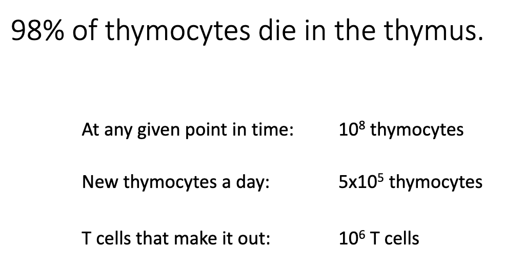
What proportion of thymocytes survive thymic selection?

  • 98% of thymocytes die.

  • Thymocyte numbers:

    • ~100 million total at any time.

    • ~50,000 new thymocytes enter per day.

    • Only ~1 million successfully exit the thymus.

  • Reasons for death:

    1. Failure to make productive VDJ rearrangements.

    2. TCR cannot bind MHC.

    3. TCR binds too strongly to self-peptides → negative selection.

<ul><li><p>98% of thymocytes die.</p></li><li><p><strong>Thymocyte numbers:</strong></p><ul><li><p>~100 million total at any time.</p></li><li><p>~50,000 new thymocytes enter per day.</p></li><li><p>Only ~1 million successfully exit the thymus.</p></li></ul></li><li><p><strong>Reasons for death:</strong></p><ol><li><p>Failure to make productive <strong>VDJ rearrangements</strong>.</p></li><li><p>TCR cannot bind <strong>MHC</strong>.</p></li><li><p>TCR binds too strongly to <strong>self-peptides</strong> → negative selection.</p></li></ol></li></ul><p></p>
46
New cards

Summarize T cell development in order.

  1. Bone marrow: T cell precursors form

  2. Thymus entry: proliferation (DN)

  3. β & α chain rearrangement → TCR

  4. Positive selection (cortex): keep MHC-binders

  5. Lineage choice: CD4 or CD8

  6. Negative selection (medulla): remove self-reactive

  7. Naïve single-positive T cells exit to lymphoid tissues

<ol><li><p><strong>Bone marrow:</strong> T cell precursors form</p></li><li><p><strong>Thymus entry:</strong> proliferation (DN)</p></li><li><p><strong>β &amp; α chain rearrangement</strong> → TCR</p></li><li><p><strong>Positive selection (cortex):</strong> keep MHC-binders</p></li><li><p><strong>Lineage choice:</strong> CD4 or CD8</p></li><li><p><strong>Negative selection (medulla):</strong> remove self-reactive</p></li><li><p><strong>Naïve single-positive T cells</strong> exit to lymphoid tissues</p></li></ol><p></p>