MICROPARA LE 4: MYCOLOGY
1/72
There's no tags or description
Looks like no tags are added yet.
Name | Mastery | Learn | Test | Matching | Spaced | Call with Kai |
|---|
No analytics yet
Send a link to your students to track their progress
73 Terms
fungi
microscopic eukaryotic organisms
first classified under kindome plantae, later separately classified under kingdom fungi
due to the presence of unique rigid cell wall, which is chemically diffrent from the bacterial cell wall
non photosynthetic
similar appearance w/ plants
80K species described: 400-medically important, <50 responsible for more than 90% of fungal infections of humans and animals
FUNGAL CELL WALL
rich in carbohydrates: polymers of acetylglucosamine “CHITIN”, forming a thick layer protecting inner organelles from the adverse external environment
CELL MEMBRANE
contains ERGOSTEROL; organized nucleus mores often reproduce by asexual spores (but can also produce sexually)
produce multi-celled hyphae or single-celled yeasts
classified according to morphology and taxonomy
fungal cell wall
boxes = antifungal drugs
distinct about ungi is the presence of CHITIN
fungi look like plants macroscopically, but looke more like animal kingdom microscopically (they are closely related to animals than plants)
morphological classification of fungi
yeast
moulds
dimorphic
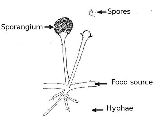
hyphae
tubular-like structure that compose a mold colony
help in the interexchange of cytosol and organelles between adjacent cells
may be septate or pauciseptate
may be pigmented or not
septae
cross walls present in some hyphae; singular: septa
nonseptae
lacking septa
hyaline
lacking pigment
molds do not have pigment; they are colorless and transparent
dematiaceous
containing dark pigment
chlamydoconidia
large, round spores in or on hyphae
conidia
asexual spores produced by molds with septa
mycelium
a colony, made up of rope-like filaments called hyphae
spores: conidia
produced in a CONIDIOPHORE
macroconidia/microconidia
spores or hyphae may be pigmented or not
not all can be found in every fungal species
chlamydospore
spores that grow on the hyphae itself (circles)
arthroconidia
bigger segments on the hyphae compared to the septae
sporangiospores
found int he sporangia connected to the hyphae
conidia are connected tot he conidiophore connected to the hyphae
sporangiophore
hyphae specialized in bearing sporangia
rhizopus and mucor
aspergillus and penicillium
these closely resemble conidiospores and conidia
mould
1. Eukaryotic Organisms
Moulds have complex cells with a true nucleus and membrane-bound organelles.
2. Multicellular (Filamentous)
Most moulds grow as filamentous hyphae, which form a network called mycelium.
These hyphae may be septate (with cross-walls) or coenocytic (without cross-walls).
3. Heterotrophic (Saprophytic or Parasitic)
Moulds cannot make their own food.
They absorb nutrients from dead organic matter (saprophytic) or from living hosts (parasitic).
4. Reproduction (Sexual and Asexual)
Asexual reproduction is most common and occurs through spores (like sporangiospores or conidia).
Sexual reproduction involves specialized spores like zygospores, ascospores, or basidiospores, depending on the fungal class.
5. Rapid Growth
Under favorable conditions (moist, warm, nutrient-rich), moulds can grow and spread quickly.
6. Cell Wall Made of Chitin
Unlike plants (which have cellulose), mould cell walls are made of chitin, a strong, flexible polysaccharide.
7. Thallus Structure
The vegetative body of a mould is called a thallus, which is composed of mycelium (mass of hyphae).
8. Colonial Appearance
Moulds grow in colonies that may appear fuzzy, cottony, or powdery, often in colors like green, black, white, or grey depending on the species.
9. Common Habitat
They thrive in moist, warm environments — soil, decaying matter, stale food, walls, etc.
10. Importance
Positive: Used in antibiotics (e.g., Penicillium), cheese production, decomposition.
Negative: Can cause spoilage, allergies, and fungal infections (e.g., Aspergillus, Rhizopus).
yeast
unicellular eukaryotic organisms (unlike molds which are multicellular)
appear smooth and mucoid on the media
aerobic organisms, but growth is enhanced in ANAEROBIC CONDITIONS
acquire energy from an organic compound by oxidation
a single cell produces ASEXUALLY through budding
the third “new cell” produces a daughter cell and each cell has a nucleus, cytoplasm, reserve food bodies and a vacuole
vacuole is one distinctive feature of the yeat with the budding
dimorphic fungi
exists in both mycelial and yeast forms in varying temperatures
37C
yeast like colonies grow best in this temperature
25C
mould like colonies grow best in this temperature
pathogenic
yeast forms are ____
saprophytic
mould forms are ___; loves to feed on dead plants and animal remains - this is why fungi are big factors and contributors to decomposition
Genus | Common Name | Use/Impact |
|---|---|---|
Penicillium | Blue/green mould | Antibiotics, cheese |
Aspergillus | Black mould | Allergies, aflatoxins |
Rhizopus | Bread mould | Spoilage, fermentation |
25C
at ___, the forms are mycelial, or they look like molds
37C
at ___, the forms look like yeast
opportunistic fungi
these are the different forms of fungi that we can see, and the fungi that can cause diseases or mycosis
opportunistic fungi
first, yeast cells. we have the parent yeast cell; it buds to produce daughters. during budding it can produce pseudohyphae or a germ tube
pseudohyphae - chains of elongated yeast cells that remains attached after budding
germ tube - a tube like outgrowth from a yeast cell that does not have constriction at the base. it is a sign of yeast transitioning to a more invasive form, mould.
there are also the mould forms: candida and hyphae or hypha
cryptococcus is somewhat unique because of the presence of capsules
a genus of yeast-like fungi
cryptococcus neoformans - medically important which causes infections in the brain or lungs
zygomycetes
lower fungi with non-septate hyphae produce sporangiospores (asexual spores)
the spres are inside the sporangium, so they are called sporangiospores
ascomycetes
produces septate hyphae and ascospores. (sexual spores are present inside the sac or ascus)
inside the ascus, there are the ascus spores
basidiomycetes
produce septate hyphae and basidiospores (sexual spores are present inside the basidium)
the basidium looks like long, goblet cells, at the top are the basidiospores
the goblets are called basidium.
basiosopores are the name of the spores found on the basidium
deutromycetes
fungi imperfecti
produces septate hypahe and cannot be classified into sexual or asexual because their sexual state is unknown
share common features with ascomycetes
most medically important fungi belongs to this group
taxonomic classifications of spores
1. Zygomycetes
Definition: Zygomycetes are a class of fungi that reproduce sexually through the formation of zygospores, which are thick-walled resting spores formed by the fusion of two similar-looking gametes (isogamous). They also reproduce asexually using sporangiospores inside sporangia.
Structure: Their hyphae are usually coenocytic (no cross-walls).
Example: Rhizopus stolonifer — commonly known as black bread mould.
2. Ascomycetes (Sac Fungi)
Definition: Ascomycetes are fungi that reproduce sexually by forming ascospores inside a sac-like structure called an ascus. They also reproduce asexually through conidiospores (conidia).
Structure: They have septate hyphae (with cross-walls).
Example: Aspergillus species — some cause lung infections; others are used in food fermentation.
3. Basidiomycetes (Club Fungi)
Definition: Basidiomycetes reproduce sexually by forming basidiospores on a club-shaped structure called a basidium. These fungi are often large and visible (like mushrooms).
Structure: Their hyphae are septate, and they often form fruiting bodies (basidiocarps).
Example: Agaricus bisporus — the common edible mushroom you find in groceries.
4. Deuteromycetes (Fungi Imperfecti)
Definition: Deuteromycetes are fungi without a known sexual stage. They are classified based on their asexual reproduction, usually through conidia. Many medically important fungi belong here.
Structure: Usually have septate hyphae.
Example: Candida albicans — a dimorphic fungus that causes candidiasis in humans.
zygomycetes - rhizopus stolonifer
A class of fungi that reproduce sexually by forming zygospores, which are thick-walled spores resulting from the fusion of similar gametes. They also reproduce asexually through sporangiospores produced inside sporangia. Their hyphae are usually coenocytic (lacking cross-walls).
Example: R______ s________ (black bread mould)
ascomycetes - aspergillus spp.
Definition: Fungi that reproduce sexually by forming ascospores within a sac-like structure called an ascus, and asexually through conidiospores (conidia). They have septate hyphae.
Example: A_________ spp. (some species cause lung infections or are used in fermentation)
basidiomycetes - agaricus bisporus
Definition: These fungi reproduce sexually by forming basidiospores on a basidium (a club-shaped structure). They typically form large fruiting bodies like mushrooms and have septate hyphae.
Example: A_______ b________ (common edible mushroom)
deutromycetes - candida albicans
Definition: A group of fungi with no known sexual stage, classified based on asexual reproduction, usually through conidia. Most have septate hyphae and include several medically important fungi.
Example: C______ a________ (causes candidiasis)
sexual spores
zygospores - found between hyphae
ascospores - found inside the ascus; if ascus bursts, then the ascospores will be dispersed
basidiospores - basidium looks like goblet cells; basidiospores, on top of it, what connect the basidiospores and basidium is the sterigmata; sterigmata looks like small feet or pods connecting the spores to the basidium
asexual spores
vegetative spores
formed by budding (yeast cells)
formation of speta in hyphal filament (moulds)
folding and thickening of hyphal filaments (resulting in thick-walled spores)
aerial spores
conidiospores
microconidia
macroconidia
sporangiospores
labels
conidiospore
phialides
vesicle
conidiophore
septate hyphae
advantages of fungi
decomposition - carbon cycle = plants grow
fermentation
food
pharmaceuticals - fungal metabolites are used in antibiotic drugs like penicillin = came from penicillium spp.
model research organisms - fungi are the basis of genetic research becaue of their simle eukaryotic form. before they proceed to the multi-celled, complicated-complex forms like humans, they start with the simpler ones, like fungi and bacteria
disadvantages of fungi
tree disease
crop disease
food spoilage - bread molds, food molds, expiration date molding
wood and timber degradation
mycoses or fungal infection
mycoses
they are fungal infections.
diseases caused by fungi
causes of mycoses to humans
inhalation of spores - enter through the lungs, gets localized, causes respiratory infection
inoculation of spores - through cuts, localized i the skin
alteration of normal flora - due to overconsumption of heavy dose of antibiotics (alter or kills normal flora that acts as a physical barrier, this enhancing entry of pathogenic fungi)
suppressed immune system - patients undergoing chemotherapy, in steroids due to transplantation, HIV, or diabetics
4 subtypes of mycosis
superficial mycosis
subcutaneous mycosis
deep mycosis
opportunistic mycosis
superficial mycosis
externally localized on th layers of the skin, hair, nails and grow well on dead layers
classified into two: surface mycoses and cutaneous mycoses
tinea versicolor
caused by MALASSEZIA GLOBOSA
is a yeast form of fungi that live on the skin i few numbers but multiply dururing adverse conditions leading to skin infections
they can exist as a normal flora, but during adverse conditions, and with te several causes mentioned earlier, it can lead to skin infection
tinea versicolor
pathogenesis: malassezia globosa
transmission
transmitted through contact, through adherence and invasion of fungi
become apthogenic when they change morphology from yeast to mycelium (mold colony)
dimorphic in nature
pigmentation
due to azelaic acid, which is the econdary metabolite produced by fungi. it reacts on the melanin pigments that is found on the skin, thus producing pigments (patches)
s/s
mild patches on cehst, back, neck, and arms
patches lead to pigmentation
discoloration starts spreading in untreated conditions
dryness of the skin
itching of the skin
tinea versicolor
lab diagnosis: malassezia globosa
specimen
skin scrapings of the lesions (superficial layers is enough)
direct microscopy
KOF (potassium hydroxide) wet mount. skin scraping is fixed on slide with 20% KOH, and then the skin scraping is mixed into that wet mount, covered with a cover slip and tehn short unbranched hyphae can be observed
culture:
saubrouraud’s dextrose agar @ 32-37C
round and smooth colonies
after the culture, it is then mounted on lactophenol cotton blue wet mount to further see the morphology of the malassezia species
treatment for tinea versicolor
topical applicaiton of antifungal ointments
WHITEFIELD’S OINTMENT - composed of benzoic acid, salicylic acid, ciclopirox olamine, and tincture of iodine
sulfur containing ointment
oral antifungals - triazole, itraconazole, ketoconazole. used in more severe cases.
tinea nigra
caused by HORTAEA WERNICKII
dimorphic fungi taht exists in both yeast and hyphal forms
responsible for asymptomatic mycoses
saprophytic in nature and found on dead and decayed materials
tinea nigra
pathogenesis: hortaea wernickii
entry
inoculation of fungus through cuts and wounds
enter, localize, and cause superficial infection on palms and foot soles
from the name itself, you can deduce that the color is darker, compared to tinea versicolor
halotolerant
it is tolerant to ionic stress, or the ability of tolerance is the ability of an organism to grow at salt concetrations higher than those for growth
able to survive in human tissues by ccumulating and utilizing melanin. it can survive on areas of the skin that actually are more prone to sweat, like palms and foot soles
because of its appearance, there are times that this mycoses is confused witht he type of skin cancer or melanoma
s/s
mild patches on palms and foot soles
patches appear brownish or black in color, irregular in shape
scale-like skin growth
itching
if asymptomatic, you cannot see any of these symptoms. but there is funal present still on the skin
tinea nigra
lab diagnosis: hortaea wernickii
specimen
skin scrapings of the lesions (superficial)
direct microscopy
KOH wet mount, budding yeast cells on branched hyphal elements
culture
SDA, at 32C -37C, 3 weeks: dematiaceous colonies that become velvety with age
in culture, they appear mycelial bold-like forms, unlike the malassezia that appears yeast-like
very slow forming fungal colony (coin-sized)
lactophenol cotton blue
further testing of isolated colonies
this is the wet mount usinf LPCB because it is blue and it can be seen more clearly, compared to the previous slide which contains KOH wet mount
these fungal colonies are that much clearer
you can see the form is fully mycelial
previous slide was not that clear because there is a mixture of yeast cells and mycelial hyphal elements - like in the middle transformation
since the LPCB gets its specimen formt he colony grown in culture, you can see the form is now full mycelial or hyphal
tinea nigra
treatment: hortaea wernickii
topical applicaiton of antifungal ointments
good hygiene
avoiding exposure to moist and dirty places
piedra
this affects the hair
white p___
black p___
white piedra
caused by TRICHOSPORON BEIGELII
asexually reproductive
part of normal flora
yeast-like fungi that change to septate hyphal filaments
dimorphic
white piedra
pathogenesis
trichosporon beigelii
close contact with the infected person’s towel, soap, comb, etc.
white nodules: spores transmitted by contact
symptoms
acclimation of white lump of yeast cells on the hair follicles of the head, beard, even pubic hair
you can directly egt this and place t on the wet mount and immeadiately see the yeast cells
hair loss
itching
black piedra
caused by PIEDRAIA HORTAE
asexual
transmitted through contact
black piedra
pathogenesis
found on thehair shafts of the beard and scalp
symtoms
brown to black nodules firmly attached to the hair shaft
nodules: ascostroma (asci and ascospores)
mostly asymptomatic
hair breakage in extreme cases
black piedra
lab diagnosis
specimen: hair
direct microscpoy: KOH wet mount hair nodules
culture: sabourand dextrose agar
LPCB: further testing of isolated colonies can be done through LPCB
treatment
topical application of antifungal ointments
imidazoles and selenium sulfide
amphotericin b ointments
using separate towels, soaps and combs
subcutaneous mycosis
causes disease in living tissue leading to tissue damage
three types:
mycetoma
chromoblastomycosis
rhinosporidiosis
mycetoma
caused by ACTINOMYCETES (actinomycetoma) or filametous fungi: EUMYCETOMA
persistent subcutaneous granulomatous infection affecting the foot, and as infection progresses, the bones
aka MADURA FOOT
not just causde by fungi but also bacteria such as the ACTINOMYCETES
pathogenesis
transmission
inoculation of fungus through cuts or wounds from soil
enters tissue, localize, and replicatie inside living cells
spores from clumps inside cells resulting in granules
granules may vary in color, depending on contributing agents
s/s:
itching
swelling in the area of itching
pus formation
ulceration and nodules
pus expulsion
irrigation in area of infection (watery irrigation)
disfiguration of the leg
mycetoma
lab diagnosis
specimen: pus exudates
direct microscopy
gram staining - for identification of actinomycetes (gram positive)
KOH wet mount hair nodules for identification of eumycetes (fungi)
culture
after the direct microscopy, if it is found to be a bacteria, then it can be grown on BA or NA
if fungi, then it is grown on sauboraud’s dextrose agar SDA
treatment
surgery and removal of abcess at early stage with proper administration of oral antifungal and antibacteruak drug therapy (to prevent the further spread of the fungi)
ACTINOMYCETOMA - antibacterial drug; rimfampicin, dapsone and sulphonamides
EUMYCETOMA - oral antifungal; itraconazole and ketoconazole
treatment should be prolonged for complete eradication of pathogens.
chromoblastomycosis
caused by five different vegetative fungi (pathogens):
phialophora verrucosa
fonsecaea compacta
fonsecaea pedrosoi
rhinocladiella aquaspersa
cladophialophora carrionii
cause persistent infection that slowly progress and form granulomatous lesion
leads to accumulation of ketinocytes in epidermal layer resulting in sloughing skin
caused by ACTINOMYCETES (actinomycetoma) or filamentous fungi - EUMYCETOMA
aerobic, filamentous fungi commonly found ind soil
chromoblastomycosis
pathogenesis
chronic subcutaneous infection
transmission: cuts or wounds, primarily in leg region
upon entry, fungi spreads adn invades tissue very slowly (due to growing capacity)
very resistant to immune cells due to its melanin cell wall
affects tissue:
forming hyperplasia of epidermis producing nodules with pus formation
distinct feature: painless
drains lymphatics
severe: damage organs
it can take years undetected. it starts with nodules, if not treated, then it can drain the lymphatics, and cause organ damage
s/s:
itching are of entry
swelling with pus formation
nodule formation as infecting drains to lymph
ulcerations
chromoblastomycosis
lab diagnosis
specimen: pus cells or skin scrapings
methods:
KOH wet mount
histopathological analysis: check multinucleated giant cells with granules and sclerotic bodies inside the outside cells
culture: SDA, brown or black moldy colonies
treatment
surgery and removal of pustule and proper oral treatment heat threapy (early stages)
antifungal agents:
flucytosine
ketoconazole
itraconazole
rhinosporidiosis
caused by RHINOSPORIDIUM SEEBERI
granulomatous infection of the nose, eyes, and mouth
first classified as sporozoan parasite, but later classified as lower fungi
still shares morphological similarities with aquatic parasites
ccording to DNA sequencing, they belong to fish parasites (which is right in between animala nd fungal division)
pathogenesis
fishermen adn washermen are mostly affected due to constant contact with water habitats
mode of transportation:
entry thru cuts and wounds
also enters thru nasopharyngeal route or even eyes and external genitalia
s/s:
accumulation of large mass of cells that hangs out as separate layer
pus accumulation leafing to foul sme;;
breathing difficulty due to protrusion of layer
lab diagnosis:
skin biopsy: histopathological analysis
KOH wet mount: for endospores within sporangium
treatment:
surgery
intravenous administration and AMPHOTERICIN B and DAPSONE
deep mycosis
aka SYSTEMIC MYCOSIS
involve both pathogenic and opportunistic fungi yeast form and start invading tissue
pathogenic fungi: gain entry
oppportunistic fungi: suppresses the immune system system
histoplasmosis
systemic mycosis caused by HISTOPLASMA CAPSULATUM
saprophytic fungi mostly found in soil
dimorphic fungi
solo-celled microconidia
colony: produces moldy white colonies
affects respiratory syste causes pulmonary infection
pathogenesis
intracellular pathogen
mot: inhalation: molds -convert→ yeast
yeast cells engulfed by alveolar macrophages
yeast replicates inside and use th host cells to travel around the body and invade other areas like liver, spleen, and lymph nodes
results in pulmonary infection in men, children and immunocompromised pxs (HIV-AIDS and undergoing chemotherapy)
s/s:
dry cough
body pain
high fever
restlessness
lymphadenopathy
fatal in sever cases affecting liver, eyes, and glands
lab diagnosis
specimen: sputum, urine, throat swab, bone marrow apirator
direct microscopy: histopathological staining or geimsa staining to view intracellular yeast (biopsy specimen)
KOH will nor work
culture: SDA (for moldy colonies), blood agar (for yeast colonies
serology: complement fixation test and enzyme immunoassay
does not involve th efungi itself
involves antibody-antigen reaction
reacts to blood antigen
one of the fastest tests due to fungal colonies needing to take a few days for culture to develop
treatment
ITRACONAZOLE
AMPHOTERICIN B
relapses may occur for immunocompromised patients (prolonged treatment with itraconazole)
oppotunistic mycosis
ifection tha occurs in immunocompromised pxs
depends on the load of organism and the virulence caused to the host
CANDIDIASIS
CRYTPOCOCCOSIS
candidiasis
caused by candida albicans, candida tropicalis, candida parapsilosis and candida krusei
part of the normal flora of the skin and GIT
cells are oval-shaped and divide by budding , which forms into pseudohyphae
some species of candida albicans are DIMORPHIC
Symptoms by Affected Area:
Vaginal (Yeast Infection):
Itching and soreness in the vaginal area.
Abnormal vaginal discharge (often thick and white).
Burning sensation, especially during urination.
Redness and swelling of the vulva.
Oral (Thrush):
White or yellowish patches on the tongue, inner cheeks, gums, or throat.
Redness and soreness in the mouth.
Cotton-like feeling in the mouth.
Loss of taste.
Pain or difficulty swallowing (if it spreads to the throat).
Cracking and redness at the corners of the mouth.
Skin :
Rash (often red and itchy).
Scaling or flaking of the skin.
Small pustules (pus-filled bumps).
Skin may become cracked and sore.
Areas like skin folds, underarms, groin, and between fingers are prone to infection.
Esophageal :
Pain and difficulty swallowing (dysphagia).
Invasive :
Fever and chills.
Symptoms may vary depending on the organs or bloodstream affected.
Other General Symptoms:
Fatigue.
Bloating, gas, constipation, or diarrhea.
Brain fog (difficulty concentrating).
Skin rashes.
Nutritional deficiencies (linked to Candida overgrowth).
Cravings for sugary or high-carbohydrate foods.
In infants, persistent diaper rash can be a sign.
candidiasis
Symptoms by Affected Area:
Vaginal (Yeast Infection):
Itching and soreness in the vaginal area.
Abnormal vaginal discharge (often thick and white).
Burning sensation, especially during urination.
Redness and swelling of the vulva.
Oral (Thrush):
White or yellowish patches on the tongue, inner cheeks, gums, or throat.
Redness and soreness in the mouth.
Cotton-like feeling in the mouth.
Loss of taste.
Pain or difficulty swallowing (if it spreads to the throat).
Cracking and redness at the corners of the mouth.
Skin :
Rash (often red and itchy).
Scaling or flaking of the skin.
Small pustules (pus-filled bumps).
Skin may become cracked and sore.
Areas like skin folds, underarms, groin, and between fingers are prone to infection.
Esophageal :
Pain and difficulty swallowing (dysphagia).
Invasive :
Fever and chills.
Symptoms may vary depending on the organs or bloodstream affected.
Other General Symptoms:
Fatigue.
Bloating, gas, constipation, or diarrhea.
Brain fog (difficulty concentrating).
Skin rashes.
Nutritional deficiencies (linked to Candida overgrowth).
Cravings for sugary or high-carbohydrate foods.
In infants, persistent diaper rash can be a sign.
cryptococcosis
caused by:
CRYPTOCOCCUS NEOFORMANS
CRYPTOCOCCUS GATTII
mostly found in soil, feces of birds
yeast-like fungi with resistant polysacchardie capsules
can be cultures using fungal or bacteriological media, 24 hrs at 37C
colony: produces mucoid white colonies (due to capsule)
PATHOGENESIS
● Mode of transmission: inhalation of fungal cells
● Enter respiratory system, affecting circulating
cells and compromises immune system by
resisting immune reactions
● Multiply and infect other parts of the body,
especially the CNS causing meningoencephalitis
(inflammation of brain tissues and meninges)
● Symptoms:
○ Fever
○ Headache
○ Body pain
○ Flu symptoms
○ CNS affected in severe causes, causing
encephalitis
LAB DIAGNOSIS
● Specimen collected: Sputum, Blood (severe
systemic cases), Throat swab, CSF (suspected
encephalitis)
● Direct microscopy
○ India Ink: special staining used specifically for
showing the capsule of Cryptococcus spp.
● Serology
TREATMENT
● Amphotericin B and flucytosine
○ Combination of both for severe cases (ie:
immunocompromised patients)
○ Prolonged treatment provided to avoid
recurrent infections