Theme 1 Module 1: The structure of a cell
1/26
Earn XP
Description and Tags
The learning objectives in this module are to: Identify that we are mosaics of cell types derived from different genetic origins. Recognize the 4 core macromolecules that make up all living cells. Describe the composition and function of cell membranes. Explain how fluidity and transport mechanisms can influence the movement of substances across the cell membrane.
Name | Mastery | Learn | Test | Matching | Spaced | Call with Kai |
|---|
No analytics yet
Send a link to your students to track their progress
27 Terms
If every human starts as a single fertilized egg, how do we end up with an estimated 10 trillion cells as adults—and what exactly counts as a “cell” when we try to answer this question?
Slide 7: How many cells are in your body?
Every one of us began as a single cell just like this cell here - the result of the union of an egg and sperm.
As we watch this movie of a fertilized egg under the microscope, we can see mitotic cell division creating two cells, then four cells. Eventually, continued cell division allows us to reach an estimated 10 trillion cells as an adult.
Each cell shares the same genetic heritage as the original fertilized cell; however, these cells differ in function.
They may be muscle cells, skin cells, kidney cells and neurons. These are all eukaryotic cells, a classification that means the cells have a nucleus that contains most of the genetic material in the cell. But wait, if we are going to do this experiment, to count the number of cells in our body, we need a common definition of the term cell. What do we mean by the term "cell"? Do we need to identify and count only eukaryotic cells, or do we need to identify prokaryotic cells as well? What is the difference? Does this matter? If we look at the cells of our body, what do we find?
What are prokaryotic cells? Are all the cells on/in us carry genetic heritage with our own eukaryotic cells?
We carry a lot of passengers who do not share a common genetic heritage with our own eukaryotic cells. Most are prokaryotic cells, a categorization that includes bacterial cells. Prokaryotic cells are cells that do not contain a true nucleus.
what many bacterial cells are in the human body? How much of our mass does it make up?
Slide 9:
There are an estimated 10 times as many bacterial cells in the human body as our own eukaryotic cells. These cells make up 2 to 3 % of our body mass.
If you weigh 150 pounds, then 3 to 4.5 pounds (or 1.4 to 2 kg) of this weight comes from bacterial cells. Most of these cells are harmless, and in fact they are an important part of our bodies. They provide essential functions throughout our physiological systems. Some of these cells are potentially harmful but are contained or managed by our immune system to keep them under control.
What is microbiome? Microorganisms? how many microbes on and in a healthy body?
Slide 10: Human microbiome
The term microbiome is used to describe the populations of microbiotic organisms (microorganisms or microbes) within our bodies. Microorganisms are, by definition, organisms that are not visible to the naked eye, but only visible under a microscope.
The microbiome includes prokaryotic bacteria, but also small eukaryotic organisms. Bacterial cells are found in nearly every part of our body. They are on our skin and on our teeth; they are in our digestive system and respiratory tract.
Microbiome: A population of microorganism or microbes
Slide 11:
There may be about 10,000 distinct species of microbes on and in a healthy human body. Let's look at some examples.
What is Streptococcus salivarius?
Slide 12:
Streptococcus salivarius is a normal inhabitant of the upper respiratory tract and oral cavity.
It is a member of a collection of bacteria that contribute to the formation of dental plaque. It is also one of the first microbes to colonize a germ-free newborn's oral cavity and gastrointestinal tract
what is Staphylococcus haemolyticus?
Staphylococcus haemolyticus resides on our skin. If it stays there it is harmless, but it can be pathogenic if it gets inside the body. Infection commonly leads to activation of the immune system.
what is Bacteroides thetaiotaomicron?
Slide 14:
Bacteroides thetaiotaomicron is a predominant intestinal bacteria.
It makes enzymes that are useful in the breakdown of plant materials that we ingest, such as oat fibre.
Since we find both eukaryotic and prokaryotic cells in our own bodies, we will discuss the basic structures and macromolecules of all cells, keeping in mind throughout the course, the similarities, differences and relationships between both the eukaryotic and prokaryotic worlds.
What is a cell?
A cell is a membrane-bound structure containing macromolecules.
A cell is a membrane-bound structure containing macromolecules, These macromolecules fall into four classes that are used in the cell
nucleic acids make up the hereditary material of the cell and are found in the DNA; chromosome/nucleoid. The information encoded in the DNA is converted into functional products of cells (RNAs and eventually proteins);
proteins make up the structural elements of the cell (flagellum on a bacterium) and perform metabolic activities (ribosomes that synthesis enzymes);
polysaccharides or carbohydrates are also important structural components of many cells (bacterial capsule or plant cell wall) and sources of stored energy (foods made from wheat or rice).
phospholipids are the primary component of the cell membrane – an essential component of our definition of a cell.
What is one of the most important properties that a cell must have?
One of the most important properties that a cell must have is the ability to separate its internal environment from the external surroundings.
This is important to protect the cell from damaging toxins, to ensure that the cell allows entry of important compounds, and disposal of metabolic wastes. How is the cell able to do this? As we progress through this lecture, we will find answers to this question by answering a subset of other questions:
What components form the structure of a cell membrane?
How do these components "behave" when they form membranes?
What kind of barrier is the cell membrane: what can pass through the cell membrane and what cannot?
the cell membrane is made up of lipids, how are they structured?
The cell membrane of a cell is made up of lipid macromolecules. Each of these macromolecules has hydrophobic and hydrophilic properties that allow "stacked" lipid bilayers to form. These lipid bilayers have a central "water-hating" hydrophobic core, and peripheral "water-loving" hydrophilic surfaces. With the structural importance of separating the cell interior from the exterior, you would probably expect that the cell membrane would be thick and rigid (like the wall that separates your internal home from the exterior world), but the opposite is true! Cell membranes are very thin, ranging from 5-10nm in thickness. So thin, in fact that 10,000 membranes stacked on top of each other is equal to the thickness of a piece of paper!
Phospholipids are the main component of a cell membrane.
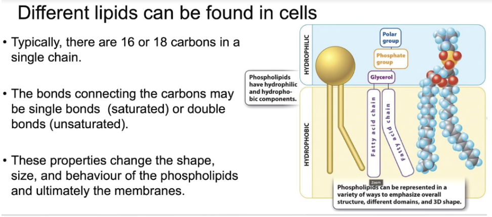
Each phospholipid consists of a glycerol molecule that is linked to a phosphate and 2 fatty acids, often called fatty acid tails.
Tail: These hydrophobic tails make up the hydrophobic core of the bilayer that we just saw.
Head: The hydrophilic head groups (glycerol plus phosphate) form the hydrophilic surface that faces the aqueous environments outside and inside the cell.
Phospholipids are said to be amphipathic because they have both a hydrophobic and a hydrophilic part.
Phospholipid tails consist of a pair of hydrocarbon chains. Typically, there are 16 or 18 carbons in a single chain. The bonds connecting the carbons may be single bonds (saturated) or double bonds (unsaturated). These properties change the shape, size, and behavior of the phospholipids and ultimately the membranes.
How is cholesterol structured?
Another important component of cell membranes are steroids such as cholesterol. These lipids are characterized by a 4-hydrocarbon ring structure. Like phospholipids, these types of lipids also contain a hydrophilic head group and a hydrophobic tail.
What are the 2 structures of lipids?
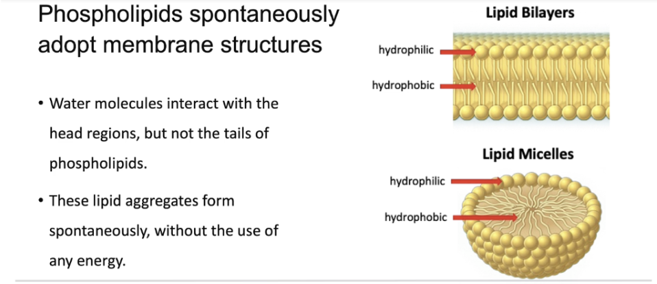
Due to their amphipathic nature, phospholipids form unique structures that are critically important for the cell. Water molecules interact with the head regions, but not the tails of phospholipids. As a result, tails will interact with tails to form hydrophobic cores and the hydrophilic heads interact with water in the extracellular or intracellular environment.
In addition to forming lipid bilayers that surround the cell and make up the membranes of various cell organelles, phospholipids can aggregate and form lipid micelles. These kinds of structures become important for absorption of fat-soluble vitamins and complex lipids in the human body. What is remarkable is that these lipid aggregates form spontaneously, without the use of any energy.
Describe how Cell membranes are '“fluid”
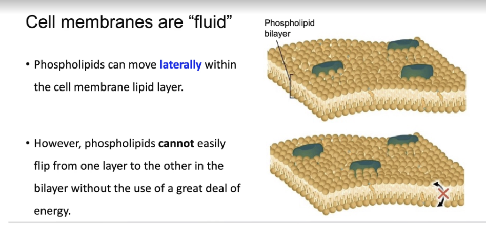
If we look in closer detail at the cell membrane, what we find is that phospholipids are not stationary because membranes are fluid.
In other words, they can move laterally within one layer of the lipid bilayer that is, they can move left/right, forward/backward.
However, phospholipids cannot easily flip from one layer to the other in the bilayer without the use of a great deal of energy.
How can fluidity of membranes be affected? (4 factors)
Factor 1) The number of carbons in a hydrocarbon chain varies: typically, there are 16 or 18 carbons. The longer chains pack together more tightly than the shorter chains, reducing the fluidity of the membrane.
Factor 2) Double bonds within a hydrocarbon tail produce kinks or bends in the chain. This has the effect of pushing neighboring phospholipids further apart and increasing fluidity. In this figure, we can see an example of how unsaturated fatty acid tails can lead to "kinks" in hydrophobic tails that affect overall permeability.
Factor 3) External environmental factors such as temperature can also influence membrane permeability.
Higher temperatures promote fluidity, while lower temperatures decrease fluidity.
Interestingly, cold-adapted organisms tend to have more UNSATURATED phospholipids in their membranes that help to maintain fluidity.
Factor 4) Steroids such as cholesterol are found in the membranes of every cell in our body, making up about 50% of the molecules found in the bilipid membranes. A bilipid membrane containing just phospholipids is actually TOO fluid. Cholesterol molecules constrain fluidity of the membrane by packing closely to neighbouring phospholipids.
At low temperatures, phospholipid bilayers behave like many other fats; they begin to solidify. At these lower temperatures, cholesterol helps to maintain fluidity by keeping the phospholipids apart from one another.
Cholesterol acts as a fluidity buffer in cell membranes, making them less fluid at high temperatures (restraining phospholipid movement) and preventing them from becoming too rigid at low temperatures (disrupting tight packing)
Recall that the ability of phospholipids to move is often referred to as….
membrane fluidity
What is a lipid raft?
Slide 24:
This animation is a short clip from the movie called. The Inner Life of a Cell. This is a wonderful animation that best represents the many diverse and dynamic properties of the membranes of a cell. Here we focus on a close-up of the phospholipid bilayer. Recall that the ability of phospholipids to move is often referred to as membrane fluidity.
We can also see that within membranes there are domains that demonstrate different degrees of fluidity, some domains are more fluid while others are less fluid.
In this case, the region of lower fluidity is called a lipid raft and it can sequester or hold macromolecules together in the membrane. These rafts are found to gather proteins involved in the same metabolic pathway or a collection of receptors on the surface of the cell. What factors contribute to this variation in fluidity? We can see that the region of the lipid raft is taller, this is due to the longer phospholipid hydrocarbon tails. The phospholipid tails are straight rather than kinked because they are saturated. This allows the phospholipids to pack together lowering the fluidity making it possible to hold the macromolecules in the raft. A higher concentration of cholesterol decreases fluidity within the microdomain of the lipid raft.
How can fluidity change the permeability of a membrane?
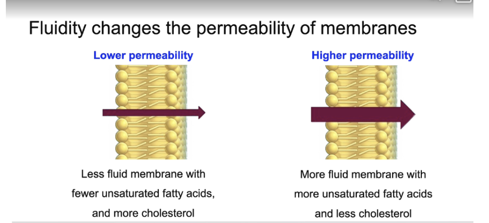
Macromolecules can move laterally within the cell membrane. The fluidity of the cell membrane also allows for transmembrane movements which includes movement of substances from the exterior to the interior of the cell and the reverse. Keep in mind that changing the fluidity will alter how much and how quickly a substance will move across the membrane. As the figure illustrates, regions of the membrane high in saturated fatty acids such as we saw with the lipid raft are less permeable than regions of a membrane that contain a high amount of unsaturated fatty acids.
What substances can pass through the phospholipid bilayers through diffusion?
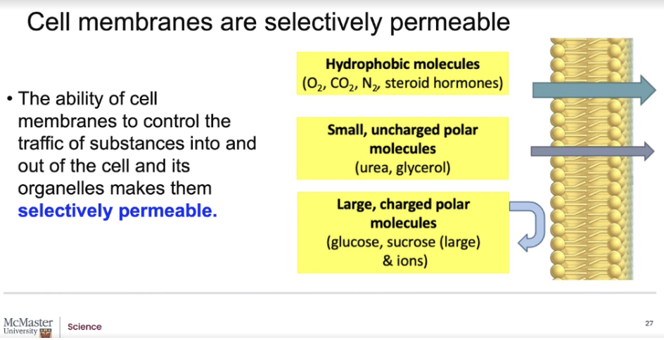
The ability of cell membranes to control the traffic of substances into and out of the cell and its organelles makes them selectively permeable.
Small molecules and ions can cross the membrane along a concentration gradient (that is, from areas of high to low concentration) through the process of diffusion.
In general, small and non-polar molecules or hydrophobic molecules can pass through the phospholipid bilayers relatively quickly, but CHARGED and LARGER POLAR substances have great difficulty in moving across the lipid bilayer of the cell membrane...if at all! So how do we get lipid-insoluble molecules and ions across the cell membrane?
Does the cell membrane only have lipid macromolecules?
No!
Cell membranes are not exclusively made up of lipid macromolecules. They are actually a "mosaic" of lipids, proteins and carbohydrates. Cell membranes always contain proteins, and most also contain carbohydrates that can be attached to lipids or proteins. As illustrated in this short movie, we see a bare lipid bilayer with attached sugar residues, glycolipids, and interspersed steroid molecules such as cholesterol. Added here are various types of protein channels that span the entire width of the lipid bilayer. These protein channels allow efficient transport of substances across the membrane.
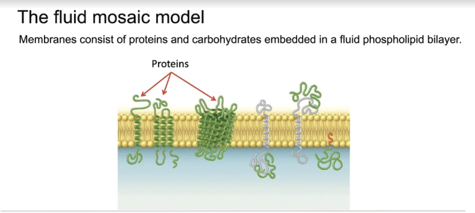
As seen in this figure, membrane-associated proteins can be attached to the cell membrane on the interior or the exterior of the cell, or actually embedded in the phospholipid bilayer. When considering movement of molecules in and out of the cell, it is the transmembrane (or integral membrane) proteins embedded within the cell membrane that enable the transport of hydrophilic molecules across the cell membrane. The Fluid Mosaic model states that membranes consist of proteins and carbohydrates embedded in a fluid phospholipid bilayer. A scientific model is a testable idea about the way that a system works. A model makes predictions about natural phenomena. Here, the Fluid Mosaic model predicts certain properties of the cell membrane including the ability of membrane components to move laterally. These predictions are consistent with experimental observations made over the last 30 years.
Unit 5, We have seen that substances can move through a membrane, can they be transported across a membrane?
What are the 3 types of transport?
Types of transport
1) Passive/simple diffusion: from areas of high to low concentration (down a gradient)
The first type of transport is passive or simple diffusion, and it involves the movement of small molecules in the direction of a concentration gradient. The process involves the molecule coming in contact with and crossing the phospholipid bilayer into or out of the cell.
2) Passive transport- facilitated diffusion: from areas of high to low concentration (down a gradient)
Passive transport is also the movement of small molecules with the concentration gradient but involves proteins embedded in the cell membrane and does not require energy
3) Active transport: from areas of low to high concentration (against a gradient) and fueled by the energy from the hydrolysis of ATP
The third type of transport is active transport, this is the movement of molecules against a concentration gradient. It involves proteins embedded in the cell membrane which require energy from adenosine triphosphate or ATP to drive the transport. We will now take a closer look at each of these three transport mechanisms.
Now we will look more into the types of passive diffusion.
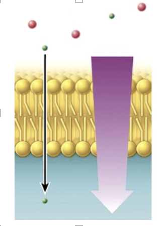
The first type of passive transport generally involves the movement of lipid-soluble molecules, gases, uncharged polar molecules and even some water across the cell membrane without the need of any energy. This is called Passive or SIMPLE diffusion. The process involves the molecule coming in contact with and crossing the phospholipid bilayer into or out of the cell. As seen in this figure, the substances are moving from an area of higher concentration (the exterior of the cell) to an area of lower concentration (the cytosol or interior of the cell).
Simple diffusion:
A solute molecule comes in contact with and crosses the lipid portion of the plasma membrane into the cell.
Movement along a concentration gradient (higher to lower).
No energy is required
Slide 33:
The most common way for substances to move passively across the cell membrane generally requires the help of transmembrane transport proteins that span the width of the phospholipid bilayer. This type of transport mechanism comes in handy since the phospholipid bilayer is relatively impermeable to ions and most hydrophilic molecules. Like passive diffusion, facilitated diffusion of substances along a concentration gradient through transmembrane transporters requires no input of energy.
Facilitated diffusion (form of passive transport):
Requires protein molecules that assist in the transmembrane movement of solutes.
Movement along a concentration gradient (higher to lower).
No energy required
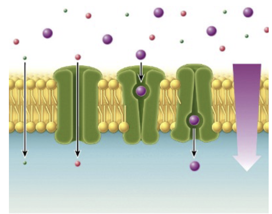
Let's review the main types of passive transport with a video. Remember that passive transport requires no expenditure of energy. With Passive or simple diffusion, gases, small molecules and even some water cross directly through the phospholipid bilayer from an area of higher to lower concentration to move into or out of a cell. With facilitated diffusion, transport proteins provide a hydrophilic core that allows large hydrophilic molecules and most water to move across the cell membrane. Once again, recall that this is a form of passive transport and it does not require the use of energy due to movement along a concentration gradient. Passive transport is the mechanism by which most water is able to cross the cell membrane. Let's take a closer look.
What is Osmosis?
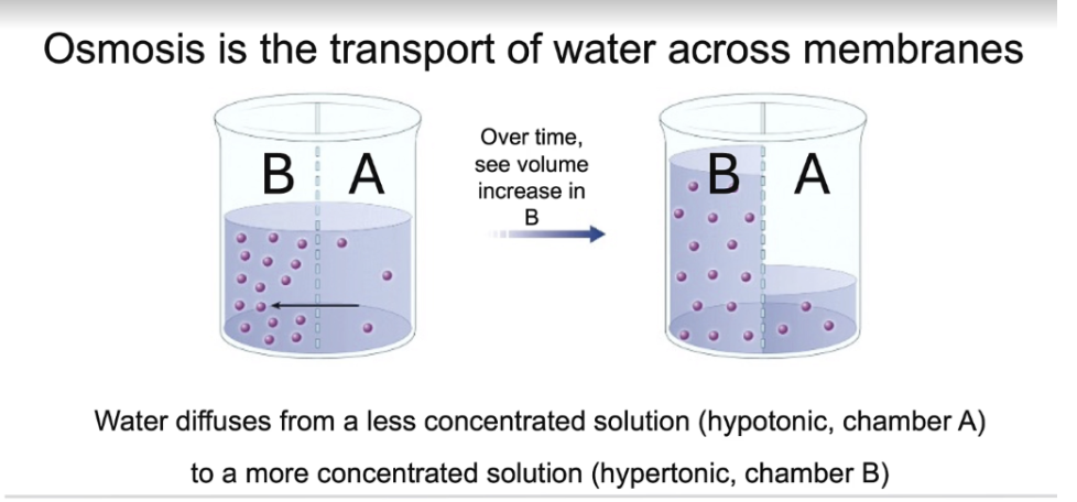
The movement of water across a membrane is a type of passive transport called osmosis. When considering the movement of water during osmosis, the rule of thumb is to focus on the concentration of solutes in solution on either side of a selectively permeable membrane. Seen in this figure is a beaker with a semi-permeable membrane that allows movement of water from one side to the other, but not solutes. As we see, in the starting condition, there is a higher concentration of solute molecules in chamber B compared to chamber A. As a result, with time, there is a net movement of water from chamber A to chamber B in the process of equilibrating the osmotic concentration on either side of the membrane.
What are Aquaporins?
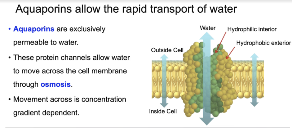
The same principle applies for movement of water across selectively permeable biological membranes. Although small amounts of water can move slowly across the cell membrane, water is able to move across at a much faster rate due to the presence of aquaporin transmembrane proteins. These protein channels exclusively allow water to pass through a hydrophilic protein core through the process of osmosis at over 10 times the rate that water takes to move directly through the phospholipid bilayer. As shown in previous slides, a general rule of thumb is that there is no use of energy and that the movement of water is always down a concentration gradient. This is from an area of higher water concentration to an area of lower water concentration, in order to equilibrate osmotic concentration on both sides of the membrane.
The effects of osmosis on a cell can be observed if we consider 3 possible scenarios.
The effects of osmosis on a cell can be observed if we consider 3 possible scenarios.
1) Isotonic environment (no net movement) : In the first scenario, a cell is in an environment or has an extracellular fluid concentration with the same osmolarity as its interior. This is referred to as an isotonic environment. In this situation, there is no net movement of water, and the cell can retain its cell shape and optimal cellular activities. This can be seen in video 1, where red blood cells in an isotonic solution retain their concave, disk-like shapes.
2) Hypotonic environment (into cell) : In scenario 2, If we place red blood cells in a hypotonic environment, this is now an extracellular fluid that has a lower solute concentration than the inside of the cell, and there is a net movement of water down its concentration gradient. This results in excessive movement of water into the cell and leads to cell "swelling" or even bursting. When cells burst in this manner, “ghost cells" are obtained as seen in the figure.
3) Hypertonic environment (out of water) : In the final scenario, a cell that is placed in a hypertonic environment, that is an environment with a higher solute concentration on the exterior of the cell relative to the interior of the cell can lead to the net loss of water from the cell interior towards the exterior environment. This is seen in video 3, where very quick membrane shrinking and shriveling is evident in these red blood cells. Researchers take advantage of the process of osmosis, experimentally, to open up cells to obtain important molecules or membranes for study.
What is Active Transport? When is it used?
Slide 38: transport can occur against a concentration gradient:
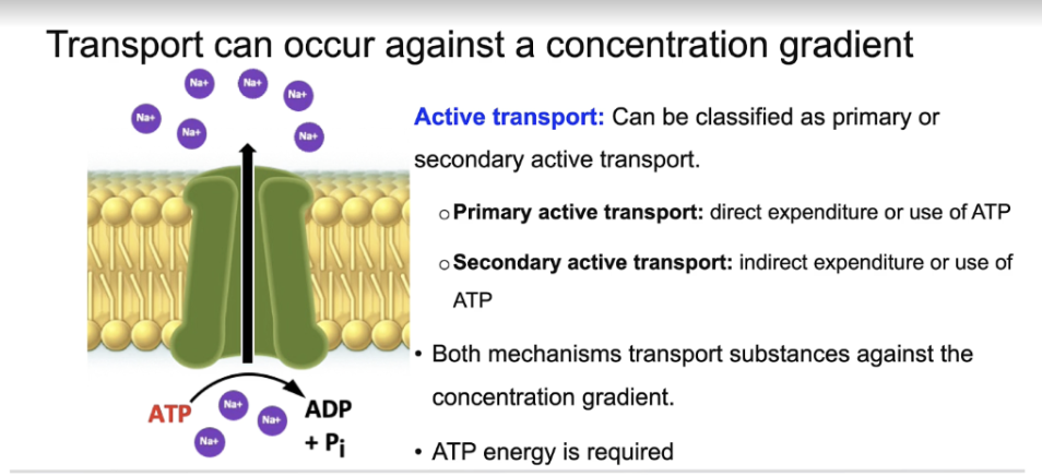
While there is a great deal of passive transport of substances across the cell membrane, that is not always the case. Many cells are able to import or export molecules or ions against an established concentration gradient. In contrast to passive transport, this type of movement requires energy which is generally provided by the hydrolysis of ATP molecules on the intracellular side of the cell.
1) Primary active transport: If the transmembrane transport protein is directly affected by the energy released from ATP hydrolysis and thus under goes a conformational change to "pump" the substance across the membrane against a concentration gradient, this is often referred to as PRIMARY active transport.
2) Secondary Active transport: If neighboring transmembrane transport proteins take advantage of electrochemical gradients established by the primary active transport pumps to move their own solutes against a concentration gradient, then this is often referred to as secondary active transport.
Describe the Sodium Potassium Pump?
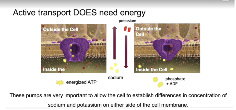
The sodium potassium pump that is embedded in many cellular membranes is a classic example of a primary active transport mechanism. These pumps are very important to allow the cell to establish differences in concentration of sodium and potassium on either side of the cell membrane, which is very important for many biological processes. While passive transport requires no expenditure of energy, in active transport, energy is expended to move substances AGAINST their concentration gradients. This energy comes from the hydrolysis of ATP. In most cells, there is generally a greater concentration of sodium in the extracellular fluid relative to the interior of the cell, and there is also a greater concentration of potassium in the interior of the cell relative to the extracellular environment of a cell in our body. This concentration difference is maintained by the ACTIVE role played by the sodium-potassium pump.
The sodium potassium pump works in the following way:
For every 3 sodium ions that are pumped out of the cells, 2 potassium ions are pumped from the extracellular environment into the interior of the cell. Since this a movement that occurs against the concentration gradient of both sodium and potassium, the energy molecule ATP gives up an energetic phosphate group to the transport protein. It is this energetic phosphate group that binds and causes a conformation change in the shape of the transport protein, and therefore allows the release of the sodium ions into the extracellular fluid in exchange for potassium. When the sodium- potassium pump binds 2 potassium ions that will enter into the cell, the phosphate group is released from the transport protein, the protein returns to its original shape, and simultaneously releases the potassium ions into the cytoplasm of the cell.
It’s moving against the concentration gradient because:
Concentration gradient means substances naturally move from high concentration → low concentration (like diffusion).
For sodium (Na⁺): There’s more sodium outside the cell than inside, so sodium would naturally move into the cell. But the pump pushes sodium out, which is the opposite direction.
For potassium (K⁺): There’s more potassium inside the cell than outside, so potassium would naturally move out. But the pump brings potassium in, again the opposite direction.
Since both movements go against their natural flow, the cell must use energy (ATP) to make it happen. This is why it’s called active transport.