Lecture 54: General Skeletal Disease Processes 1
1/39
There's no tags or description
Looks like no tags are added yet.
Name | Mastery | Learn | Test | Matching | Spaced |
|---|
No study sessions yet.
40 Terms
What is a condition characterized by an increase in bone density resulting from a failure in bone resorption by osteoclasts and fragile bones?
Osteopetrosis
What has occurred in this Angus bovid bone? note the metaphysis and diaphysis are filled with dense bone
osteopetrosis
What is osteogenesis imperfecta?
heritable connective tissue disorder due to mutation in type 1 collagen →
bone fractures (resulting from osteopenia)
joint laxity (defective composition of tendons)
dental abnormalities including fractured teeth (defective dentin)
blue sclerae (reduced thickness of this tissue)
Where are the lesions associated with osteogenesis imperfecta limited to?
bone, teeth, and eyes
What is the hereditary disorder of bone growth that occurs as a result of primary lesions in growth cartilage?
chondrodysplasia
What clinical effects are seen in animals with chondrodysplasia and what breeds are typically affected?
short-legged with normal sized heads because the boens of the calvaria (but not maxilla and mandible) arise from intramembranous, rather than by endochondral, ossification in dachshunds, pekingese, and basset hounds
What is spider limb chondrodysplasia?
condition seen in Suffolk and Hampshire sheep due to mutation in fibroblast growth fact 3 (FGF-3) → disorganized vertebral column, twisted spines, long/bent/splayed limbs
What is affecting this Hampshire sheep?
spider lamb chondrodysplasia
What causes osteochondrosis?
failure of endochondral ossification → focal retention of cartilage that is not replaced by bone and can become necrotic
What are the predilection sites for the development of osteochondrosis in pigs, horses, dogs, cattle, and poultry?
pigs: distal femur, humerus
horses: distal femur, distal tibia, talus, articular processes of the cervical vertebrae
dogs: humerus, distal femur, and talus
cattle: talus, distal femur
poultry: proximal tibia
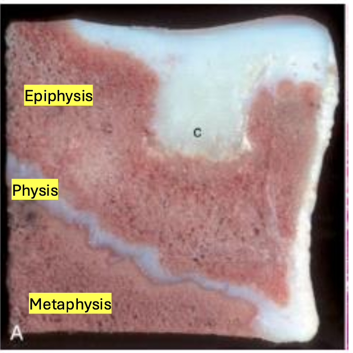
What is the earliest form of OC characterized by a locally extensive region of chondronecrosis within the epiphyseal cartilage, but the subchondral bone is NOT affected?
osteochondrosis latens
What type of OC is characterized by the necrotic, retained epiphyseal cartilage extending to border of bone and is grossly visible?
osteochondrosis
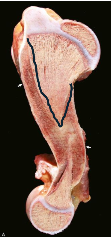
What is affecting this equine distal femur?
osteochondrosis manifesta
What is osteochondrosis dissecans (OCD)?
clefts in the necrotic cartilage with subsequent fracture of the overlying articular cartilage → some degree of separation from underlying bone → cartilaginous or osteochondral flap (chip or joint mouse) → interfere with movement of joint, pain, joint effusion
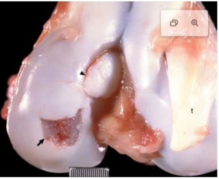
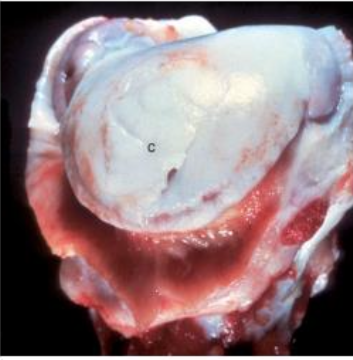
What is affecting this porcine distal femur?
osteochondrosis dissecans
What is affecting this canine proximal humerus? Note the synovium is hyperemic and hyperplastic.
OCD
What is cervical vertebral stenotic myelopathy (wobbler syndrome)?
neurologic disease of horses and dogs that occurs secondary to spinal cord compression due to abnormally developed cervical vertebrae - narrowing of spinal canal
What dogs and horses are more commonly associated with wobbler syndrome?
horses: male thoroughbreds, TN walking horses, warmbloods
dogs: large and giant breeds, dogs with osteoarthritis, vertebral malformations, or disc degeneration/rupture
What are the lesions associated with CVSM?
articular processes: osteochondrosis, asymmetry or enlargement of articular processes, osteoarthritis
malalignment of vertebral column (subluxation on flexion of neck)
tipping/thickening of dorsal lamina
What are the three types of axial skeletal malformations?
lordosis: abnormal ventral curvature of the vertebral column
kyphosis: abnormal dorsal curvature of the vertebral column
scoliosis: lateral deviation of the spinal column
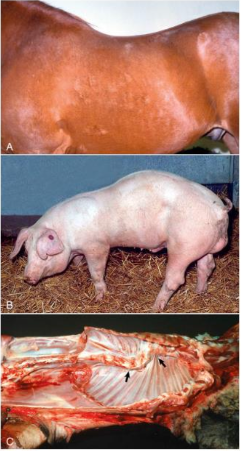
What malformations are affecting each animal?
horse: lordosis
pig: kyphosis
cadaver: scoliosis
What are two types of appendicular skeletal malformations?
polymelia: increase in total number of limbs
polydactyly: increase in number of digits
What is a reduction in bone mass/density without fracture?
osteopenia
What is reduction in bone mass/density with fracture?
osteoporosis
What are the causes of osteoporosis/osteopenia?
• Calcium deficiency
• Starvation – can impact bone homeostasis because of lack of protein and mineral from diet
• Physical inactivity – disuse results in increased bone resorption and decreased bone formation
• Reduced estrogen or androgen
• Chronic glucocorticoid administration
What is Rickets/osteomalacia and what is the difference between the two?
decreased bone mineralization with subsequent bone deformities and fractures
rickets: growing skeleton of young animals, bone and epiphyseal cartilage
osteomalacia: in adult skeleton, confined to bone
What are the causes of rickets/osteomalacia?
deficiencies of vitamin D or phosphorus, can see in chronic renal disease
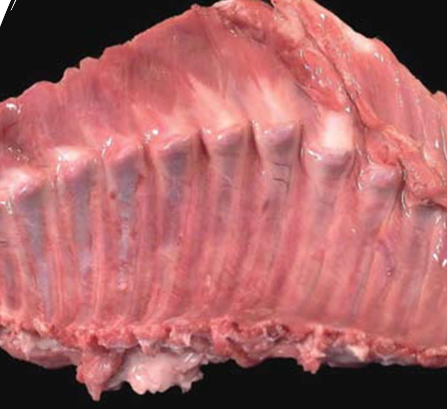
What is this an example of? note the knobs on the inner surface of ribs.
rickets
What are the macroscopic lesions of rickets?
• Knobs on inner surface of ribs (rachitic rosary)
• Spontaneous fracture
• Enlargement/Flaring of growth plates (failure of cartilage matrix mineralization and endochondral ossification)
What condition is characterized by increased osteoclastic resorption of bone (decreased bone mass) and replacement by fibrous tissue, which results in a weakened bone structure?
fibrous osteodystrophy (FOD)
What are the most common causes of FOD in animals?
secondary hyperparathyroidism:
nutritional: dietary factors decreased concentration of serum Ca → parathyroid glands respond by increasing output of PTH
nutritional: young animals fed ration deficient in calcium or high in phosphorus (phosphorus interferes with intestinal Ca absorption)
renal: CKD → high phosphorus and low vitamin D stimulates PTH to normalize calcium levels
What are the macroscopic lesions of fibrous osteodystrophy?
bone is more pliable → rubber jaw if mandible/maxilla affected, distortion of teeth
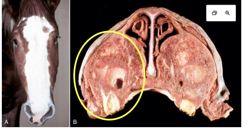
What was affecting this horse? note the swelling of the facial crest and the fibroosseous tissue in the maxillae.
nutritional fibrous osteodystrophy
What is the most common cause of hematogenous osteomyelitis in dogs?
staphylococcus intermedius
What term refers to an infection of the intervertebral disk with concurrent osteomyelitis of contiguous vertebrae?
discospondylitis
What two mycotic agents can spread hematogenously to bone to produce granulomatous and pyogranulomatous osteomyelitis?
coccidioides immitis and blastomyces dermatitidis
What protozoal agent can cause skeletal cardiac myositis and periosteal new bone formation in dogs?
hepatozoon americanum
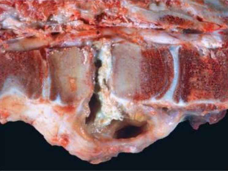
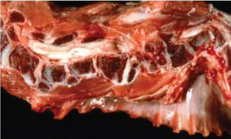
What is affecting this bovine vertebrae? note the yellow white exudate and the sclerotic bone surrounding the exudate.
bacterial osteomyelitis → vertebral abscess
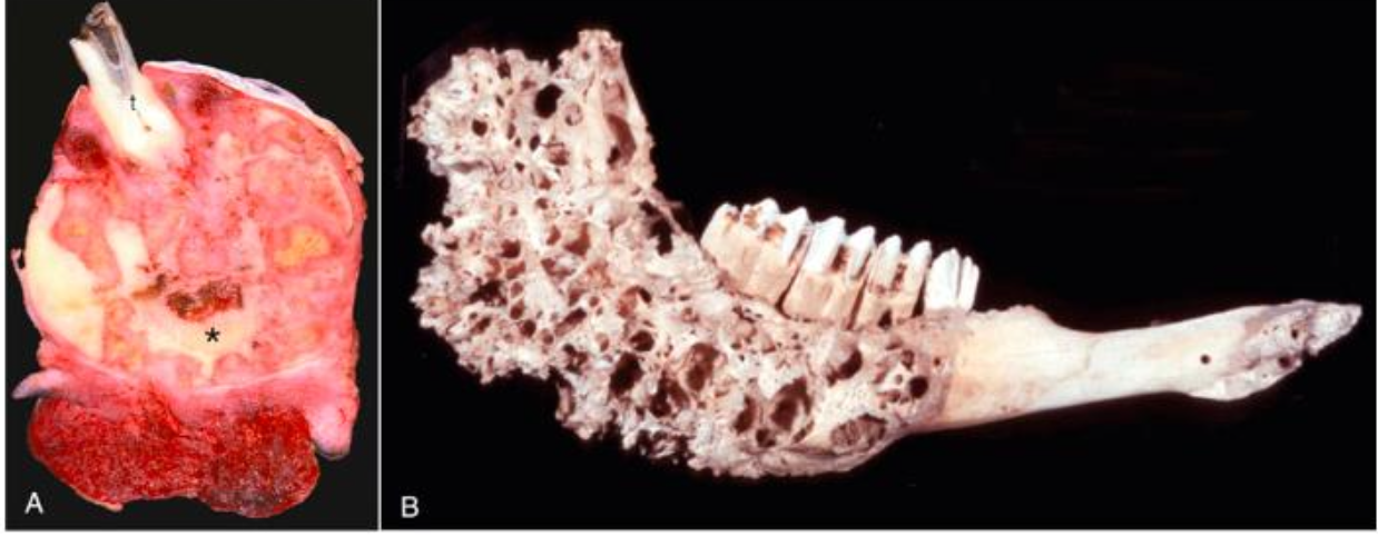
What was affecting this bovine jaw? note the nodules of tan, pyogranulomatous inflammation and porous bone.
bacterial osteomyelitis caused by actinomyces bovis = lumpy jaw
What is the most common bacterial isolate from canine discospondylitis?
staphylococcus pseudointermedius