Intro & Cell Property and Transmission (Exam 1)
1/50
There's no tags or description
Looks like no tags are added yet.
Name | Mastery | Learn | Test | Matching | Spaced |
|---|
No study sessions yet.
51 Terms
Which part of the nervous system is encapsulated by bone?
CNS
Do spinal nerves exit above or below the vertebrae in the cervical region?
Above (which is why there is a C8)

Do spinal nerves exit above or below the vertebrae in the thoracic region?
Below

Do spinal nerves exit above or below the vertebrae in the lumbar region?
Below
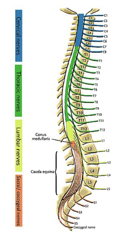
Do spinal nerves exit above or below the vertebrae in the sacral region?
Below

Where does the spinal cord end?
Around L1-L2
What structure is the tip of the spinal cord?
Conus medullaris
Why is the cervical and lumbar regions of the spinal cord thicker than the rest?
Because it has a lot of tissue to innervate.
What structures are located in the dorsal root ganglion?
Cell bodies for afferent (sensory) nerves.
Where are the cell bodies for efferent nerves located?
In the ventral horn (gray matter) of the spinal cord
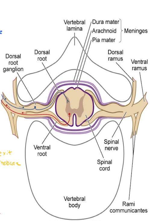
What type of nerve information is transmitted through the dorsal root of a spinal nerve?
Sensory (afferent)

What type of nerve information is transmitted through the ventral root of a spinal nerve?
Motor (efferent)

List the meninges from deep to superficial.
Pia mater (inner) → Arachnoid mater (middle) → Dura mater (outer)
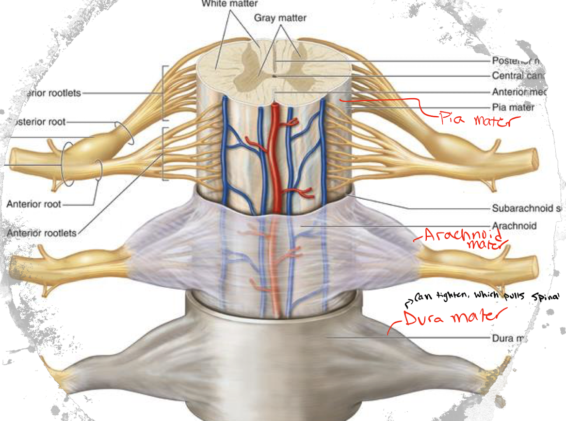
What is gray and white matter composed of, respectively?
Gray matter is composed of cell bodies.
White matter is composed of myelinated axons.
A bundle of myelinated axons traveling together in the CNS is referred to as what?
Columns/tracts/lemniscus
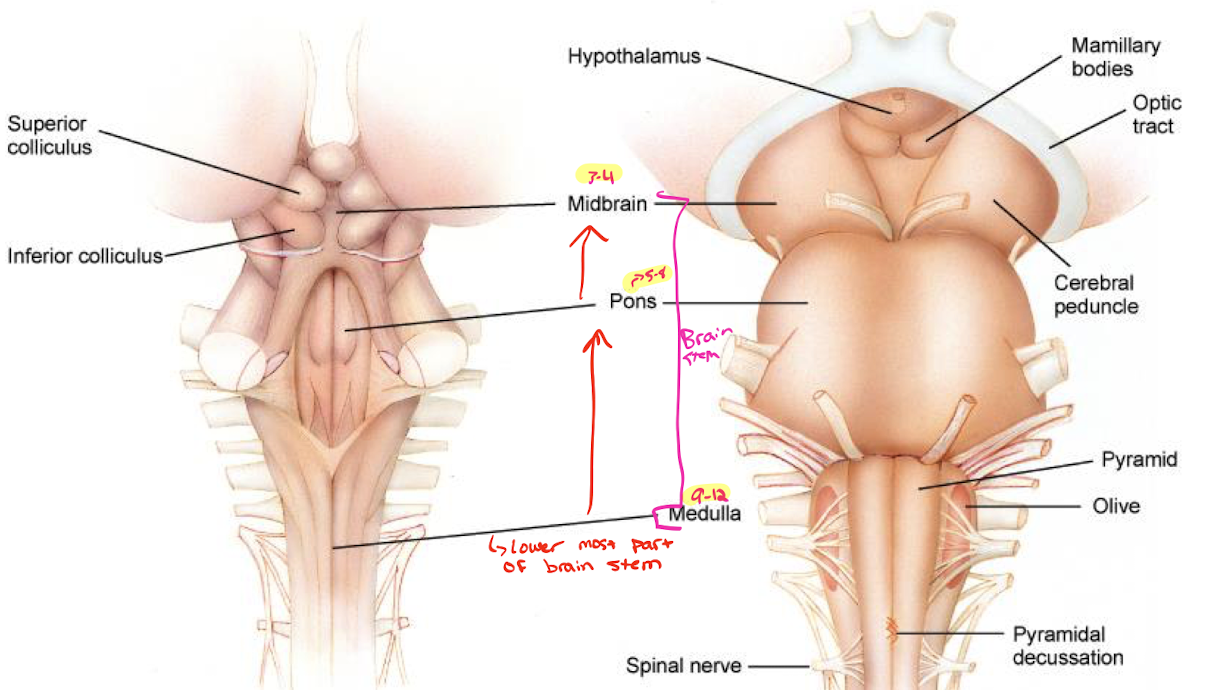
What is the function of the brain stem?
Consciousness, respiration, and core vital signs of life.
What cranial nerves exit the midbrain, pons, and medulla respectively?
Midbrain (3 and 4)
Oculomotor (CN III) and trochlear (CN IV).
Pons (5-8)
Trigeminal (CN V), abducens (CN VI), facial (CN VII), vestibulocochlear (CN VIII).
Medulla (9-12)
Glossopharyngeal (CN IX), vagus (CN X), spinal accessory nerve (CN XI), and hypoglossal (CN XII).
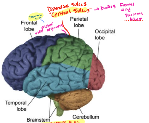
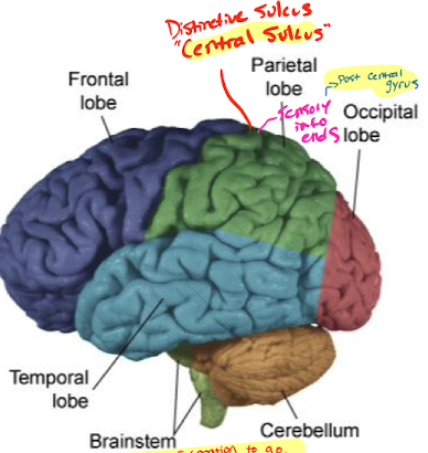
What sulcus divides the frontal and parietal lobes?
Central sulcus.
Where does motor information originate in the brain?
Precentral gyrus (just anterior to the central sulcus in the frontal lobe).
Where does sensory information end in the brain?
Post central gyrus (just posterior to the central sulcus in the parietal lobe).
What is the function of the thalamus?
Relays information and sends it to the appropriate location.
What is the main arterial supply to the brain?
Vertebral artery and internal carotid artery.
What are the 4 main components of a neuron?
Dendrites
Soma
Axon
Presynaptic terminal
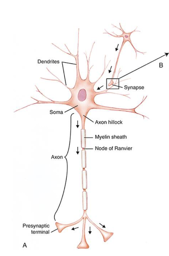
Explain saltatory conduction.
A process in which electrical impulses travel along a myelinated axon by “jumping” between gaps of the myelin sheath called nodes of Ranvier.
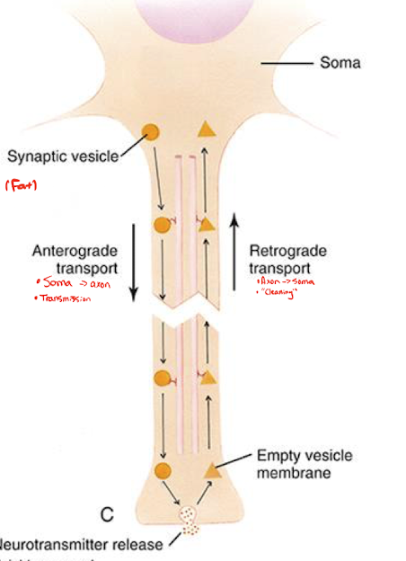
Explain anterograde and retrograde transport.
Anterograde transport: movement of materials from the soma to the presynaptic terminal.
Retrograde transport: movement of materials from the presynaptic terminal to the soma.
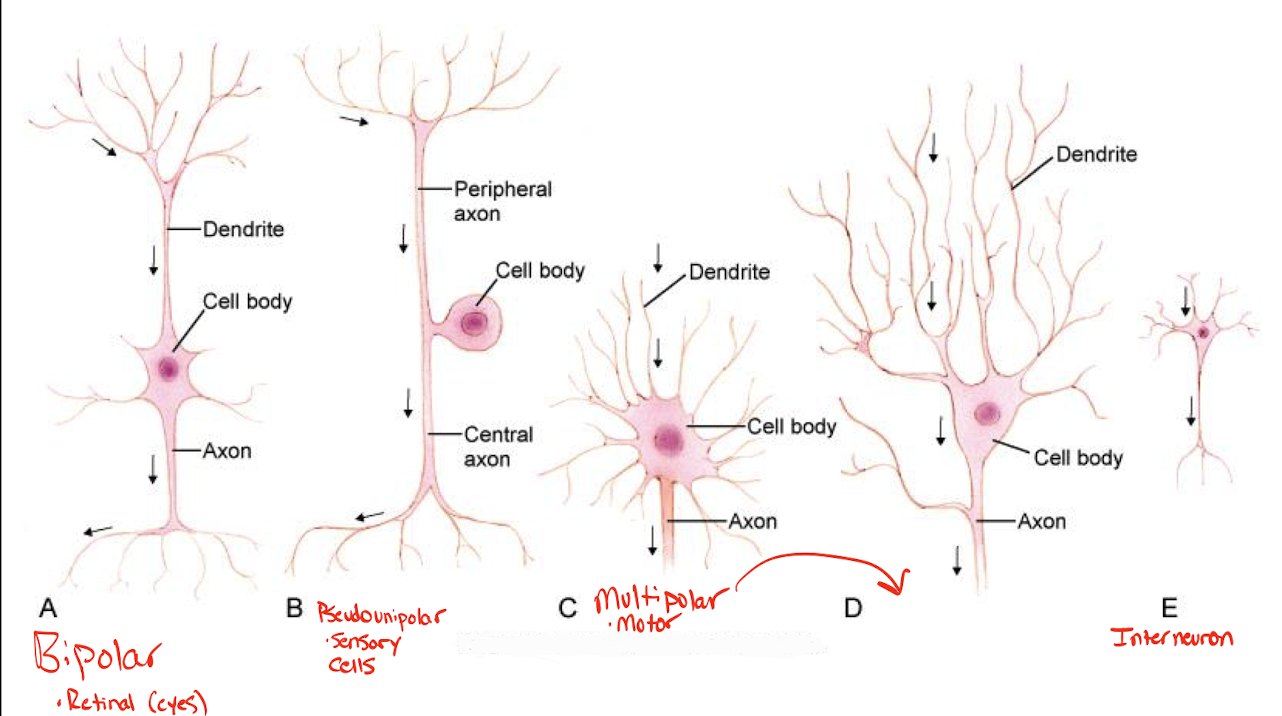
Describe the 3 different structural classifications of neurons. Provide an example of each.
Multipolar: one axon and multiple dendrites.
Motor neurons.
Bipolar: has one axon and one dendrite extending from opposite sides of the neuron.
Retinal neurons.
Pseudounipolar: contains a single process that extends into two branches: one that extends to the CNS and another that extends to the sensory receptor.
Sensory neurons.
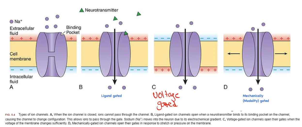
What are the 4 membrane channels that are present on cells and explain each?
Modality-Gated Channels: Opens in response to mechanical force, temperature changes, or chemical stimuli.
Ligand-Gated Channels: opens or closes in response to a specific molecule (called a ligand) binding to the channel. The ligand can be a NT, hormone, or other signaling molecules.
Voltage-Gated Channels: opens or closes in response to changes in membrane potential across the cell membrane.
Leak Channels: Allows diffusion of ions at a sow continuous rate. Not gated and no stimulus is required.
When does depolarization of a cell occur?
When the membrane potential becomes less negative than the resting potential.
What are local potentials?
Initial changes in membrane potential. They spread a short distance along a membrane. If sufficient enough, it can depolarize and get an AP.
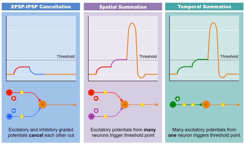
Explain the difference between spatial and temporal summation.
Spatial Summation: when multiple presynaptic neurons fire simultaneously (or close in time) at different locations on the postsynaptic neuron. Their combined postsynaptic potentials sum.
Temporal Summation: When a single presynaptic neuron fire multiple times in quick succession. The postsynaptic potentials add up over time.
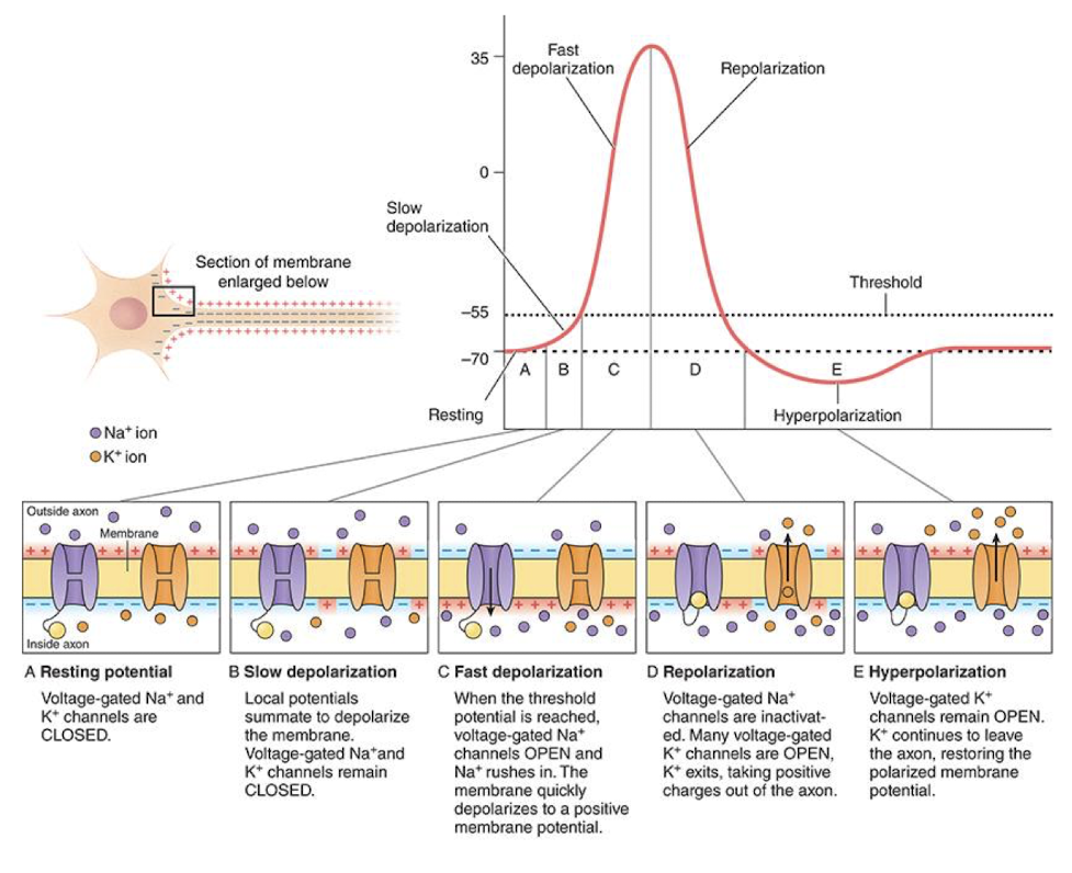
Explain the different phases of an action potential.
Resting Potential: Voltage-gated Na+ and K+ channels are CLOSED.
Slow Depolarization: Local potentials summate to depolarize the membrane. Voltage-gated Na+ and K+ channels remain CLOSED.
Fast Depolarization: When the threshold potential is reached (-55 mV), voltage-gated Na+ channels OPEN and Na+ rushes in. The membrane quickly depolarizes to a positive membrane potential.
Repolarization: Voltage-gated Na+ channels are inactivated. Many voltage-gated K+ channels are OPEN, K+ exits, taking positive charges outside of the axon.
Hyperpolarization: Voltage-gated K+ channels remain OPEN. K+ continues to leave the axon, restoring the polarized membrane potential.
What is the absolute refractory period? Explain its mechanism and when it occurs.
The period during which a neuron CANNOT generate another AP.
Occurs during the depolarization phase and early in the repolarization phase.
Voltage-gated sodium channels are either open or inactivated during this time. Since these channels need to rest from inactivated to closed before they can open again, no new AP can be initiated.
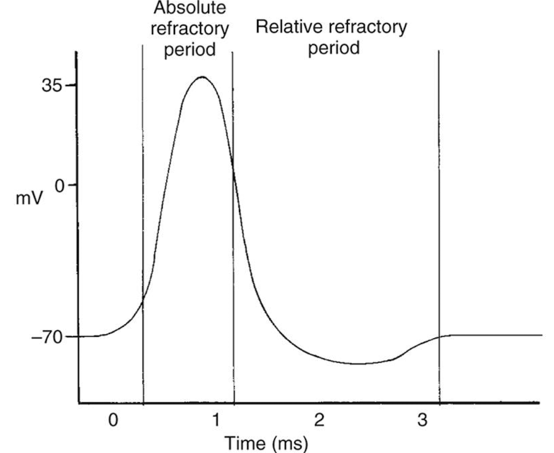
What is the relative refractory period? Explain its mechanism and when it occurs.
Period following the absolute refractory period during which a neuron CAN generate another AP, but only if the stimulus is stronger than normal.
Occurs in the later half of the repolarization phase up until the end of hyperpolarization.
Some voltage-gated sodium channels have been reset and can be reopened, but potassium channels are still open, making the membrane more negative.

What are the three macroglial cells?
Astrocytes
Oligodendrocytes
Schwann cells
What are the functions of astrocytes?
Nutritive
Clean up function within the CNS
Cell signaling using Ca2+
Which glial cell is responsible for the myelin within the CNS and PNS, respectively?
CNS: Oligodendrocytes.
PNS: Schwann cells.
What is an example of a microglial cell, and what does it do?
Phagocytes: acts as the CNS’ immune system and cleans the neural environment.
What part of the nervous system does Guillain Barre affect and what is the mechanism of injury?
What is another condition that affects this part of the nervous system?
Affects the PNS.
Guillain Barre causes an autoimmune attack on Schwann cells. This causes demyelination of the axons, impairing function.
Another PNS condition: Neuropathy.
What part of the nervous system does multiple sclerosis (MS) affect?
What glial cells are impacted by MS?
CNS.
Oligodendrocytes.
Once an AP reaches the presynaptic terminal, what voltage-gated ion channels open?
Voltage-gated Ca2+ channels open.
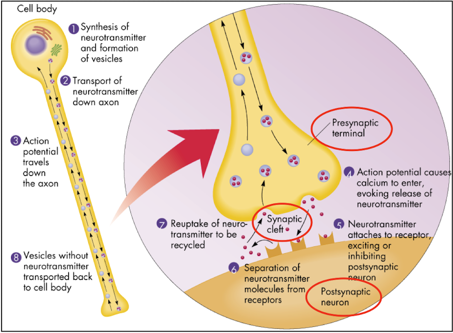
The influx of what ion into the presynaptic terminal of a neuron triggers the movement of synaptic vesicles?
Calcium
What classification of neuron is an interneuron considered?
Multipolar neuron.
Explain presynaptic facilitation.
A facilitatory neuron releases NTs that bind to the receptors on the presynaptic terminal of a second neuron. These NTs typically increase the influx of Ca2+ ions into the presynaptic terminal in response to an AP. As a result, more Ca2+ enters the terminal, which enhances vesicle fusion and NT release into the synaptic cleft.
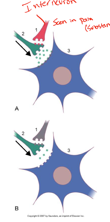
Explain presynaptic inhibition.
An inhibitory neuron releases NTs onto the presynaptic terminal of another neuron. These inhibitory NTs typically decrease the influx of Ca2+ ions into the presynaptic terminal when an AP arrives. This reduction in Ca2+ influx leads to less vesicle fusion and decreased NT release.

What are ionotropic receptors?
Are their responses fast or slow?
Ligand-gated ion channels that open or close in response to the binding of a NT.
Fast response.
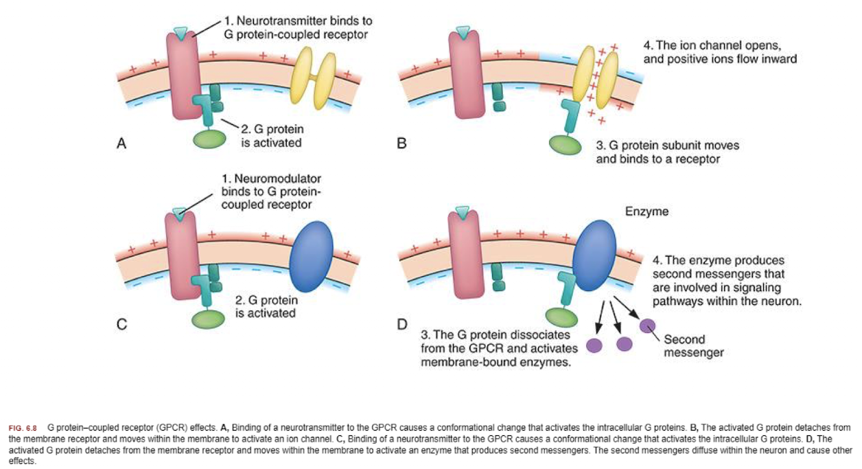
What are metabotropic receptors?
Are their responses fast or slow?
G protein-coupled receptors (GPCRs) that do not directly form ion channels. Instead, they trigger intracellular signaling pathways that indirectly affect ion channels or other cellular processes. Includes second messengers.
Slower responses with more prolonged effects.
Is acetylcholine (ACh) excitatory or inhibitory?
Excitatory.
What are the amino acid transmitters, and are they excitatory or inhibitory?
GABA: Inhibitory.
Glutamate: Excitatory.
Glycine: Inhibitory.
What are the amine transmitters, and are they excitatory or inhibitory?
Dopamine: Either.
Norepinephrine: Either.
Serotonin: Usually inhibitory.
What are the peptide transmitters, and are they excitatory or inhibitory?
Endorphins: Usually inhibitory.
Substance P: Usually excitatory.
Explain myasthenia gravis (MG).
MG is an autoimmune disorder that affects the NMJ. The mechanism involves autoantibodies that attack and block the ACh receptors on the postsynaptic membrane of the muscle, disrupting communication between the motor neuron and the muscle.
The result is less ACh that can bind to the receptors, which leads to impaired muscle contraction. This leads to muscle weakness, particularly in muscles that require frequent use, like those controlling eye movement, swallowing, and breathing.