Neuro Brain Development
1/46
There's no tags or description
Looks like no tags are added yet.
Name | Mastery | Learn | Test | Matching | Spaced | Call with Kai |
|---|
No study sessions yet.
47 Terms
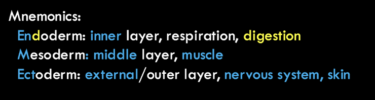
What layers do all animals have in fetal development?
3 germ layers aka groups of cells in embryo. Endoderm (inner for digestive and respiratory systems), middle layer mesodermal (middle for bone, muscle and connective tissue), and outer layer ectoderm (skin and nervous system).
How do germ layers develop?
Gastrulation during which a hollow cluster of cells called a blastula that reorganizes germ layers.
What does the ectoderm layer close in upon itself to form?
neural tube (precursor to brain and spinal cord)
What happens by the fourth week after fertilization?
One end of the neural tube will begin to show three swellings that will become the forebrain, midbrain, and brainstem and cerebellum. The other end of the tube closes and forms the spinal cord.
What developmental phases does neurons pass through?
cell birth, migration, differentiation, axon growth, and cell death
Cell birth
In the ventricular zone (inner portion of the neural tube wall) contains newly born cells (neural progenitor cells aka soon-to-be brain cells).
Migration
Neural progenitor cells migrate from ventricular zone to intermediate zone of neural tube before going to final destination of brain. They are guided by radial glial cells to their destination.
Differentiation
Once arrive at its destination it takes the shape and function of cells in that region (morphology). As it becomes that cell it undergoes differentiation.
Axon growth
Axons rely on growth cone at the tip of the developing axon to find its target. Chemoattractants (+) and chemorepellants (-) signal the developing axon which direction to move and which to avoid. Chemorepellants prevent the developing neuron from going off course. Then the growth conte temporary adheres to the cell adhesion molecules (CAMs). All of these guides where the neuron should be.
Cell Death
Apoptosis aka pruning neurons because we create more than needed. So we prune to give the other neurons a better environment to thrive.
Nerve growth factor (NGF)
protein that prevents apoptosis AND promotes neuronal growth
What happens when nerve growth factor (NGF) isn’t present?
neurons die
How much does brain weight increase from birth to adult?
around 2lbs (born: 1lb, adult: 3.2lb)
When does the brain “mature”?
As age increases, cortical thickness also increases. Varies by brain region, imaging modality, etc. Range from 20 to 32 depending on scan (ex. cortical thickness, fMRI for functional connectivity, and fractional anisotropy).
Are there regional changes in brain development and if so, how do they relate to behavior (e.g. human imaging)?
Peak cortical thickness form at different rates for different regions. Subcortical limbic regions involved in desire, rage, fight & flight suppressed by prefrontal cotex. Circuits develop with age.
How do experiences/INPUTS impact brain development (age- vs experience-related changes)?
Synaptogensis aka formation of synapses to create brain networks for brain connectivity. Explosive increase in synapses in early postnatal period then get pruned to be region specific. Experiences prune less relevant synapses and strengthen relevant ones. (neurons that fire together wire together)
What are critical periods of development?
A WINDOW OF TIME in early development in which a system is open to structuring and restructuring on the basis of imput from the environment. Before and after this period, environmental influences cannot affect the snesitivity or response of that system. (ex. ducklings following boots that look like the mother duck)
Two important parts of the developmental process
Overgrowth of synapses at birth
The brain becomes more receptive to imput
Experiences prune less relevant synapses and strengthen relevant ones
Key Componenets of the Critical Period
TIMING of the opening of this window is contrained by the maturation of underlying circuit (biological readiness). (ex. at least 10 min exposure to mother duck)
IMPUT is necessary to trigger the opening
Plasticity of the visual system is shaped by?
One’s early environment. AKA critical periods are sensitive to orientation/direction. (ex. kitten raised with goggles with only horizontal and vertical lines on each eye so as adults the eyes raised with that orientation only responded to lines of that orientation)
Orientation Columns
Neurons within one column of the cortex respond to lines of the same orientation
Ocular Dominance Columns
perpendicular to orientation columns are alternation columns of neurons that receive input from left versus right eye
Visual deprivation of one eye during critical period affects what?
reduces ocular dominance column width for that eye (eye closed then no development of ocular dominance column)
What has a weaker effect on the formation of ocular dominance columns the later it is done?
Closure because the columns become more segregated with time.
What can plasticity allow?
Brain areas to take on new functions, especially during development (ex. remove part of brain young so other parts of brain take over the function of the removed part)
What happens when you amputate a part of the body?
Inputs from neighboring areas expand into what had been that part’s somatosensory areas. Perception of touch missing (phantom limb)
What triggers critical periods? What causes them (brakes)?
Opening a critical period: GABA cells (parvalbumin cells) maturation and pivotal plasticity switch during critical period
brings order to chaos
tells excitatory cells to hush
shift from more excitatory to more inhibitory synapses
(ex. rush hr of restaurant is excitatory, midnight with no one at restaurant is inhibitory)
Are synapses that are pruned predominantly excitatory or inhibitory during development?
Excitatory (especially in prefrontal cortex throughout adolescence)
What plays a critical role in plasticity and learning?
BDNF
What causes synaptic stabilization and closing of critical period?
Myelination of axons: formation of myelin creates physical barriers to sprouting/axonal growth
Perineuronal nets: lattice of molecules consisting of proteins and sugars that wrap around parvalbumin cells as they mature —> end of critical period (barrier) it disrupts so loss of info.
Why would we want to close a critical period?
To consolidate learning and memory
Plasticity
Process by which experiences reorganize neural connections/pathways
When does long lasting functional changes in the brain occur?
when we learn new information (consolidation)
As age goes on, stages of brain development?
Imature (brakes and learning is susceptible to be lost), Plastic (critical period), Consolidated (brakes aka learned)
Why is consolidation important?
Imprinting, communication, survival of species
Do perineuronal nets protect fear memories from erasure?
Yes and loss of perineuronal nets renders fear memory susceptible to erasure but does not interfere with memory consolidation. Adults without nets have less fear.
Can we accelerate, delay, extend, prevent a critical period?
Manipulation of GABA can impact the onset of a critical period.
enhancing GABA pharmacologically with benzodiazepines can accelerate critical periods
Genetically modifying expression of GABA can delay critical periods
Early life stress can increase GABA levels —> premature opening and closing of critical periods
How might critical periods inform aging and/or disease?
Preneuronal networks (PNNs) increase in prefrontal cortex by adolescent years, so is it associated with aging? There are less PNNs when older. Loss of PNNs are associated with aging in auditory cortex.
What performance can vary widely with age?
Cognitive perfromance
Alzheimer’s disease leads to?
detectable shrinkage of the brain
Brain Pathology characteristic of Alzheimer’s disease
Increase in amyloid plaques and neurofibrillary tangles
Amyloid plaques: clusters of beta amyloid protein in extracellular spaces in brain
Neurofibrillary tangles: clusters of misfolded tau protein located inside of neurons
What does tau protein do?
Binds to microtubles to stabalize them. Microtubes are inside the neuron that allow material to move from one part of the neuron to another.
What happens to the tau protein in Alzheimer’s disease?
Prevents tau from binding to microtubes so microtubes lose stability and neurofibrillary tangles in the neuron form. These # of tangles correlate with neuronal loss and degree of patient’s cognitive decline.
Where are tangles and plaques found?
Tangles almost exclusively in hippocampus and cortex. Plaques throughout the brain.
What gene increase the risk of Alzheimer’s?
polymorphisms in ApoE gene. Three alleles (E2, E3, E4). E2 and E4 are rare in the general population but increases number of E4 increases risk for Alzheimer’s.
What factors are associated with the risk for Alzheimer’s?
Interaction of environmental and genetic factors
Potential role of PNNs in Alzheimer’s
Reduction of PNNs in Alzheimer’s brain. PNNs localize with both plaques and tangles so PNNs are either instrumental in, reaction to, or formation of them. PNNs may also provide protection against tangles.