What is Sick Sinus Syndrome (SSS)?
combination of 2 or all in the the same pt: sinus exit block, sinus pause, sinus arrest -can be on the same rhythm strip
What are sinus node arrhythmias generated by?
sinus node
What are PACs?
early ectopic impulse originating outside the SA node, but w/in the atria
What is bigeminy?
irregular beat/rhythm/complex occurring every other beat
What is trigeminal or trigeminy?
irregular beat/rhythm/complex occurring every 3rd beat
What quadrigeminal or quadrigeminy?
irregular beat/rhythm/complex occurring every 4th beat
What causes WAP?
3 different areas w/in the atria that are competing to be the pacemaker of the heart
What is atrial flutter?
rapid atrial rate caused by a reentry circuit
When interpreting atrial flutter, what must you document?
ratio of F waves to QRS complex
What is the most treated cardiac arrhythmia?
atrial fibrillation
What causes a-fib?
irregular activity of multiple atria sites that suppress SA node → loss of atrial kick
What is paroxysmal a-fib?
pt goes in and out of A-fib (converts w/o intervention)
What is persistent a-fib?
responds to intervention (pharm or electrical cardioversion)
What is permanent a-fib?
chronic, will not respond to interventions
What AV nodal reentrant tachycardia commonly referred to as?
supraventricular tachycardia (SVT)
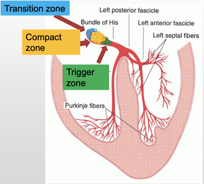
What does the transition zone of the AV node do?
receives impulses from SA node & transmits to the compact zone; responsible for most PJCs, very arrhythmogenic
What does the compact zone of the AV node do?
slows the conduction to allow for atrial contraction & ventricular filling; core of node, functions as back-up pacemaker
What does the trigger zone of the AV node do?
receives signals from compact zone and shoots them down the bundle of His
In junctional rhythms, if the signal travels up the AV node and depolarizes the atria from bottom up, how would the P wave present?
inverted
In junctional rhythms, what direction do impulses generated in the AV junction travel?
both upwards towards atria and down the His bundle to the ventricle
In junctional rhythms, if an impulse from the AV junction depolarizes the atria and ventricles at the same time, how will the P wave present?
hidden in the QRS → no P wave
In junctional rhythms, if the impulse from the AV junction never depolarizes the atria, how will the P wave present?
absent / missing
In junctional rhythms, if the impulse is infero-nodal, meaning the AV junction is generated in the trigger zone and therefore depolarizes the ventricles first, what would the P wave morphology be?
P wave will appear after the QRS complex; most likely inverted
What causes junctional escape beats?
SA node fails to fire or is firing too slowly
During a junctional escape rhythm, what is the main pacemaker of the heart?
AV node
What causes ventricular beats?
increased automaticity or failure of both SA & AV node
What are characteristics of ventricular rhythms?
contralateral T waves, wide QRS, abn looking
It is call a run of V-tach after more than ___ PVC’s in a row.
5
A pt presents w/ a 6 PVC’s in row. Is intervention needed?
yes, it is now V-tach
What causes Idioventricular rhythm (IVR)?
Purkinje network takes over as primary pacemaker
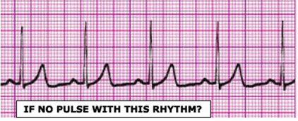
Can V-tach be present w/o a pulse?
yes; can be w/ or w/o
What causes ventricular fibrillation?
asynchronous, chaotic firing of multiple foci w/in ventricles
Why is there no cardiac output or pulse during V-fib?
there is no ventricular contraction
What is happening when there is organized electrical activity on the monitor, but no mechanical activity?
pulseless electrical activity (PEA)
What is happening during ventricular standstill?
there is still atrial contractions, but no ventricular contractions; no pulse or cardiac output
What is a 1st degree AV block?
benign PR delay resulting form slowed conduction through the AV node
What causes a 2nd degree Type1 AV block?
blockage of the conduction in the AV node
What is a 2nd degree Type 2 AV block a sign of?
underlying disease of the conduction system
What causes a 2nd degree Type 2 AV block?
intranodal blockage occuring low in the AV node and His bundle
What causes a 3rd degree AV block?
age related fibrosis, acute MI (espicially inferior)
What is the pacemaker in a 3rd degree AV block?
2 independent pacemakers: SA node & AV junction or Purkinje
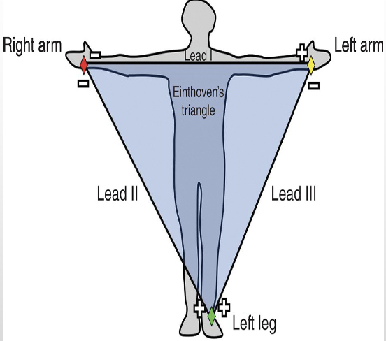
What is Einthoven’s triangle?
Leads 1-3 create triangle over the body using both shoulders and the left LE (bipolar leads)
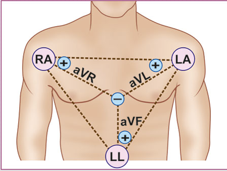
What are the augmented leads?
leads aVL, aVR, aVF: view created by ECG machine using a theoretical negative pole on the center of the hearts -Wilson’s terminal (unipolar leads)
What are the hexaxial leads?
Leads 1-3 & augmented (aVR, aVL, aVF) -these come together to form the first 6 leads of the 12 lead ECG
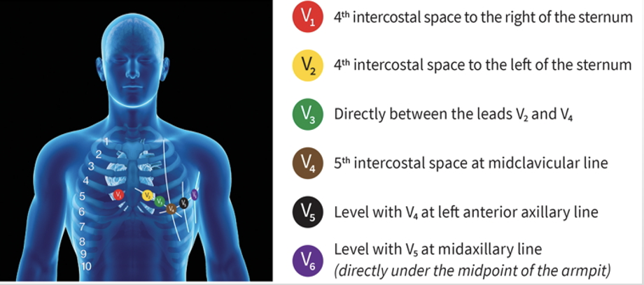
What are the precordial leads?
chest leads, unipolar, give a horizontal view of the heart; have very specific placement (see pic)
Which leads look at the septal wall?
V1, V2
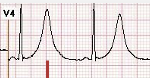
Which leads look at the anterior wall of the left ventricle?
V3, V4
Which leads look at the lateral wall of the left ventricle?
I, aVL, V5, V6
Which leads look at the inferior wall?
II, III, aVF
What are contiguous leads?
two or more leads that look at the same area of the heart
Which 2 leads do you look at to determine the axis?
I and aVF
If both lead I and aVF are positive, what is the axis?
normal
What is the axis if lead I is + and aVF is -?
LAD
What is the axis if lead I is - and aVF is +?
RAD
What is the axis if both lead I and aVF are -?
eRAD
Where should J point be in a healthy pt?
at baseline
What is often the very first sign of ischemia?
hyperacute T waves
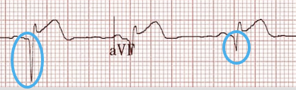
What represents infarction and actual death of cardiac tissue from previous or acute cardiac event?
pathological Q waves
What are reciprocal changes?
mirror image of a cardiac event on the opposite leads which look at the same are of the heart
Why are reciprocal changes significant?
confirmatory sign of ischemia
What area would show reciprocal changes if the ST elevation was in leads V1-4?
posterior (would need a 15 lead)
What area would show reciprocal changes if the ST elevation was in the lateral leads (I, aVL, V5, V6)?
inferior leads (II, III, aVF)
What are would show reciprocal changes if the ST elevation was in the inferior leads (II, III, aVF)?
lateral leads (I, aVL, V5, V6)
What area would show reciprocal changes if the ST elevation was posterior?
anteroseptal leads (V1-4)
What would you need to see a posterior MI on an EKG?
15 lead EKG
What are we worried about w/ inferior MIs?
RV involvement
How do we check for RV involvement during an inferior MI?
check V4R
What is a left anterior fascicular block?
failure of left anterior fascicle to conduct impulses; common abnormality during an acute MI
What is the most common STEMI mimic?
LVH
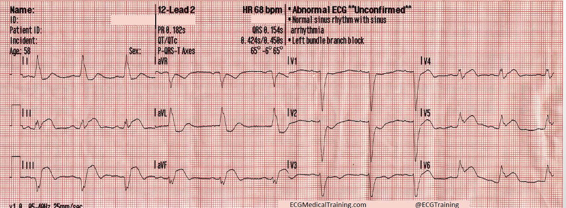
What is Sgarbossa’s Criteria used for?
dx MI in the presence of LBBB or ventricularly paced rhythm
What is Sgarbossa’s critera?
concordant ST elevation of 1+ mm in any lead w/ a + QRS complex
concordant ST depression of 1+mm in V1-V3
discordant ST elevation of 5+ mm in any lead w/ - QRS complex
What is the accessory pathway in WPW?
Kent bundle