Biol 40 Chapter 6 and 7 Review
1/57
Earn XP
Description and Tags
Lipids, Membranes, and the 1st cells + inside the cell
Name | Mastery | Learn | Test | Matching | Spaced | Call with Kai |
|---|
No analytics yet
Send a link to your students to track their progress
58 Terms
What separates life from nonlife?
Plasma Membrane: serves as a selective barrier from damaging chemicals and substances
Lipids
umbrella term for carbon-containing compounds characterized by their lack of solubility in water
Why are lipids not soluble?
this is a result of their nonpolar hydrocarbons
Hydrocarbons
molecules containing only hyrdrogen and carbon (nonpolar due to electronegativities)
Fatty acid
simple lipid made of a hydrocarbon chain bonded to a polar carboxyl functional group (-COOH)
Cis Fatty Acid
hydrogens are on the same side of the chain, creating a kink in the chain that promotes fluidity and thus increases solubility
Trans Fatty Acids
hydrogens are on opposing sides of each other resulting in a straight, rigidly packed chain that decreases solubility
Unsaturated fats
a hydrocarbon chain with 1+ double bonds (trans and cis fats fit into this category)
Saturated fats
hydrocarbon chain consisting of only single bonds (waxes)
waxes
saturated lipids that have extremely long hydrocarbon tails; forming stiff solids at room temperature
oils
highly unsaturated lipids that are liquid at room temperature
how can unsaturated lipids be converted into saturated lipids?
this is done by breaking down double bonds and adding hydrogen atoms via hydrogenation
steroids
lipids distinguished by their bulky four-ring structure
differ from each other by the functional groups attached to different carbons in hydrophobic rings
ex: estrogen, testosterone, and cholesterol (important component in plasma membrane)
fats
nonpolar molecules composed of 3 fatty acids that are linked to a 3-carbon molecule called glycerol
frequently referred to as triglycerols or triglycerides
if glycerol-linked fatty acids are polyunsaturated the resulting triglycerides are liquid at room temp (fats can store up to 2x as much chemical energy as carbohydrates due to their bonds)
fats form when a dehydration reaction occurs between a hydroxyl group of glycerol and the carboxyl group of a free fatty acid (fats are NOT polymers and fatty acids are NOT monomers)
phospholipid
consist of glycerol that is linked to a phosphate group and 2 hydrocarbon chains of either isoprenoids or fatty acids (phosphate group is also bonded to a small organic molecule that is charged or polar)
phospholipids with fatty acid tails are found in bacteria/eukarya
phospholipids with isoprenoid tails are found in domain archaea
phospholipids are a crucial part of the plasma membrane
lipid roles
store chemical energy
act as pigments to trap sunlight
vitamin in cellular processes
serve as signals between cells
waterproof coating on cells/skin
cell membranes
amphipathic
contain both a hydrophobic (nonpolar) and hydrophilic (polar) region (phospholipid’s hydrophilic phosphate head and hydrophobic lipid tail)
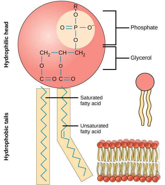
phospholipid phosphate head
hydrophilic
polar
faces solutions in lipid bilayers
phospholipid lipid tails
hydrophobic
nonpolar
face the inside of a membrane in lipid bilayers
micelles
tiny spherical aggregates created when the hydrophilic heads of lipids face outward and interact with water while their hydrophobic tails face inward and interact with each other
lipid bilayer
when lipid molecules align in paired sheets, heads facing the surrounding solution while tails face each other
what kind of molecules can and can’t pass through a selectively permeable membrane?
CAN: small, nonpolar substances are permitted across the membrane (ex: O2 or CO2)
CANT: large, polar substances are NOT permitted across the membrane (ex: C6H12O6)
What happens to permeability as temperature decreases?
Molecules begin to move slowly and become less fluid:
lipid bilayers begin to solidify and permeability will decrease
as temp increases permeability will increase
diffusion
spontaneous movement of molecules and ions across a membrane along with the concentration gradient (molecules move from areas of high concentration to low concentration)
passive transport
substances diffusing across a membrane without an outside energy source, at equilibrium this movement will not stop but continues at random
osmosis
diffusion that is specific to water
only unbound water molecules are capable of diffusion
shrinks or bursts the volume of cells
hypertonic
solution outside of the cell has a higher concentration of solute than the inside, this causes water to move out of the cell and the cell will SHRINK
hypotonic
solution outside of the cell has a lower concentration than the inside of the cell, water will move from outside of the cell to the inside, BURSTING the cell
isotonic
solute concentration is equal (no change in cell’s volume)
proto cells
vesicle-like structures that harbor nucleic acids
fluid-mosaic model
membranes are a dynamic and fluid mosaic of phospholipids and different types of proteins
ion channels
specialized pore-forming proteins
ions diffuse from high to low concentration
electrochemical gradients
channel proteins
pore-like channels in cell membranes
aquaporin - allow water across the membrane
gated channels - open or close in response to a signal (binding of a substance or change in electrical charge)
passive transport
facilitated diffusion
when transmembrane proteins assist in passive transport
carrier proteins
used to facilitate diffusion of substances (act as bridges across membranes)
active transport
transport AGAINST the gradient that requires energy (ATP)
active transport proteins = pumps
sodium potassium pump
sodium potassium pump
sodium ions are in higher concentration on the outside of the cell than on the inside (+)
potassium ions are in higher concentration on the inside of the cell than on the outside (-)
the pump sends three sodium ions (Na+) out of the cell and 2 potassium (K+) ions into the cell
secondary active transport
ATP doesn’t directly engage, but provides energy as gradient that powers movement of different solute against its gradient
Pulse Chase Experiment
Pulse: expose cells to radiation of modified amino acid for a short time
Chase: end pulse by removing radiation and replacing w/ normal molecule, follow moelcule’s track throughout the cell at set times
Purpose: mark a population of molecules over a set interval of time and follow their fate over that time
Describe how proteins enter the endomembrane system
Protein synthesis begins on a free ribosome in the cytosol, ribosome synthesizes the endoplasmic reticulum signal sequence using info fromt he mRNA
Signal sequence binds to a signal recognition particle (SRP) - a complex of RNA + protein - the SRP causes protein synthesis to stop
Ribosome + signal sequence + SRP moves to rough endoplasmic reticulum membrane where it attaches the SRP receptor
Once the lock (receptor) and key (SRP) connect, the SRP is released and protein synthesis continues through translocon
Growing protein is fed into ER lumen, ER sequence is removed
endoplasmic reticulum → golgi apparatus → plasma membrane
glycosylation
addition of 1+ carbohydrate groups — result is referred to as a glycoprotein
cis face of golgi apparatus
where the protein enters, immature
trans face of golgi apparatus
where the protein exits, matured and ready to go to plasma membrane
exocytosis
process in which vesicle membrane and plasma membrane make contact, fuse, and the vesicle lets it’s contents exit the cell
endocytosis
cell taking in material from the outside
receptor mediated endocytosis
uses receptors to bind to macromolecules outside of the cell
early endosome
where cargo is delivered
late endosome
acidified + matured; eventually a lysosome
phagocytosis
brings small cells/food particles inside of the cell through the plasma membrane, engulfing it and delivering it to the lysosome
autophagy
portions of the cytoplasm, including damaged organelles are enclosed in a membrane and delivered to the lysosome
actin filaments (microfilaments)
fibrous structures made of globular protein subunits (actin)
smallest, but most abundant (5-10%) protein in animal cells
exhibits polarity
structure: two coiled strands
function: maintain cell shape by resisting tension, move cells via muscle contraction, divide animals cells in two, move organelles + cytoplasm in plants, fungi, animals cause by MYOSIN (motor protein) + actin interaction
intermediate filaments
serve solely a structural role in eukaryotes
does not exhibit polarity
structure: cable like
function: maintain cell shape + anchor nucleus / other organelles
nuclear lamins: form a dense mesh in nuclear envelope that anchors chromosomes, nucleus, etc
microtubules
largest, assembled from subunits of 2 closely related proteins, alpha tubulin + beta tubulin, that under normal conditions exists as stable protein dimers — two parts
tubulin dimers polymerize in a head-to-tail fashion via non-covalent bonds forming thin chains — protofilaments
structure: hollow tubes
function: maintain cell shape via resisting COMPRESSION, move cells via flagella or cilia, move chromosomes in cell division, provide tracks for intracellular transport
exhibits polarity: + ends growing faster like actin!
motor proteins move vesicles along microtubules: KINESIN (every step requires energy)
microtubule organizing center (MTOC)
where the + ends of microtubules grow out of
most animals have just 1 MTOC near nucleus
centrosome: MTOC site in animal cells, centrioles: 2 bundles of microtubules
flagella
moves entire cell, consist of several microtubules that move the cell by whipping back and forth + is surrounded by the plasma membrane
cilium (cilia)
moves entire cell, short, hair like projections on eukaryotes
axoneme
the characteristic 9×2 arrangement of microtubules in flagella + cilia — 9 microtubules PAIRS + 2 central microtubules
basal body: where the 9 axoneme doublets originate
the beating of cilia requires energy
dynein
motor protein in axoneme doublet arms that require ATP to undergo conformational/shape changes
transport vesicles
dynein "arms “walk” on adjacent doublets