Lecture 4: Ventral Visual Cortex
1/21
There's no tags or description
Looks like no tags are added yet.
Name | Mastery | Learn | Test | Matching | Spaced |
|---|
No study sessions yet.
22 Terms
What is colour useful for?
Scene segmentation – as an extra cue to distinguish shadows from surfaces changes
Signalling surface – for example to tell the edibility of a banana, or to tell emotion/health.
What are the low level aspects of colour visual?
Retinal ‘hardware’
Retinal ‘software’
Anomalous colour vision.
Retinal ‘hardware’
What is retinal hardware for colour vision?
Namely the photoreceptor, note that the sensitivity of photoreceptors determines the range of wavelengths we are sensitive to. The number of differently sensitive photoreceptors gives the maximum dimensionality of our colour vision.
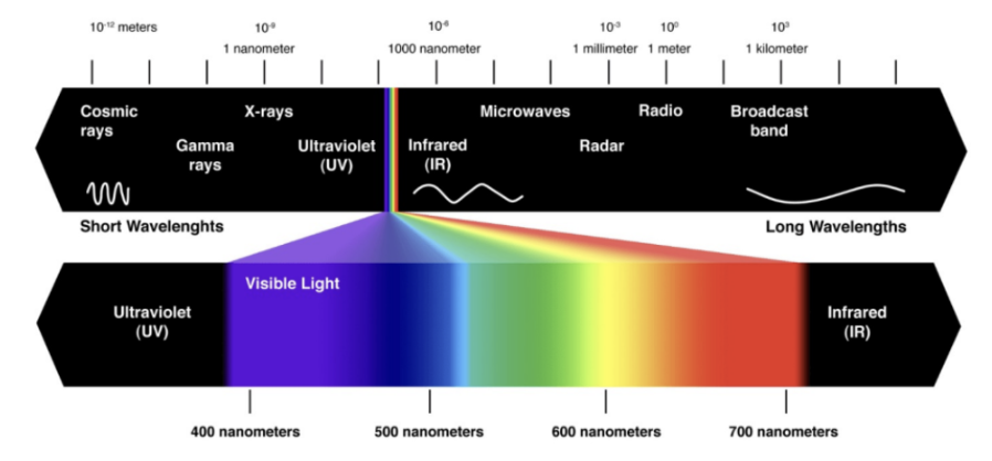
Explain wavelengths?
Different wavelengths are perceived differently for different species.
What is a Metamer?
Different stimuli that are perceptually indistinguishable, demonstrates why we don’t use wavelengths as a way of identifying colours, a colour may actually appear the same even through their wavelengths may be the same
Explain what is an L-M-S space?
A better understanding may also be sought via the L+M+S space which adds in light vs. dark as well.
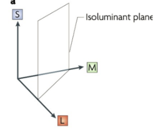
What does CMYK and why is there one more dimension?
May have an extra dimension, but this does not mean that it allows for perception.
To perceive colour we need to register the relative responses (difference) of different photoreceptors -> we lack the neural ‘software’ for 4D colour vision.
Retinal Software
Where does the M-tone come from?
Among mammals, only some primates have trichromatic vision, others are dichromats, with an S-cone and L-cone photoreceptor. • An L-cone mutation is thought to have resulted in the M-cone.
How did we already have the software to process the new cone?
Based on the parvocellular pathway we can see that we already had software for red-green colour vision.
What is the Konicellular pathway?
carries S-cone information, giving the ‘blue-yellow’ dimension to our colour vision, separate retinal ganglion cell types, e.g. small bistratified.
What is opponent coding of colour?
‘more red’ = ‘less green’ • ‘more blue’ = ‘less yellow’
This can be demonstrated via negative after-image (adaptation - prolonged looking) gives negative afterimages. Initial responses to changes in colour are higher, we pay attention to changes, rather than the state of actual events. Means our vision is relative
Describe anomalous colour vision?
Can be missing 2 x cones → translates into congenital achromatopsia
Can be missing 1 x cone → translates into a loss of colour space, rather than a missing colour.
What did Ewald Hering note?
Ewald Hering (1834-1918) noticed that some colour combinations are ‘legal’ while others are ‘illegal’ • Bluish green is legal / bluish yellow is illegal • Reddish yellow is legal / reddish green is illegal • Hypothesised the opponent coding of colour vision before retinal physiology was known.
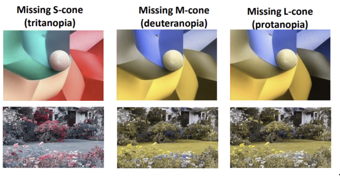
What are the different ways you can lose a dimension due to missing a cone?
Normal (trichromatic)
Missing S-cone (tritanopia) - have only yellow - blue
Missing M-cone (deuteranopia) - no red - green
Missing L-cone (protanopia) - no red - green
What are pseudochromatic plates?
have patterns that are restricted to variation along one dimension, with ‘noise’ in the other dimensions. YOU CAN SEE THE NUMBER IF YOU HAVE NORMAL COLOUR VISION.
What are Pseudoisochromatic plates?
have patterns that are restricted to variation along one dimension, with ‘noise’ in the other dimensions • Or patterns that are masked by ‘noise’ along dimensions of normal colour. YOU CANNOT SEE THE NUMBERS IF YOU HAVE NORMAL COLOUR VISION.
How is bee and bird colour vision different from ours?
Includes more sensitivity in the UV range, have a photoreceptor that detects this.
Birds have better spaced L/M photoreceptors better spaced than ours, Most birds are tetrachromats (4 dimensional colour vision) they have both the hardware and the software to see 4 x colours.
What is cerebral achromatopsia (and explain it as evidence for cerebral color processing) ?
Cerebral achromatopsia refers to a complete or partial loss of colour vision despite intact cone functioning. Damage to area V4 can result in cerebral achromatopsia
Early evidence for cortical areas comes from lesion studies in someone with cerebral achromatopsia.
Cerebral achromatopsia → lead to difficulties enjoying life and food, started to eat food that were only naturally grey
Does imaging also allow for evidence for the ventral visual cortex and it’s importance in colour perception.
Ventral visual cortex
Immediately anterior to V4 are other regions that are also strongly responsive to colour
Moving posterior to anterior, areas respond to progressively more complex features/forms (e.g. FFA, OFA, PPA), and have greater connectivity with other cortices (e.g. memory) .
What is colour constancy?
the ability of the human visual system to perceive the color of an object as remaining stable and the same, even when the lighting conditions (such as intensity and spectral composition) change significantly.
How does colour constancy work?
Because we automatically separate the scene into ‘layers’ of colour
Surface reflectance (what proportion of each wavelength a surface reflects)
Illuminants (power at each wavelength of light illuminating the scene)
Filters (spectral properties of any translucent filters)
How does the brain disentangle surface, illuminant and filter properties?
It does not do it perfectly, but the brain estimates
The illuminant properties using the average colour/white point, specular highlights and luminance/hue corrections