anat 316 pt 2 bs
1/295
There's no tags or description
Looks like no tags are added yet.
Name | Mastery | Learn | Test | Matching | Spaced |
|---|
No study sessions yet.
296 Terms
Mastication
Mastication: process by which food is crushed and ground by the teeth
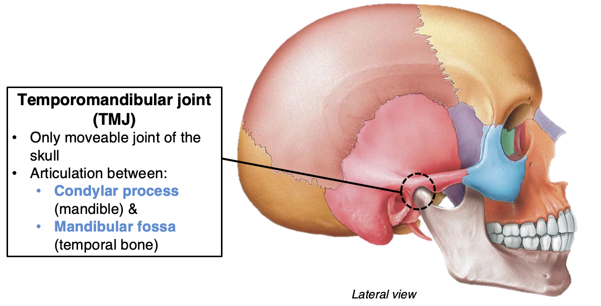
Associated with the temporomandibular joint (TMJ)
Temporomandibular Joint (TMJ)
Elevation, depression, protraction, retraction, side to side movement
Muscles of Mastication
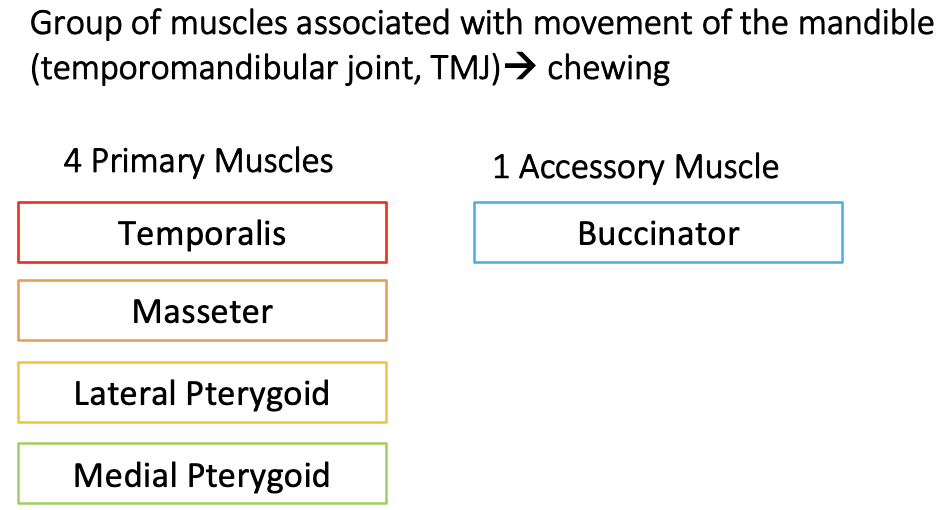
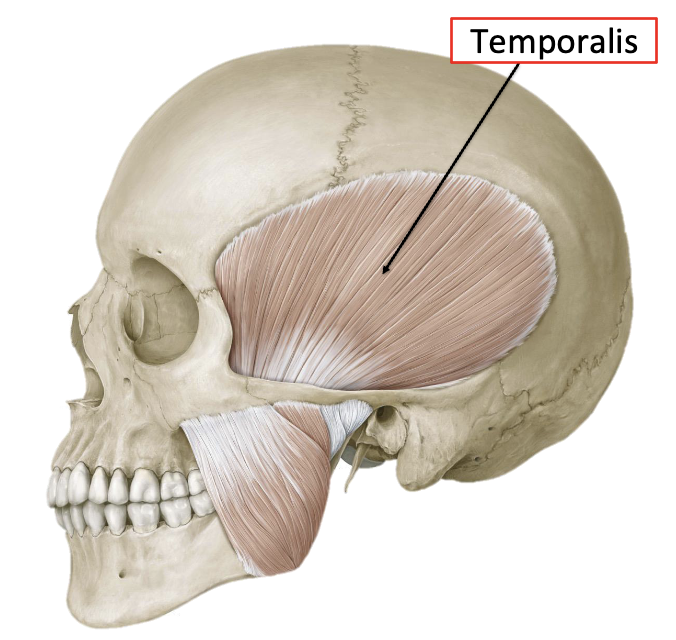
Temporalis
O: Temporal Fossa (temporal bone and surrounding areas)
I: Coronoid Process of Mandible
A: Elevate + Retract Mandible
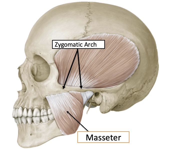
Masseter
O: Zygomatic Arch
I: Ramus + Angle of Mandible
A: Elevate Mandible (with force)
Powerhouse of mastication
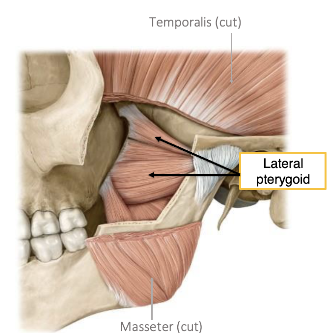
Lateral Pterygoid
O: Sphenoid Bone
I: Condyle of Mandible
A: Depression, Protraction, Side to side movement of Mandible
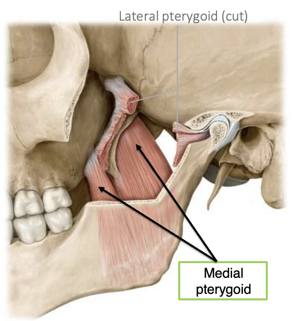
Medial Pterygoid
O: Sphenoid Bone
I: Angle of Mandible
A: Elevate Mandible
Muscles of Mastication: Innervation
Primary muscles of mastication innervated by the mandibular nerve (CNV3)
Mandibular Nerve = 3rd branch (mandibular branch) of CN V (trigeminal)

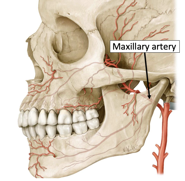
Muscles of Mastication: Blood Supply
Primary muscles of mastication supplied by branches of the maxillary artery
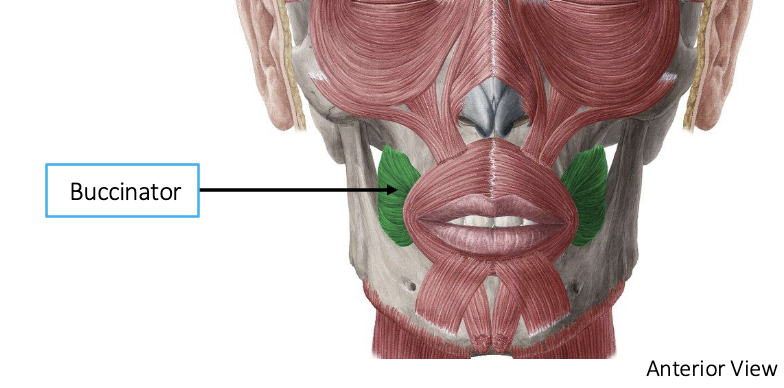
Buccinator
Important muscle in facial expression
Accessory muscle of mastication – keeps food in oral cavity
Innervated by: CNVII
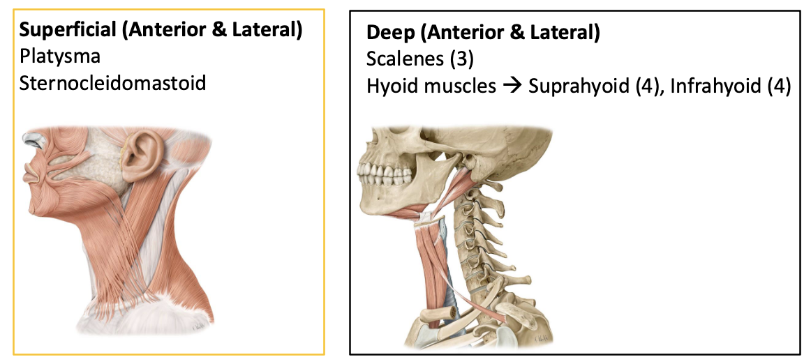
Anterior Neck Muscles
Responsible for moving the head and neck
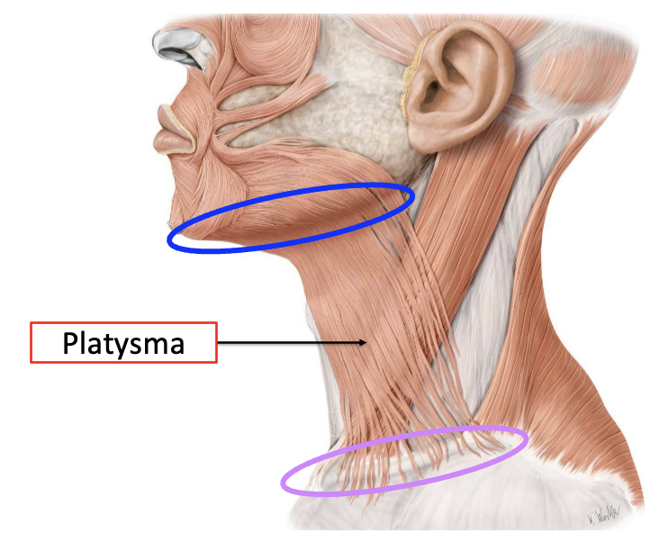
Platysma
O: Clavicle
I: Mandible
A: Depresses mandible (draws corners of the mouth inferiorly
Innervation: CNVII
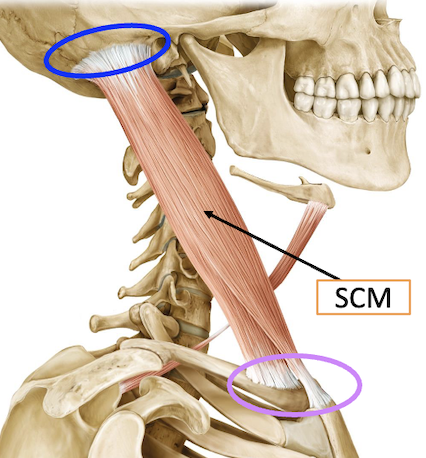
Sternocleidomastoid (SCM)
O: Sternum and Clavicle
I: Mastoid process
A:
Unilateral contraction – lateral flexion of neck, head rotation to opposite side
Bilateral contraction – head flexion (nod)
Innervation: CNXI
Scalene Muscles
Group of 3 muscles, deep within the anterolateral neck: Scalenus Anterior, Scalenus Medius, Scalenus Posterior
O: Transverse processes of cervical vertebrae
I: Superior Ribs (Rib 1, 2)
A:
Unilateral contraction – lateral flexion of neck (same side)
Bilateral contraction –anterior neck flexion (nod)
Hyoid Muscles
Supra vs Infra hyoid
Suprahyoid
Action: Elevate Hyoid bone (important for swallowing)
Infrahyoid
Action: Depress Hyoid bone (important for swallowing, speech)
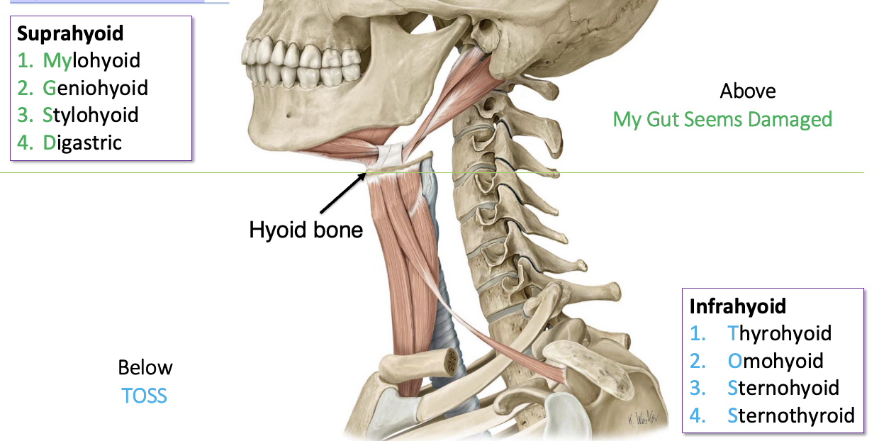
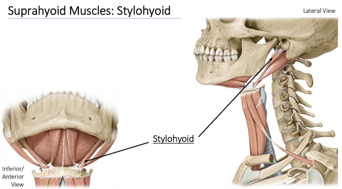
Digastric
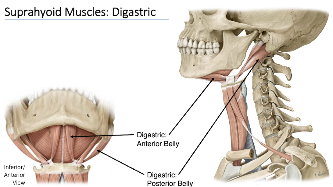
Stylohyoid
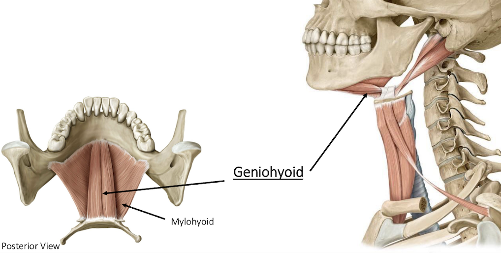
Geniohyoid
Geniohyoid is deep to Mylohyoid
O: Mandible
I: Hyoid bone
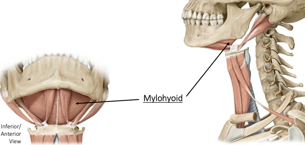
Mylohyoid
O: Mandible
I: Hyoid bone
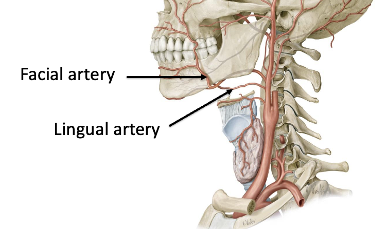
Suprahyoid Muscles: Blood Supply
Suprahyoid muscles supplied by branches of the facial and lingual arteries
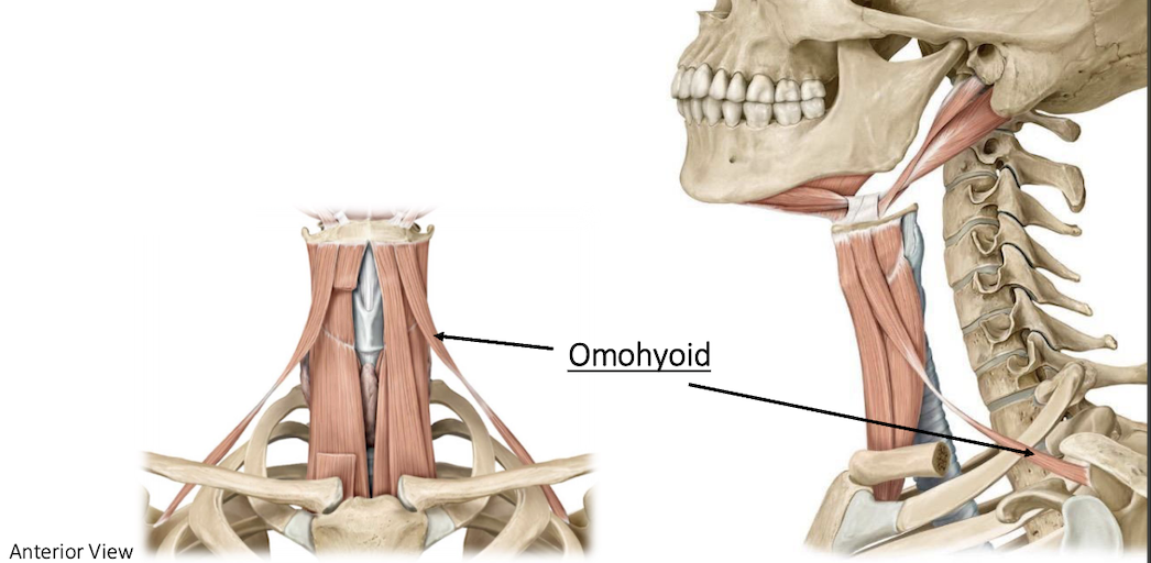
Omohyoid
Located most laterally of all infrahyoid muscles
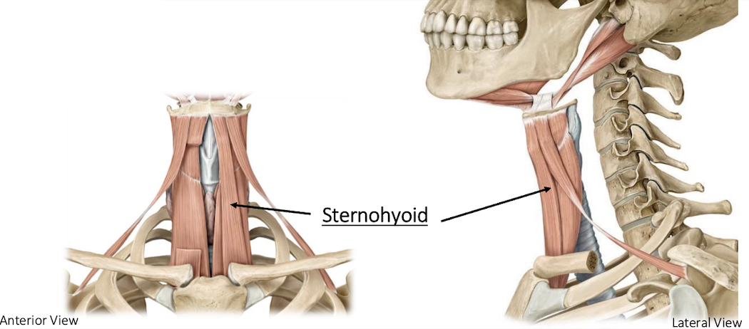
Sternohyoid
O: sternum
I: hyoid
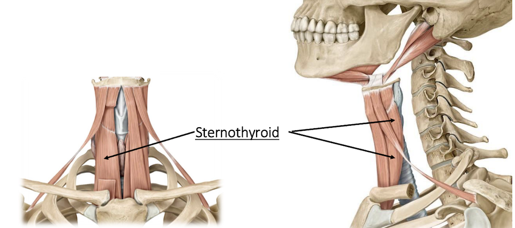
Sternothyroid
O: sternum
I: thyroid cart
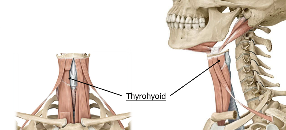
Thyrohyoid
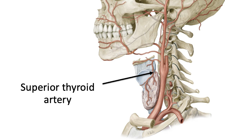
Infrahyoid Muscles: Blood Supply
Infrahyoid muscles supplied by branches of the superior thyroid artery
Posterior Neck Muscles
Responsible for supporting and stabilizing the head and neck
Superficial to Deep: Trapezius → Splenius capitis → Levator scapulae → Semispinalis capitis
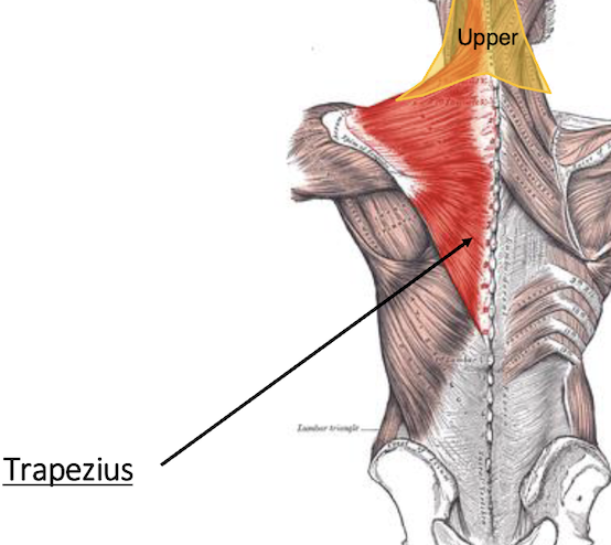
Trapezius
Superficial muscle of the back. Paired, trapezoid-shaped muscle
O: Base of skull (occipital bone), spinous processes of C7-T12
I: Scapula (acromion + spine), lateral aspect of clavicle
Action: Upper fibres extend and rotate the neck
Innervation: CNXI
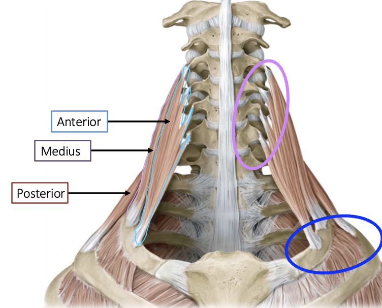
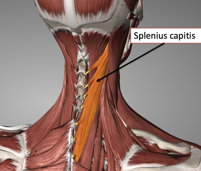
Posterior Muscles: Splenius Capitis
O: Cervical vertebrae
I: Mastoid process (temporal bone), occipital bone
A: Neck extension
Posterior Muscles: Levator Scapulae
O: Cervical vertebrae
I: Medial border of scapula (above spine)
A: Movement of the scapula

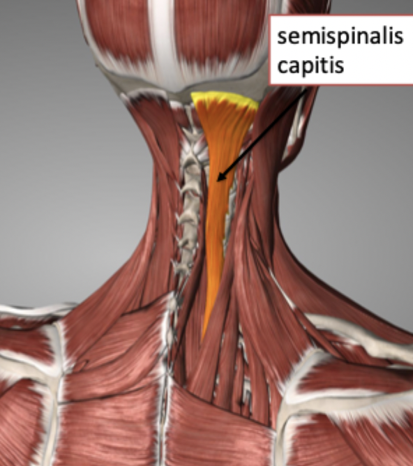
Posterior Muscles: Semispinalis Capitis
O: Cervical vertebrae
I: Occipital bone
A: Neck extension
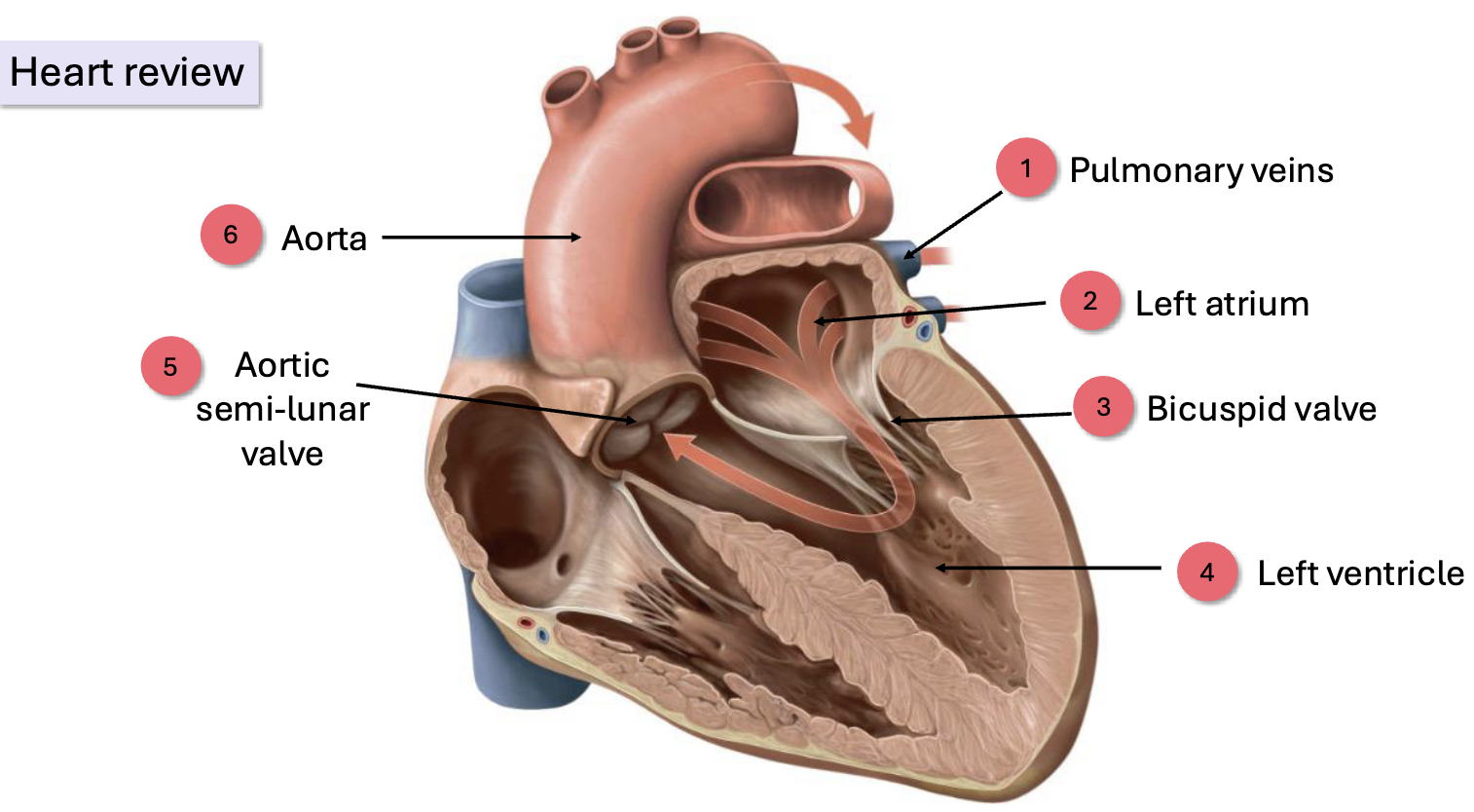
Venous Heart
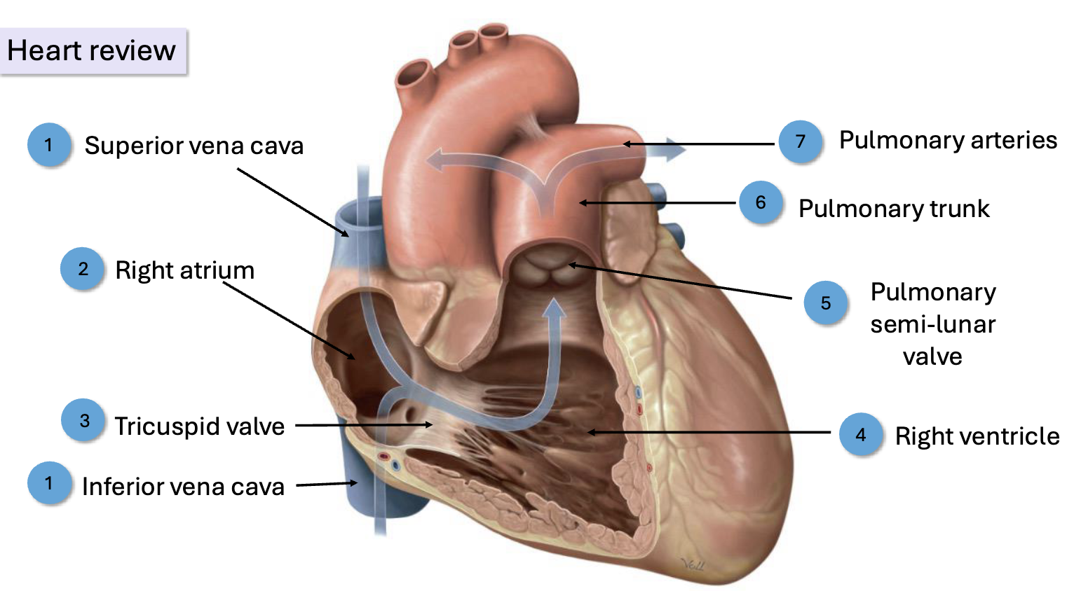
Arterial Heart
Arch of the Aorta
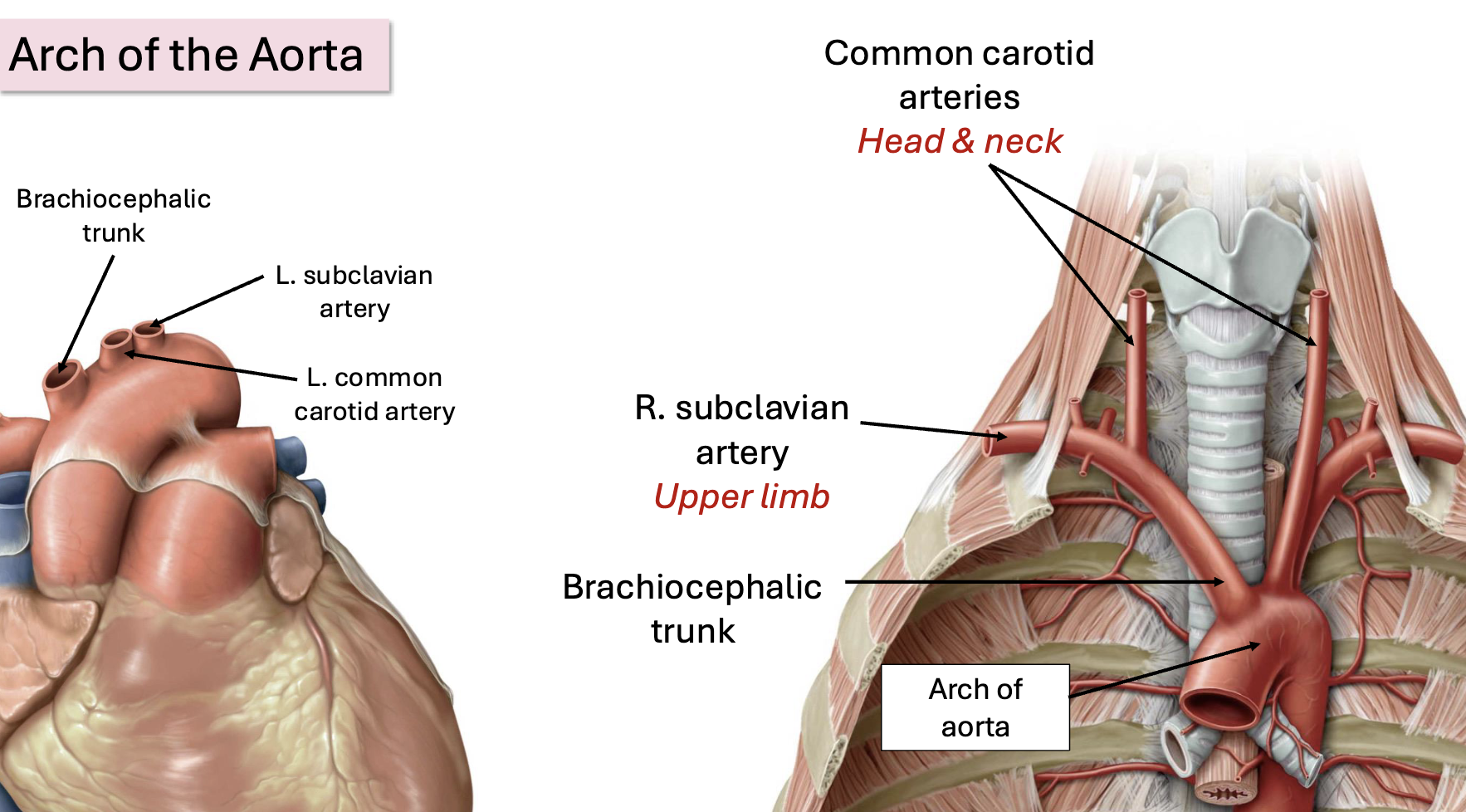
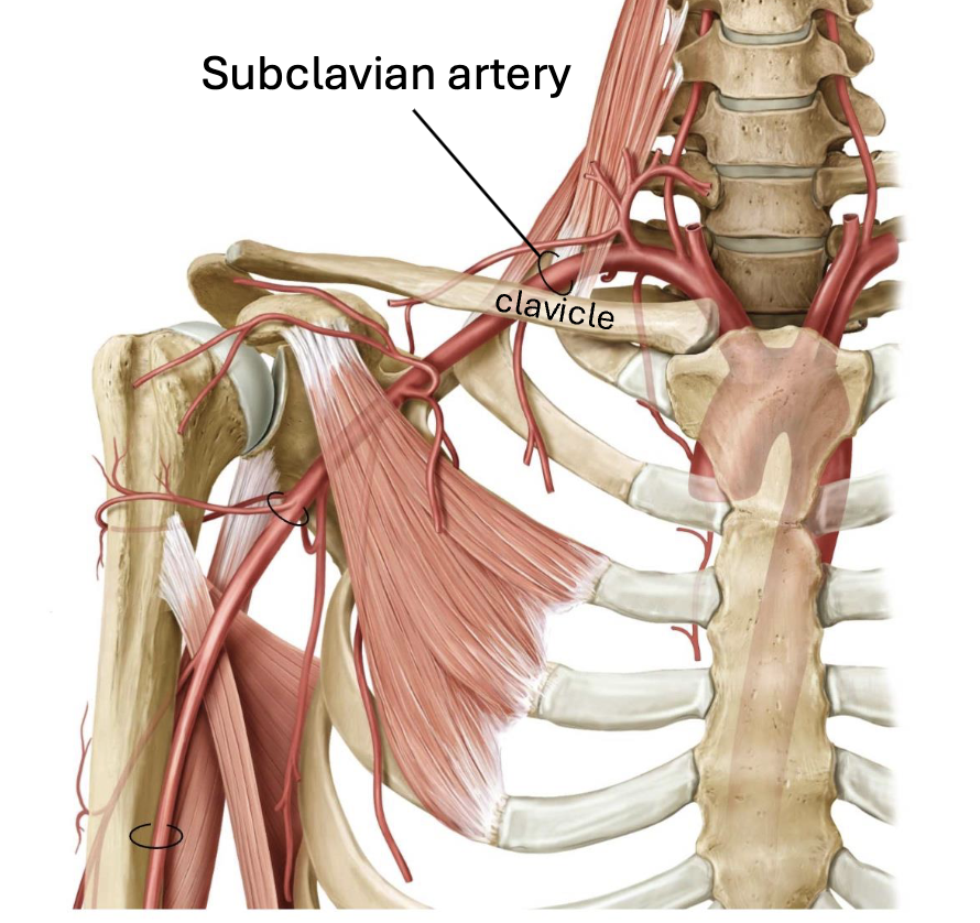
Upper limb arterial supply
Supplies blood to upper limb and branches off
Travels under the clavicle
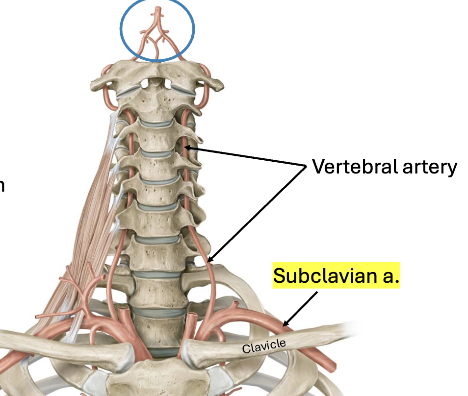
Head and Neck Arterial Supply: Vertebral arteries
Vertebral arteries: branches of the subclavian arteries
Supply: spinal cord, brainstem, brain
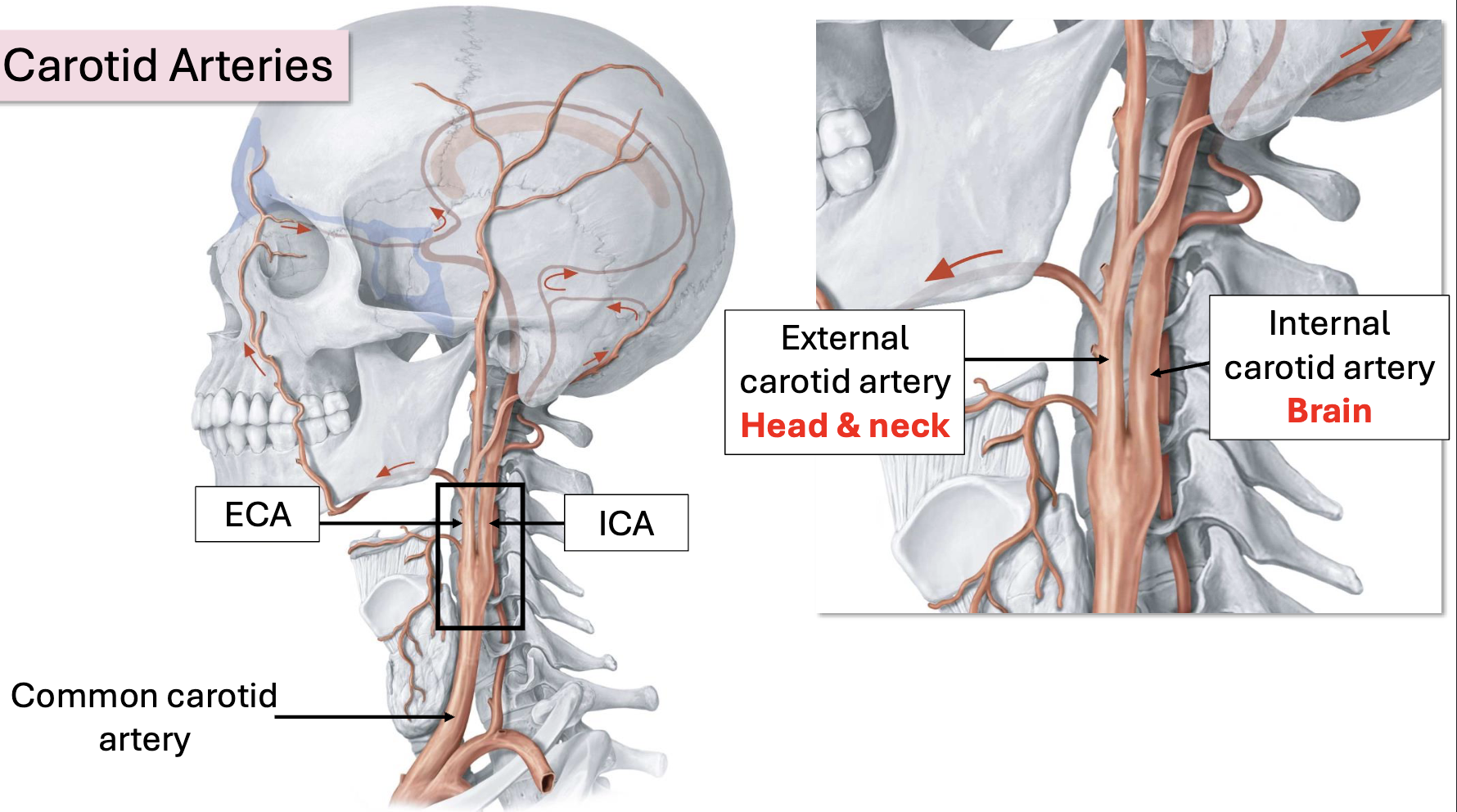
Head and Neck Arterial Supply: Carotid Arteries
ICA: Travels to brain via carotid canal
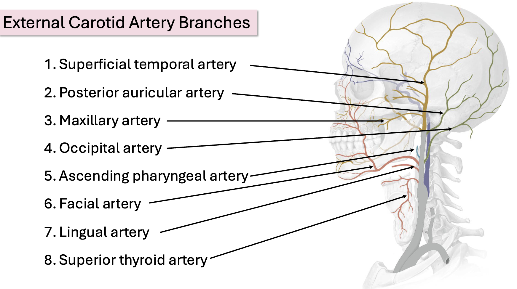
External Carotid Artery Branches
Superficial temporal artery: Supplies temple, scalp
Facial artery: Spiral like appearance. Supplies face (e.g., lips, external nose), neck (e.g., palatine tonsils)
Lingual artery: Supplies tongue, mouth
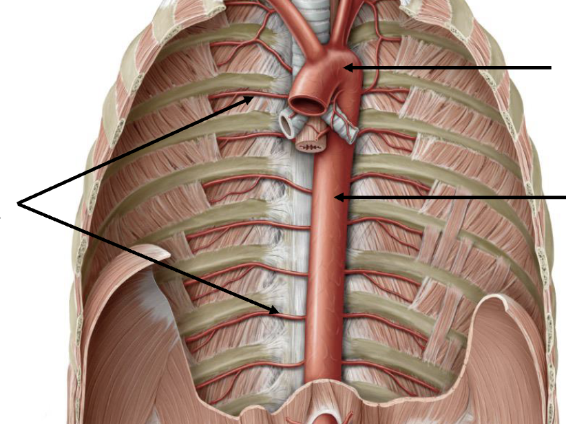
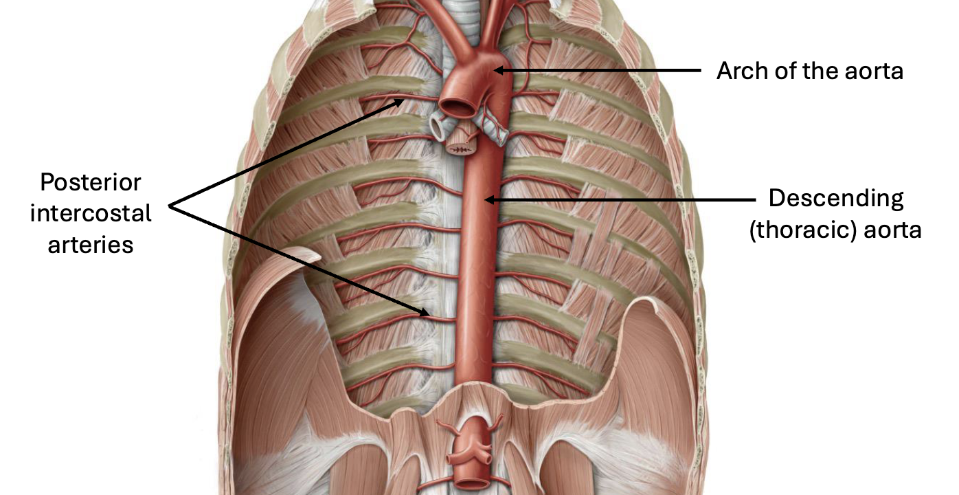
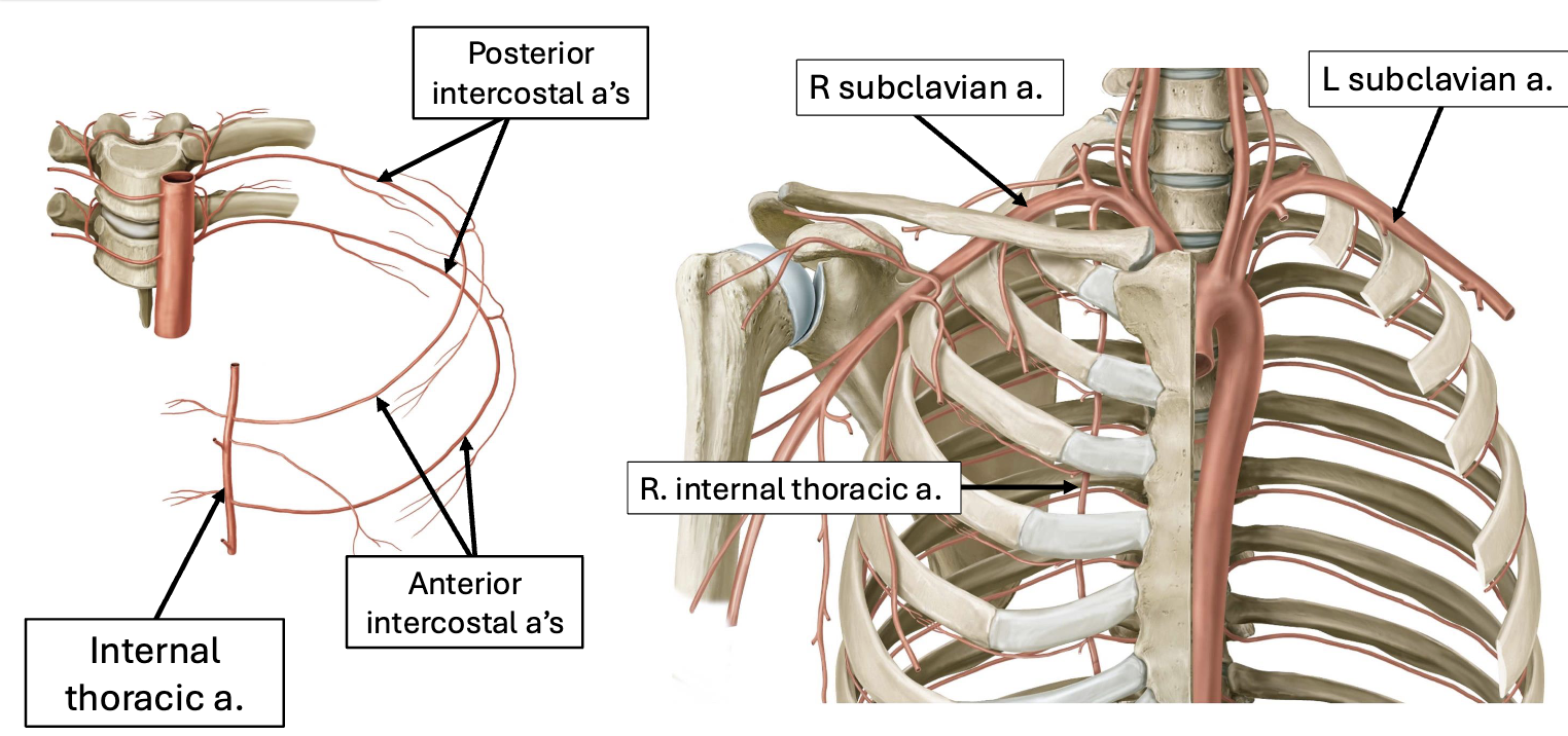
Posterior Thorax Arterial Supply
Anterior Thorax Arterial Supply
Subclavian —> Internal thoracic a. —> Anterior intercostal arteries
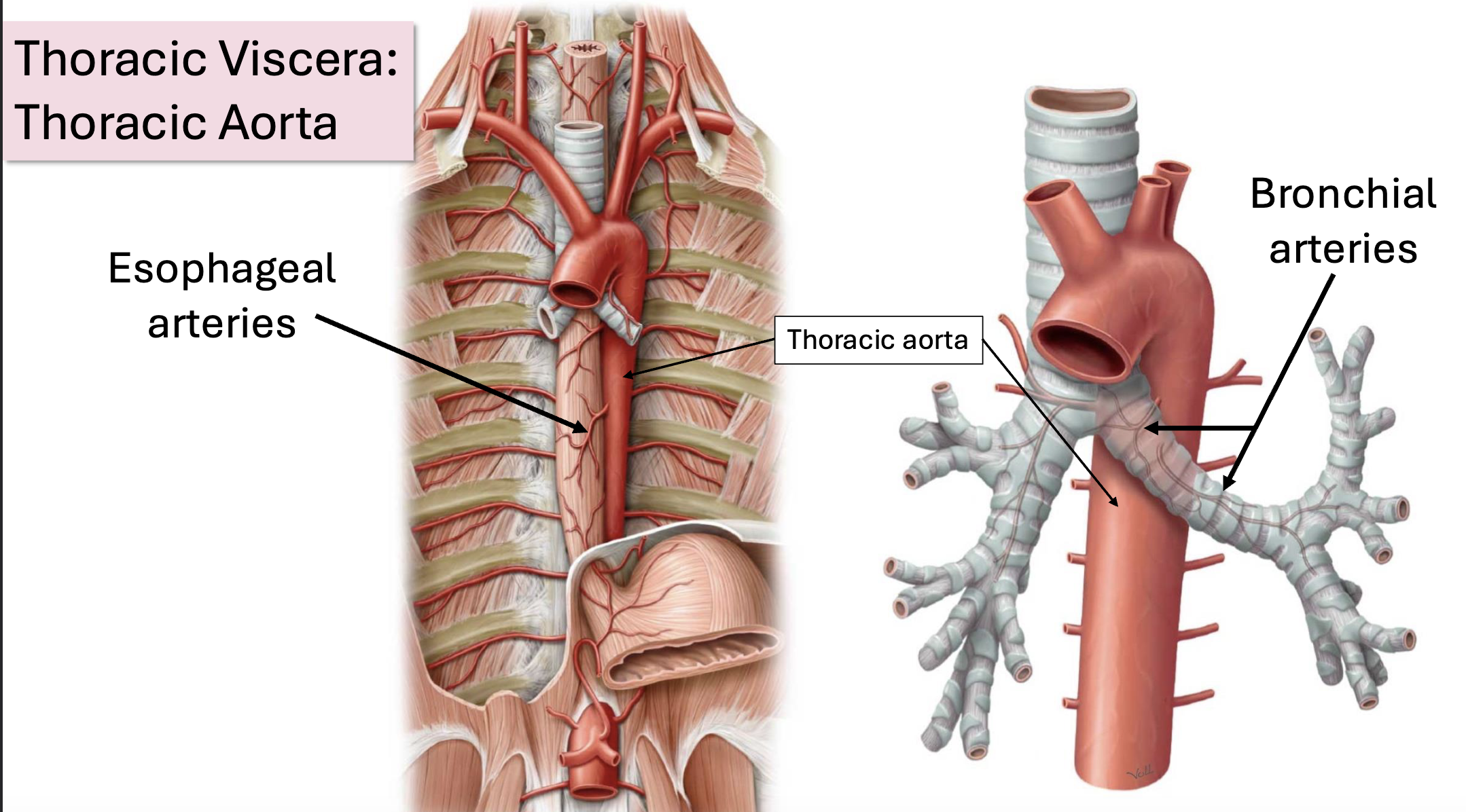
Thoracic Aorta Branches
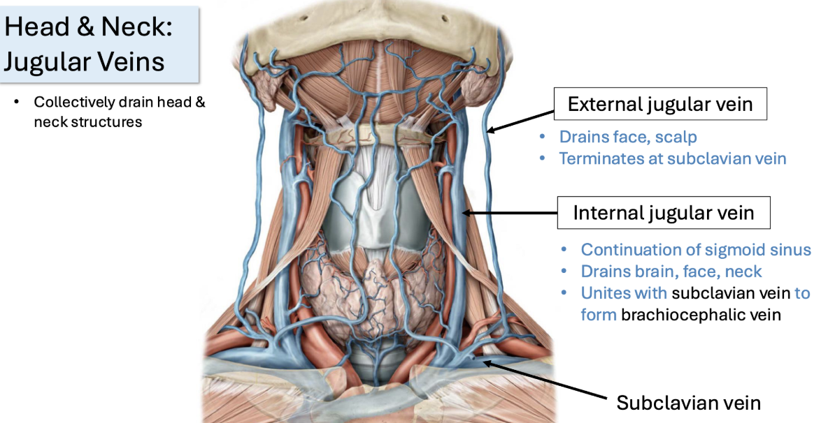
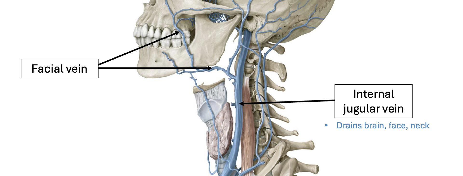
Head & Neck: Jugular Veins
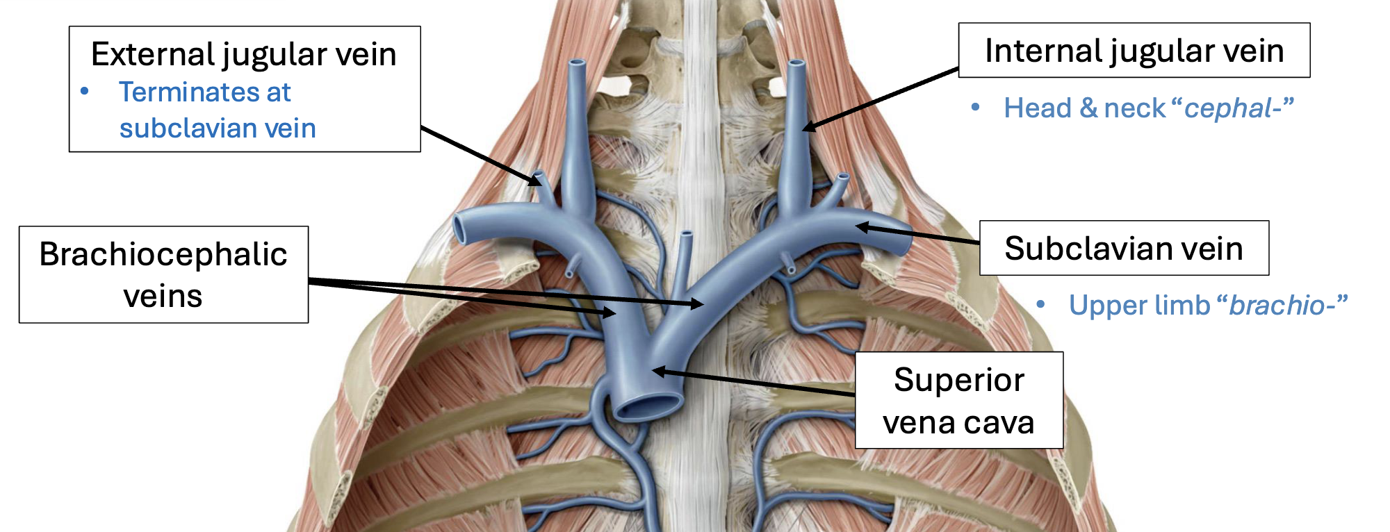
Head & Neck: Subclavian & Brachiocephalic Veins
Jugular Veins —> Subclavian Veins —> Brachiocephalic Veins —> Superior Vena Cava
Head & Neck: Facial vein
Drains into internal jugular vein
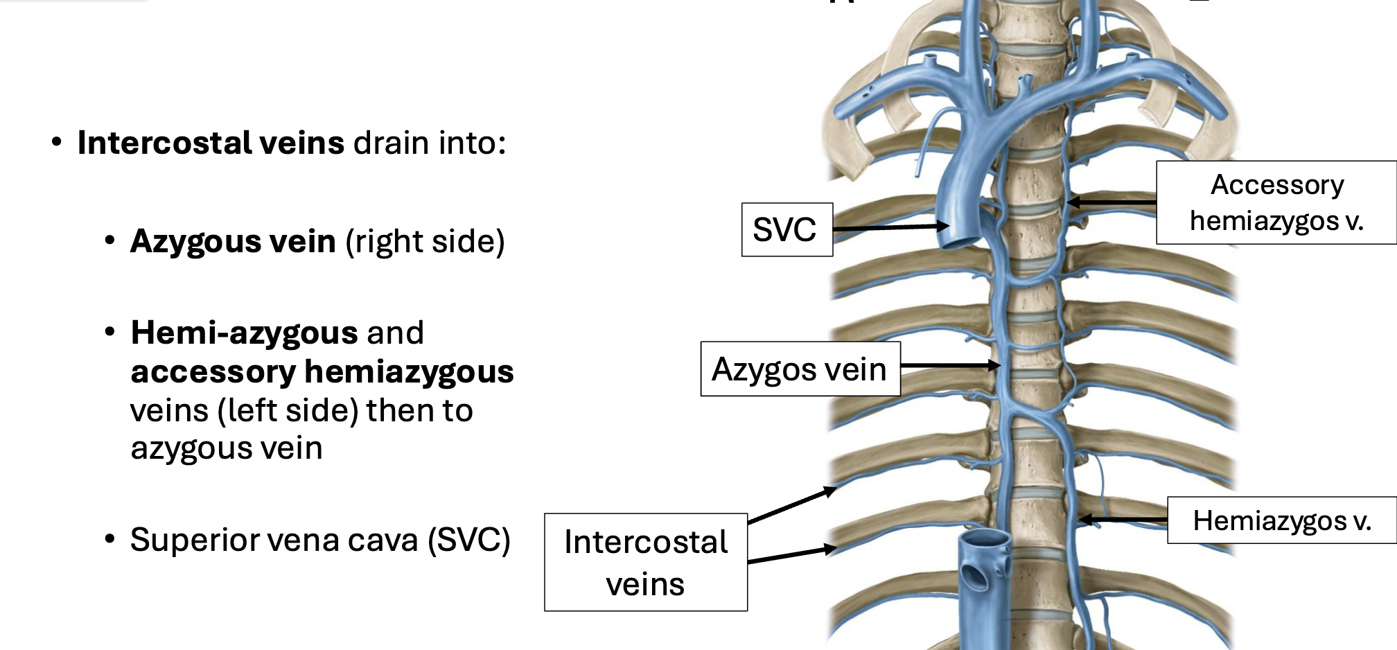
Thorax Venous Drainage
Azygous Vein —> SVC
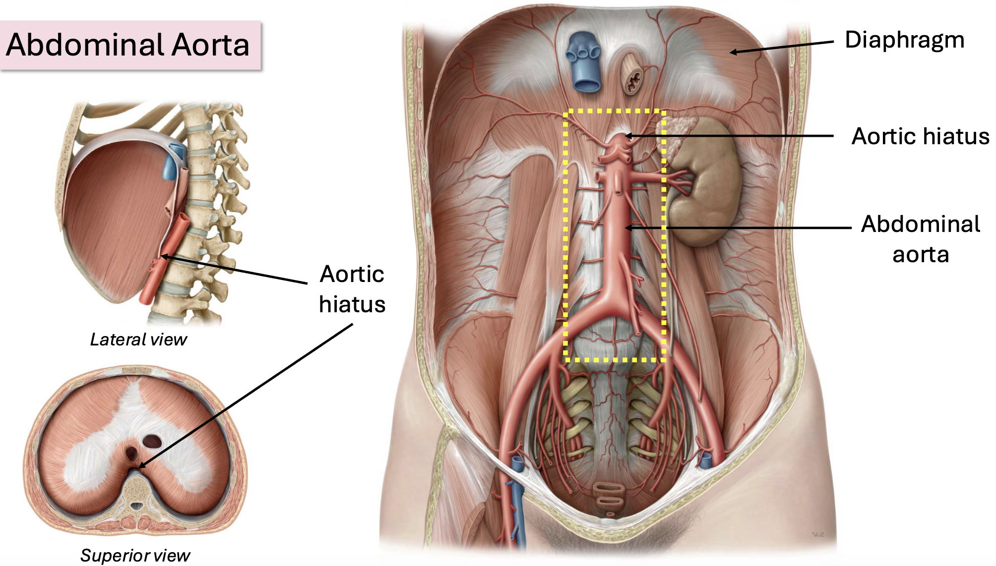
Abdominal Aorta
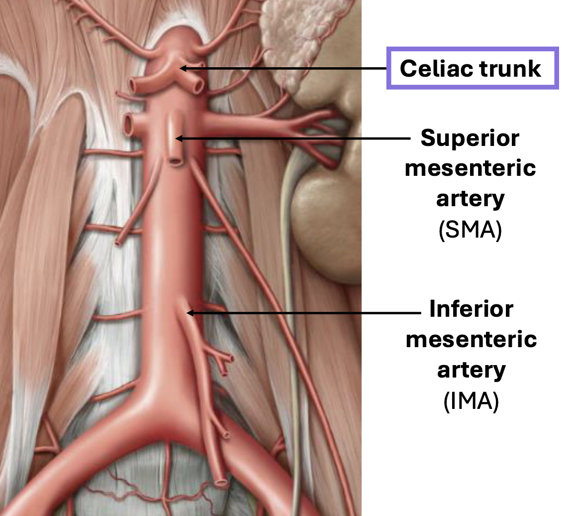
Abdominal Aorta Unpaired Branches
Celiac trunk
Stomach, pancreas, spleen, liver & gall bladder, small intestine (proximal duodenum)
SMA
Pancreas, small intestine (distal duodenum, ileum, jejunum), most of the large intestine
IMA
Distal large intestine to rectum
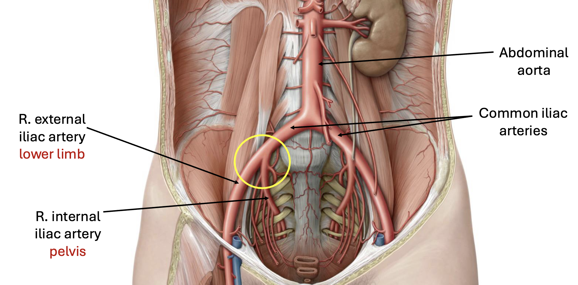
Pelvis Arterial Supply
Pelvis Veinous Drainage
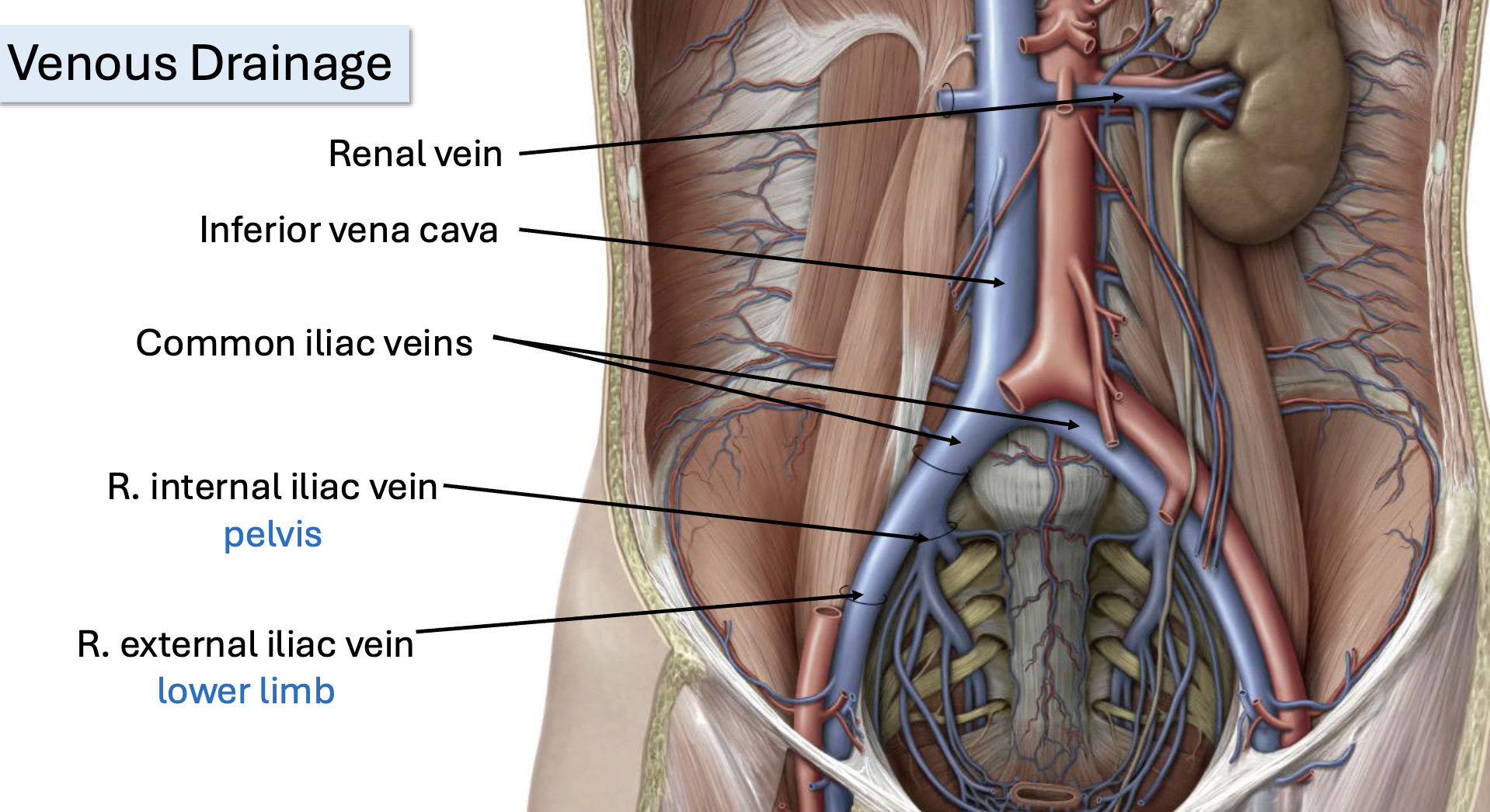
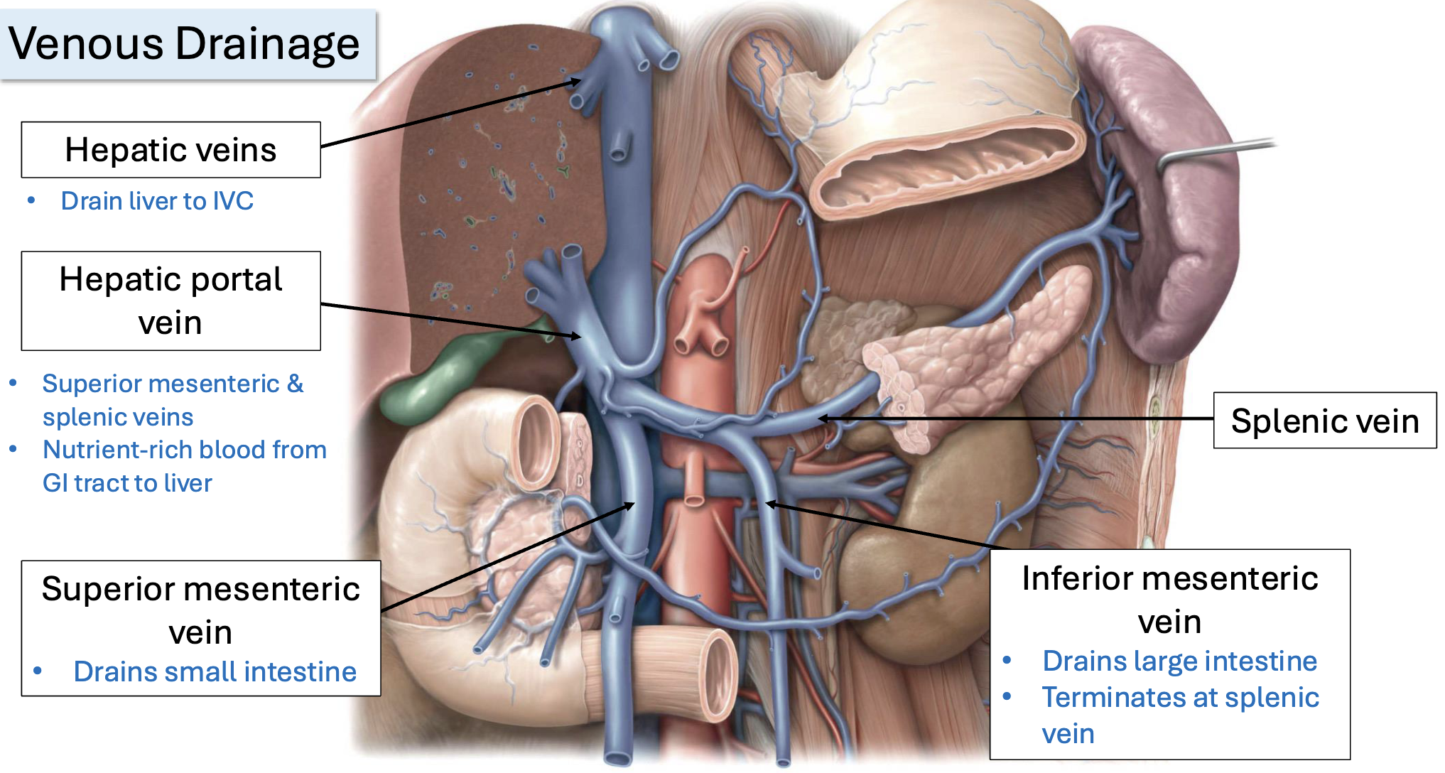
Abdominal Veinous Drainage
IVC & Abdominal Aorta

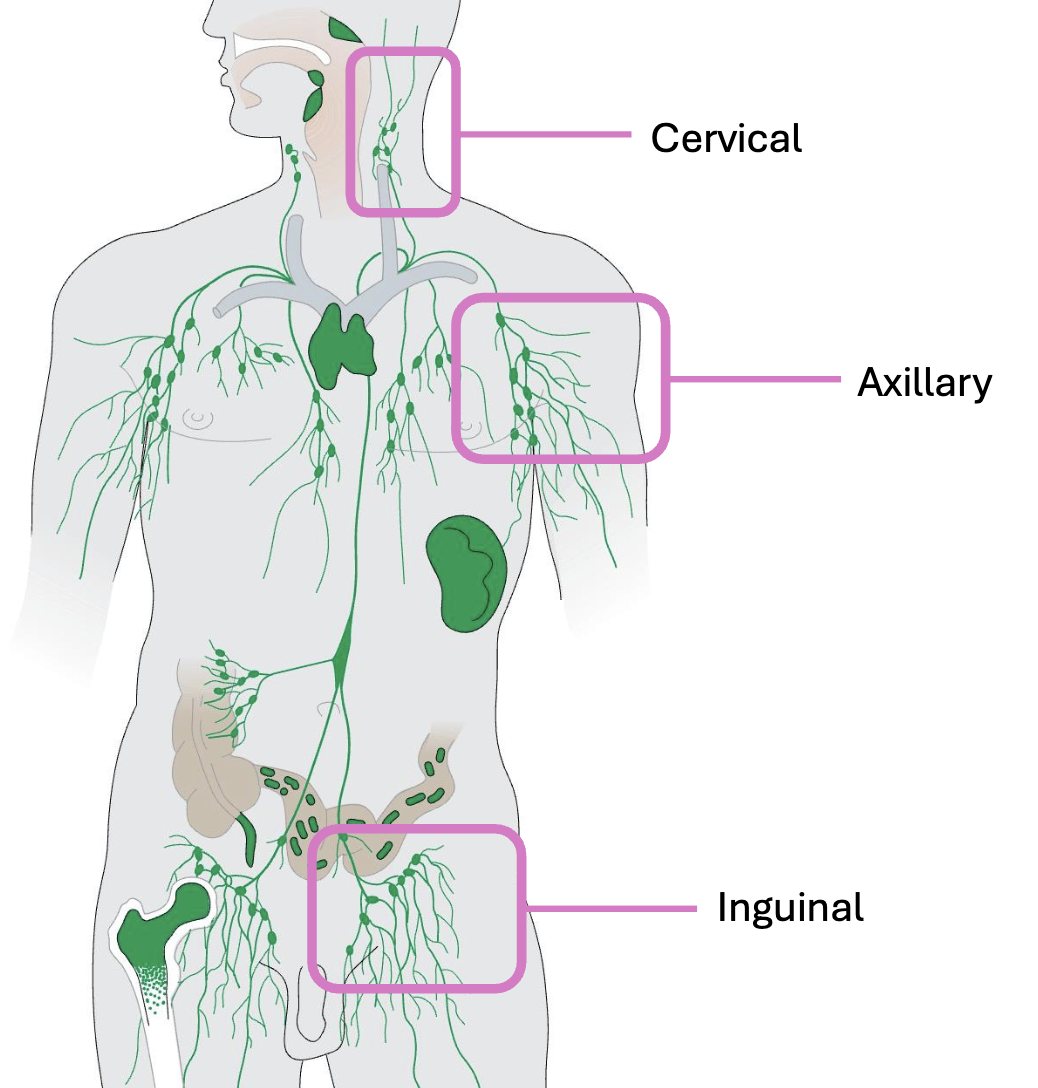
The Lymphatic System
Part of the immune system, composed of lymphatic vessels, lymph nodes & lymphatic organs (e.g., spleen, tonsils)
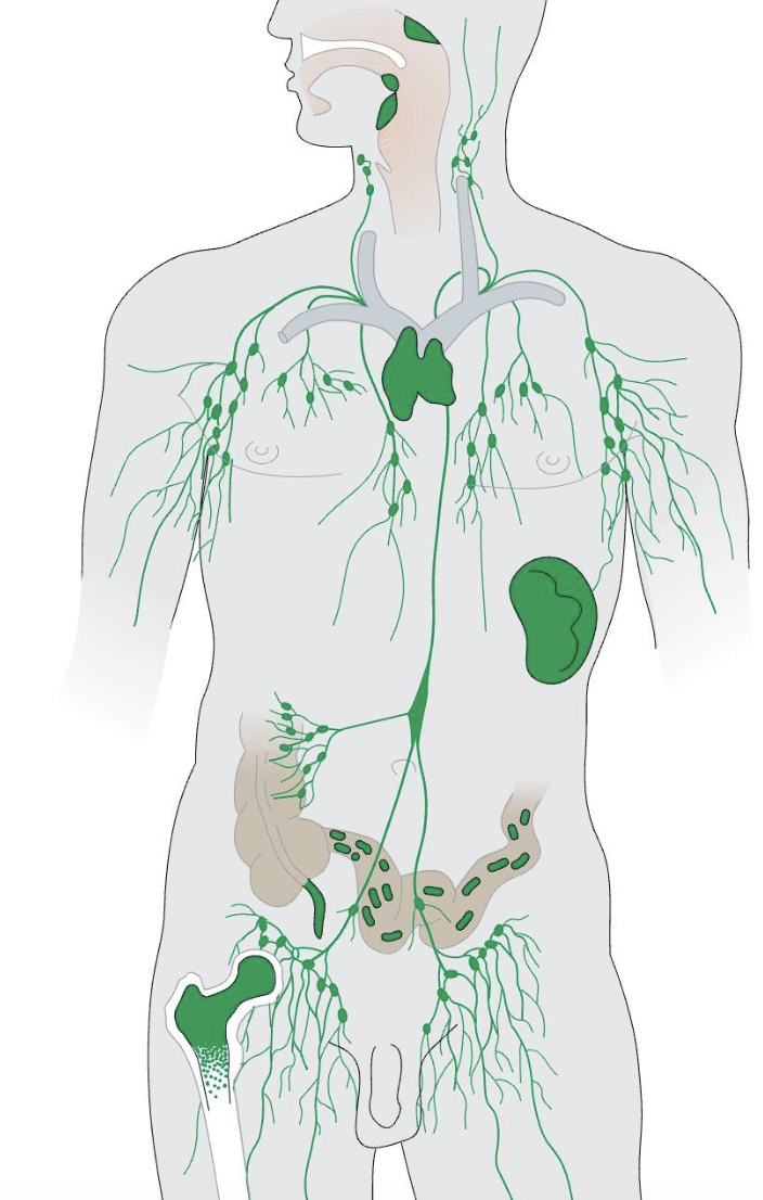
Lymph Nodes
Organs of the lymphatic system linked through lymphatic vessels
Sites of immune cells; act to filter foreign particles
The abdominal wall
Muscles that enclose the abdominal cavity
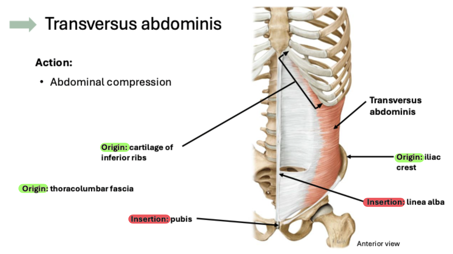
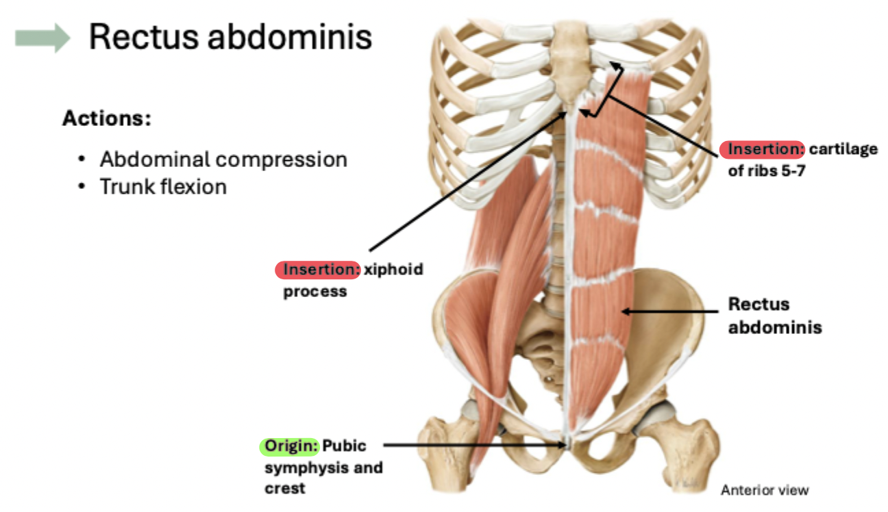
Protects abdominal viscera and stabilizes/promotes movement of the trunk
Assists in abdominal compression for forceful respiration and defecation

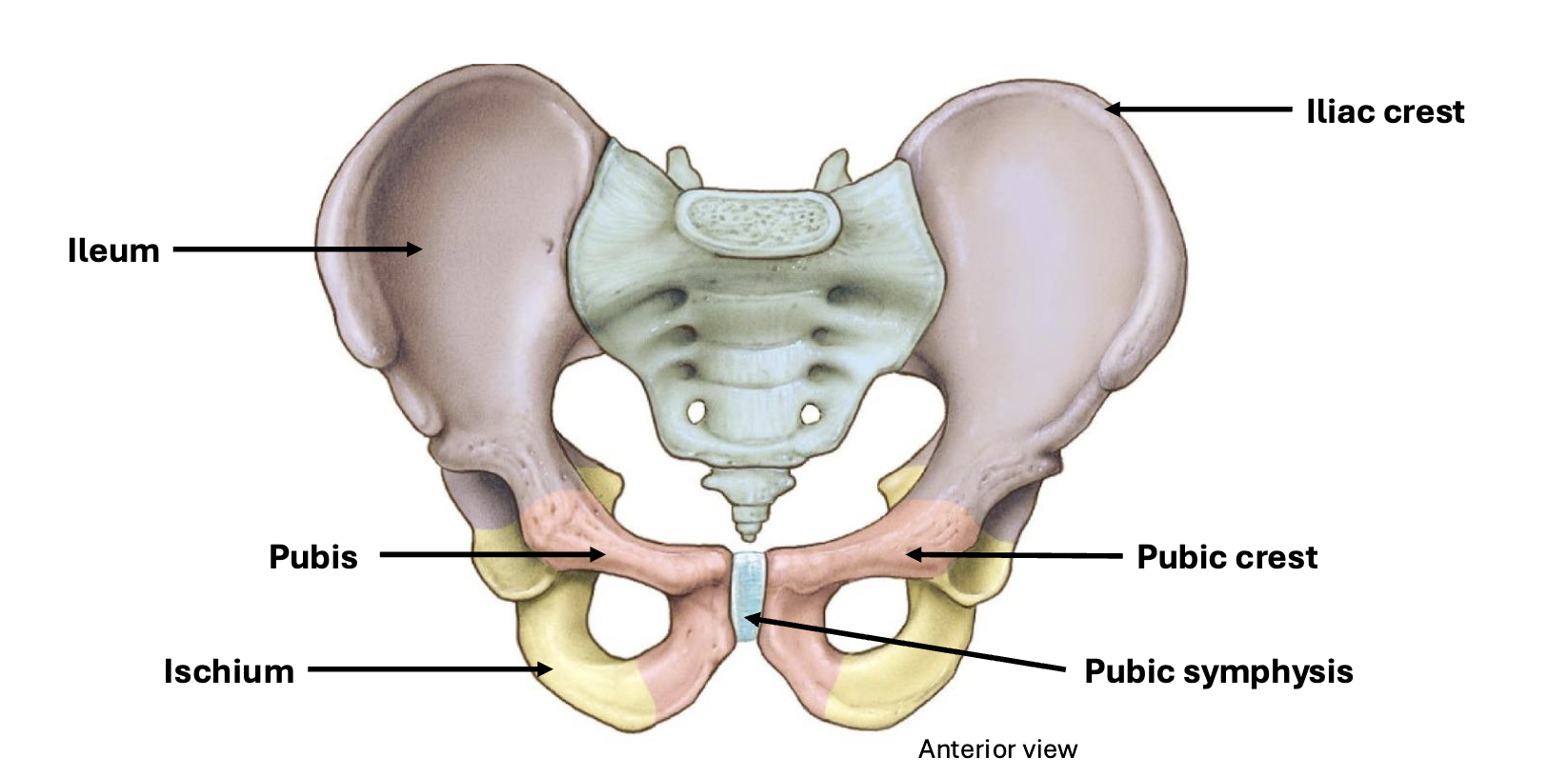
Pelvic bone anatomy

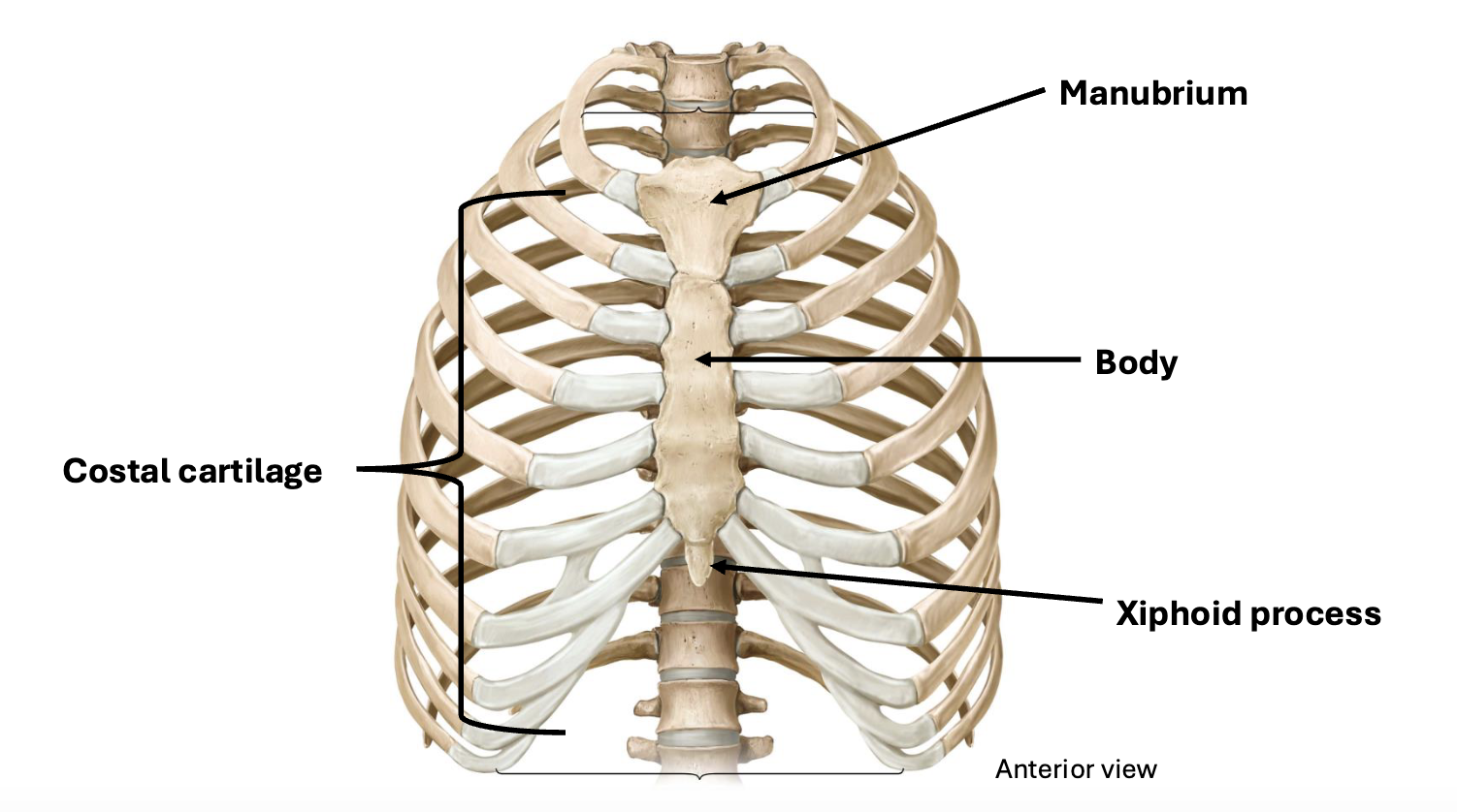
Thoracic cage anatomy
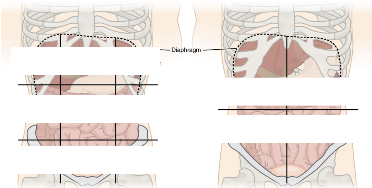
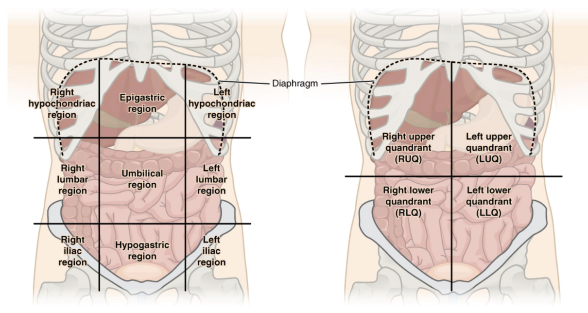
Divisions of the abdomen
Quadrants (4) and Regions (9)
Anterior/lateral abdominal wall muscles
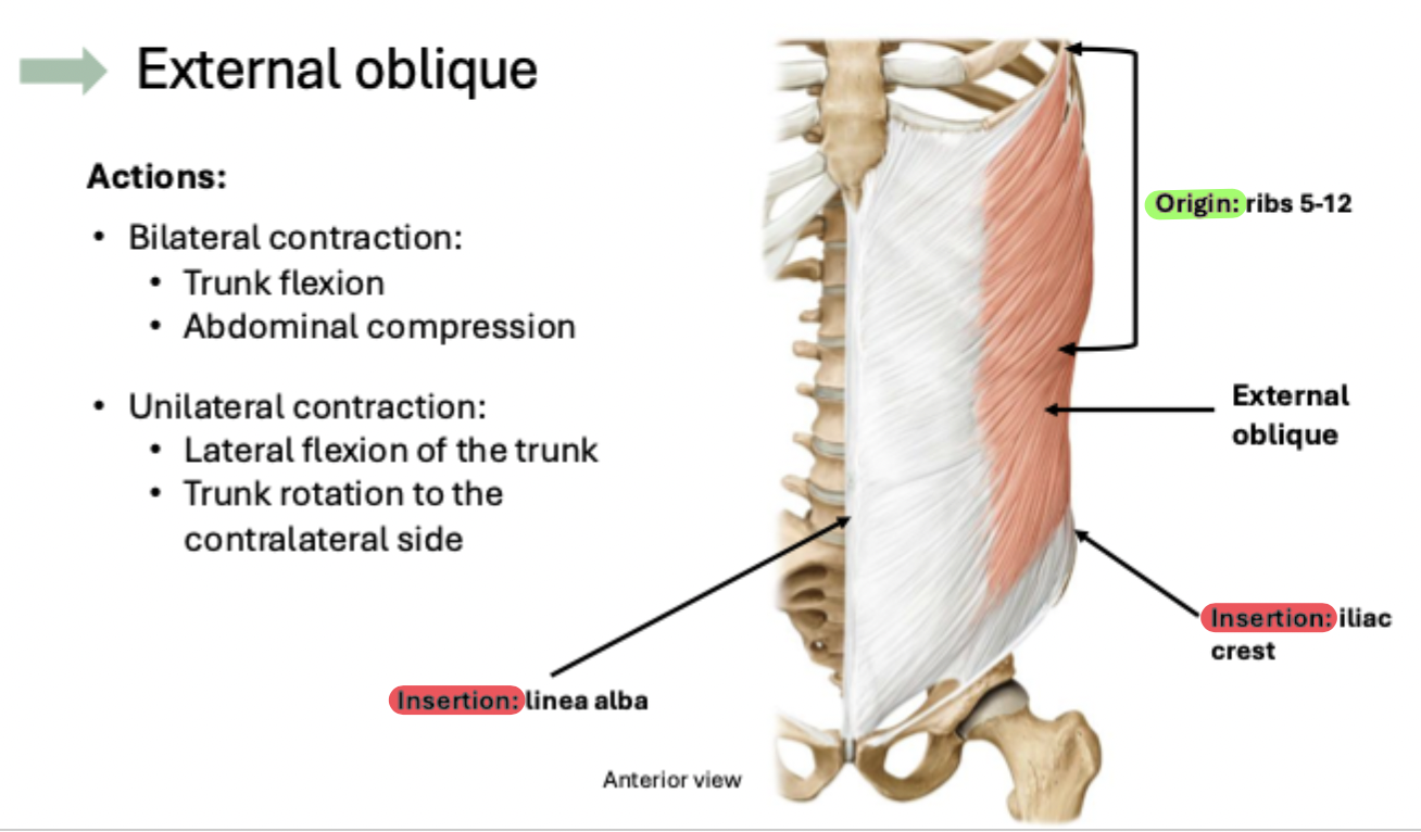
1. External oblique
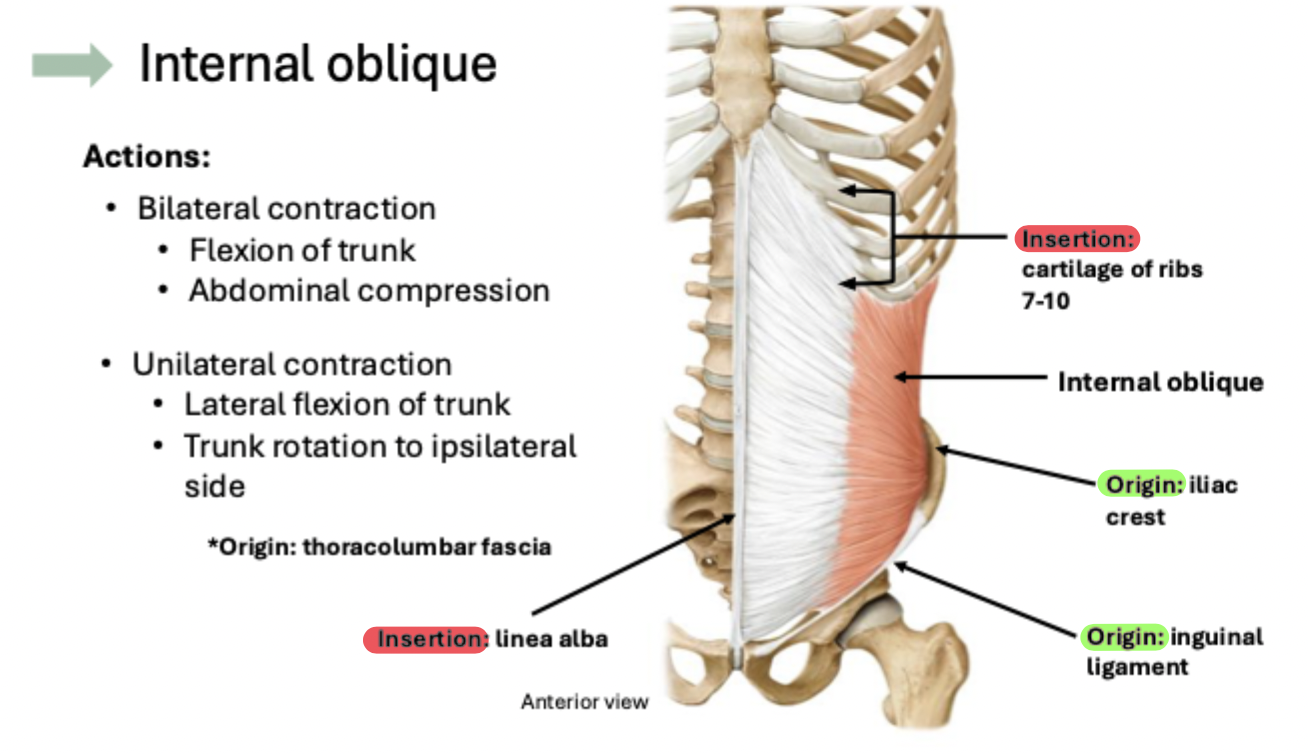
2. Internal oblique
3. Transversus abdominus
4. Rectus abdominis
Important actions
Trunk flexion, Lateral trunk flexion, Trunk rotation
External oblique
Internal oblique
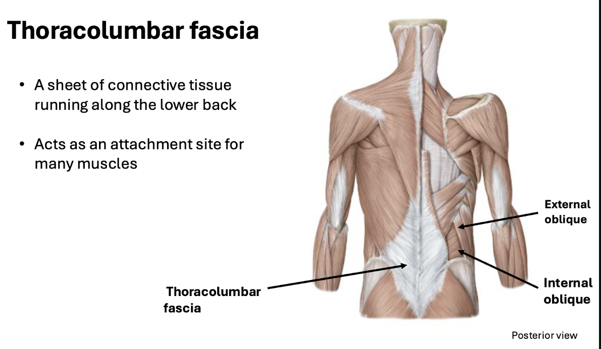
Thoracolumbar fascia
Transversus abdominis
Rectus abdominis
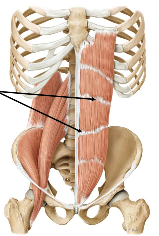
Tendinous intersections
Fibrous bands separating the rectus abdominis muscle
Allow for segmented contraction of rectus abdominis
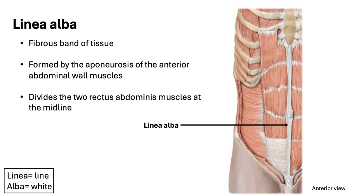
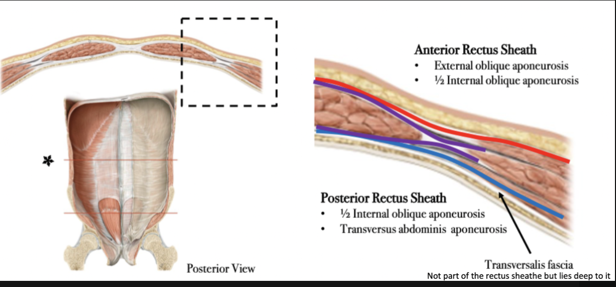
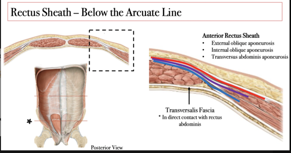
Rectus sheath
An aponeuroses of the external oblique, internal oblique, and transversus abdominis
Encloses the rectus abdominis
Converges at the midline at the linea alba
Linea alba
The arcuate line
Located just below the umbilicus and above the pubis
Marks a change in the composition of the rectus sheath
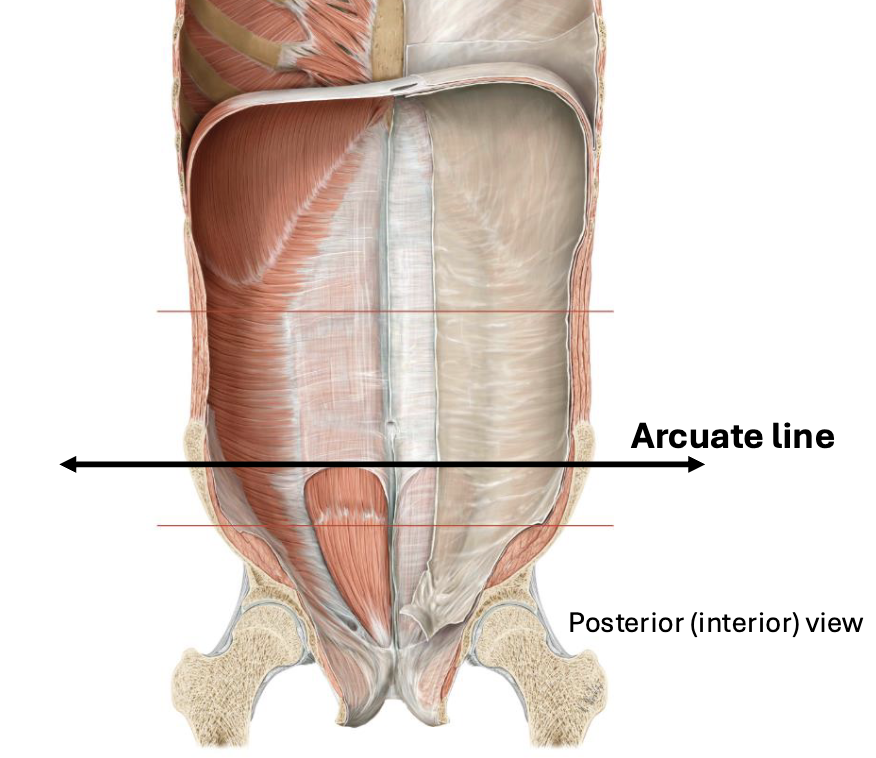
Above the arcuate line
Below the arcuate line
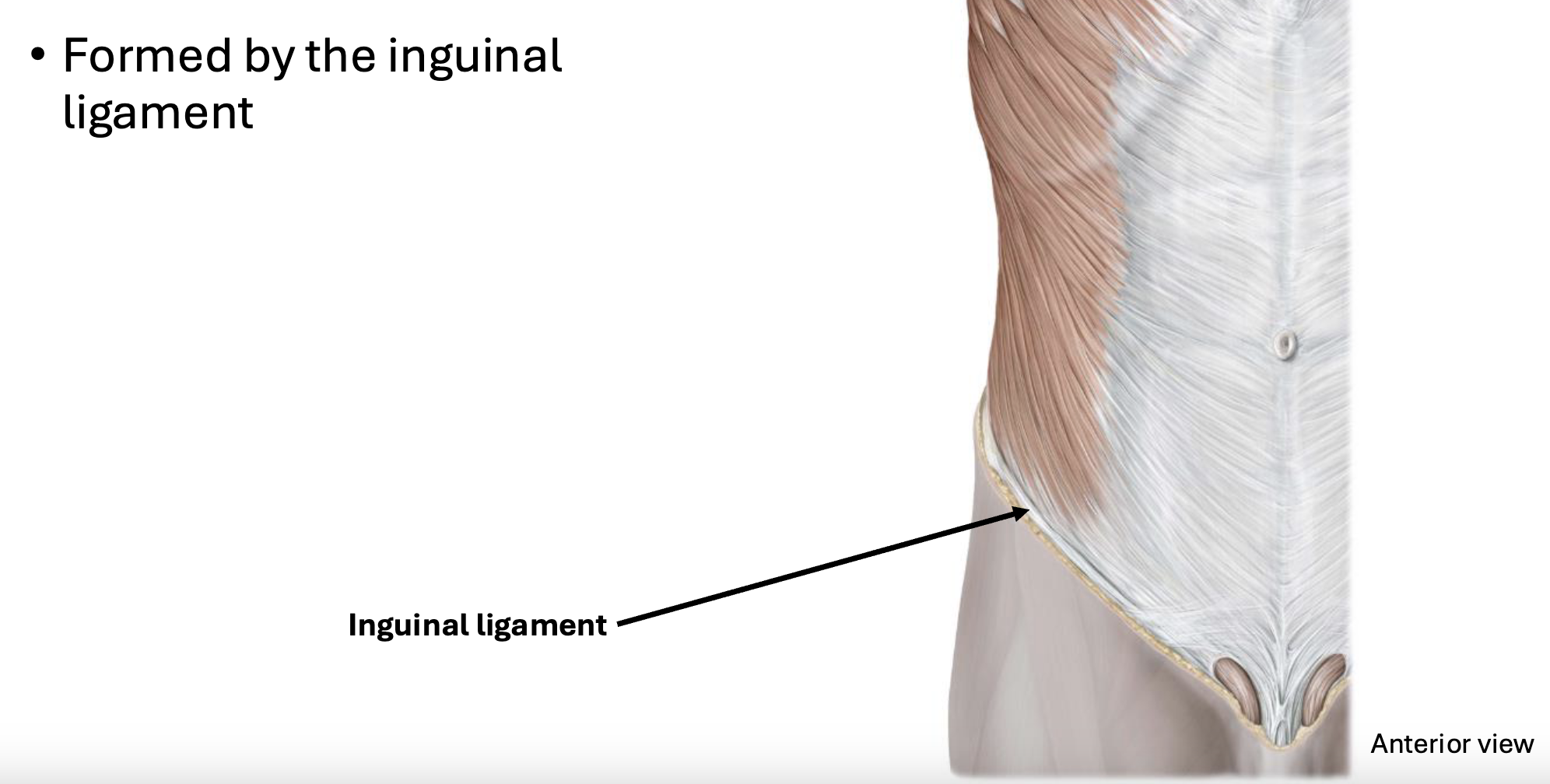
Inguinal region
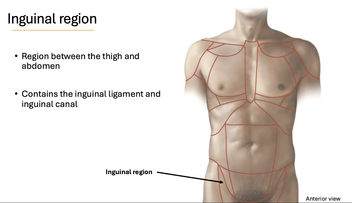
Inguinal ligament

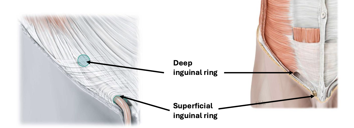
Inguinal canal
Passageway through the lower abdominal wall
Runs from the deep inguinal ring to the superficial inguinal ring
Borders of the inguinal canal
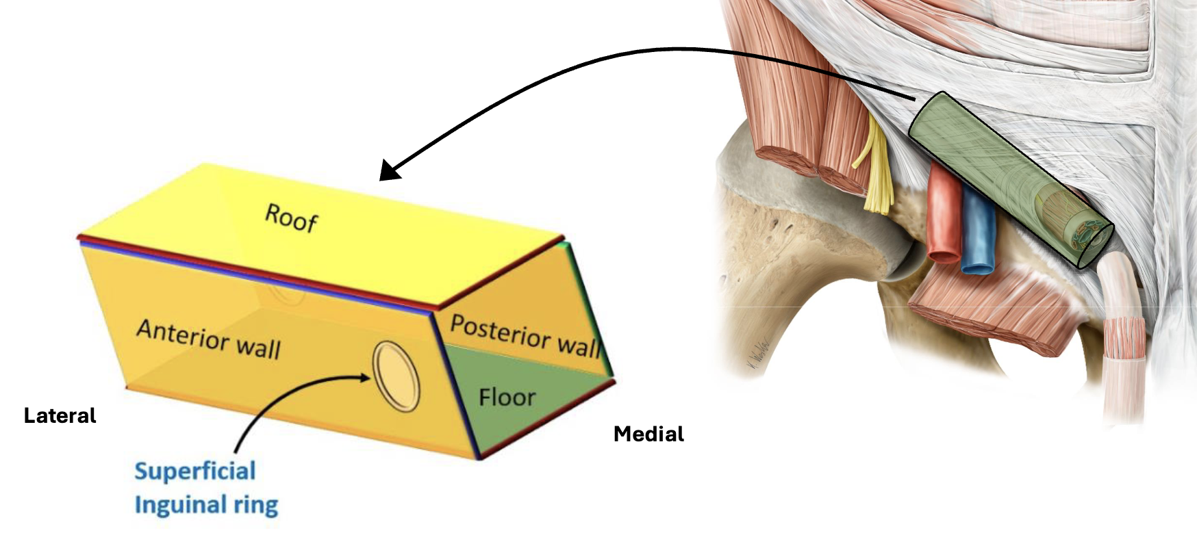
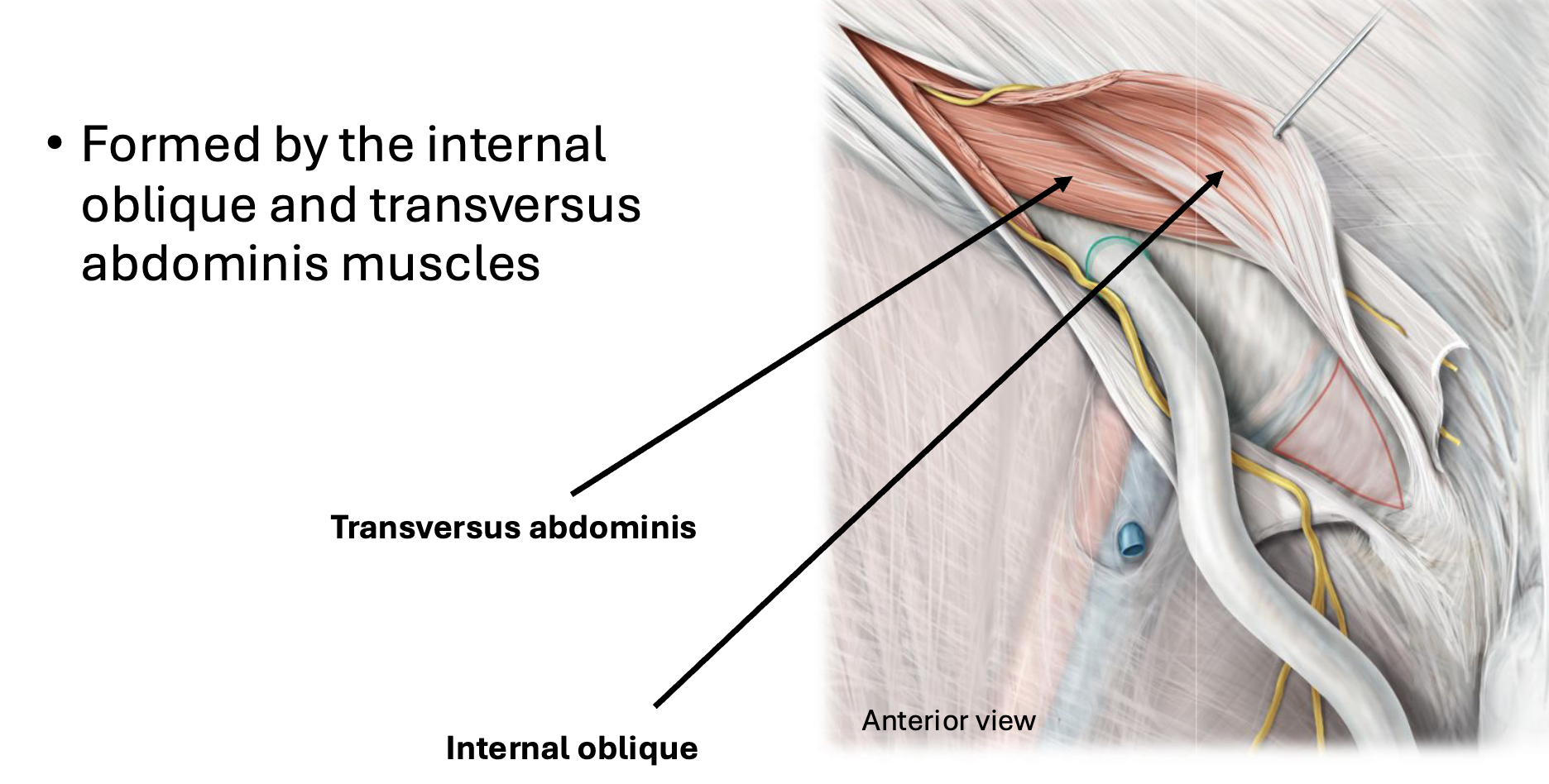
Inguinal canal Anterior wall

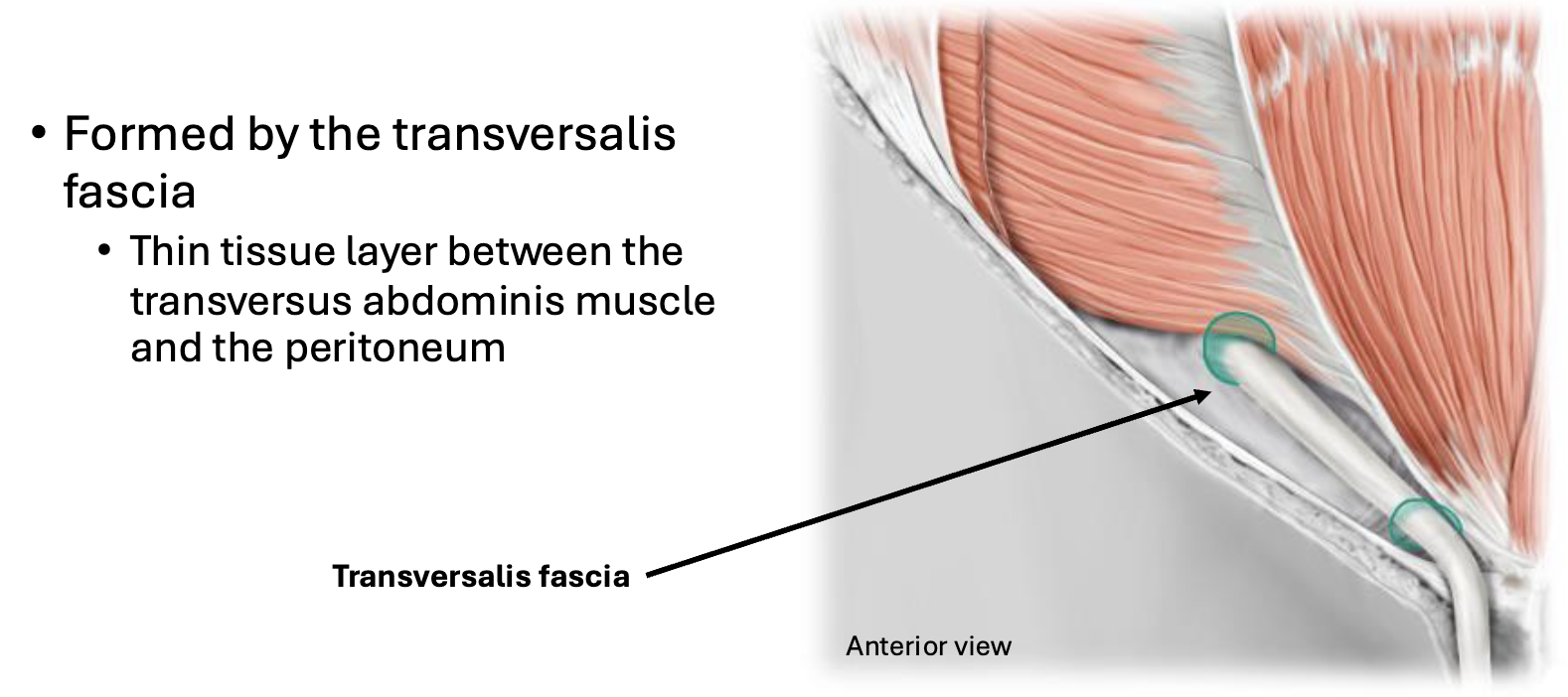
Inguinal canal Posterior wall
Inguinal canal Roof
Inguinal canal Floor
Contents: female inguinal canal
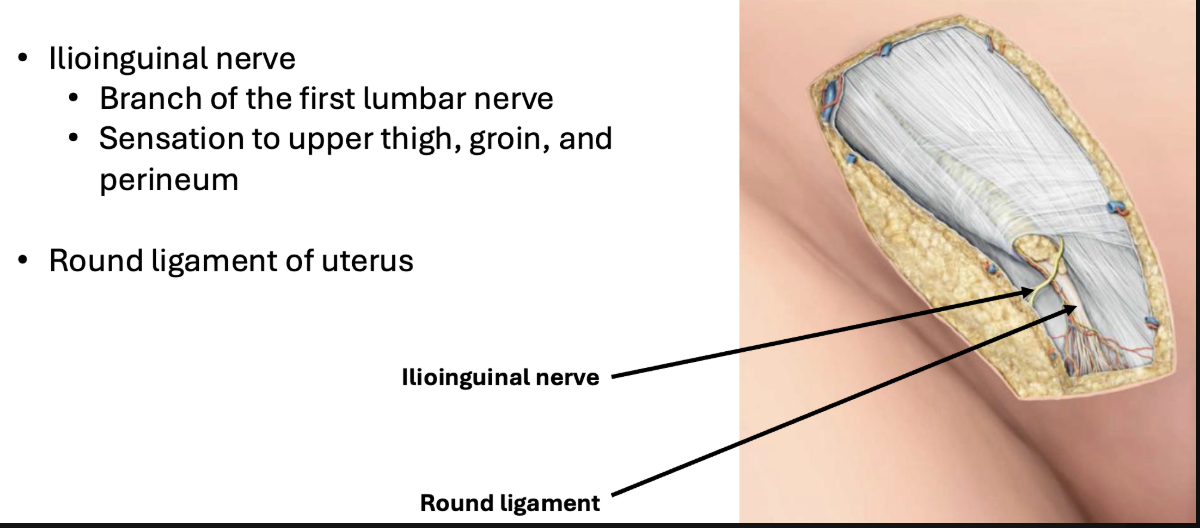
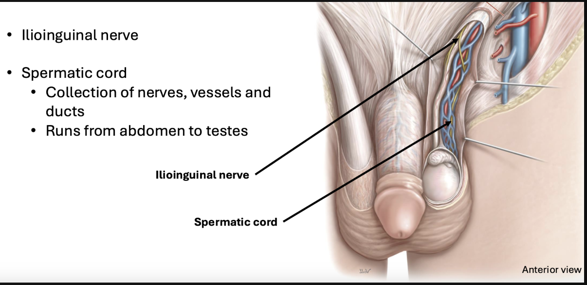
Contents: male inguinal canal
Posterior abdominal wall muscles
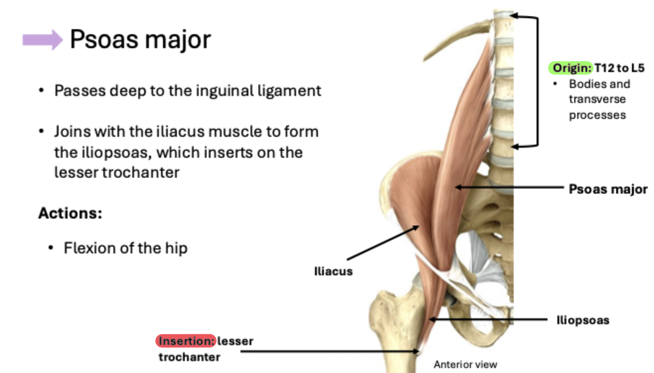
1. Psoas major
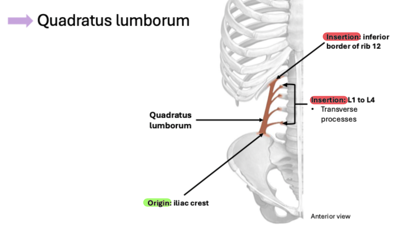
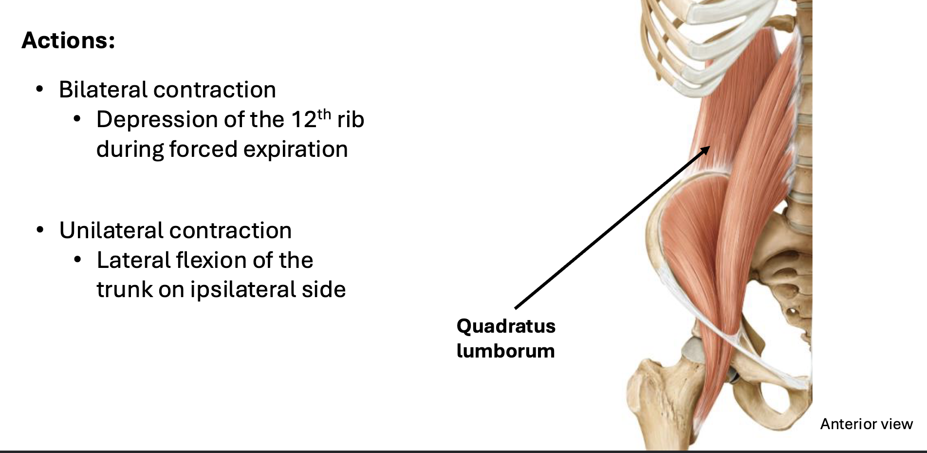
2. Quadratus lumborum
Psoas major
Quadratus lumborum
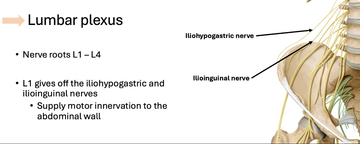
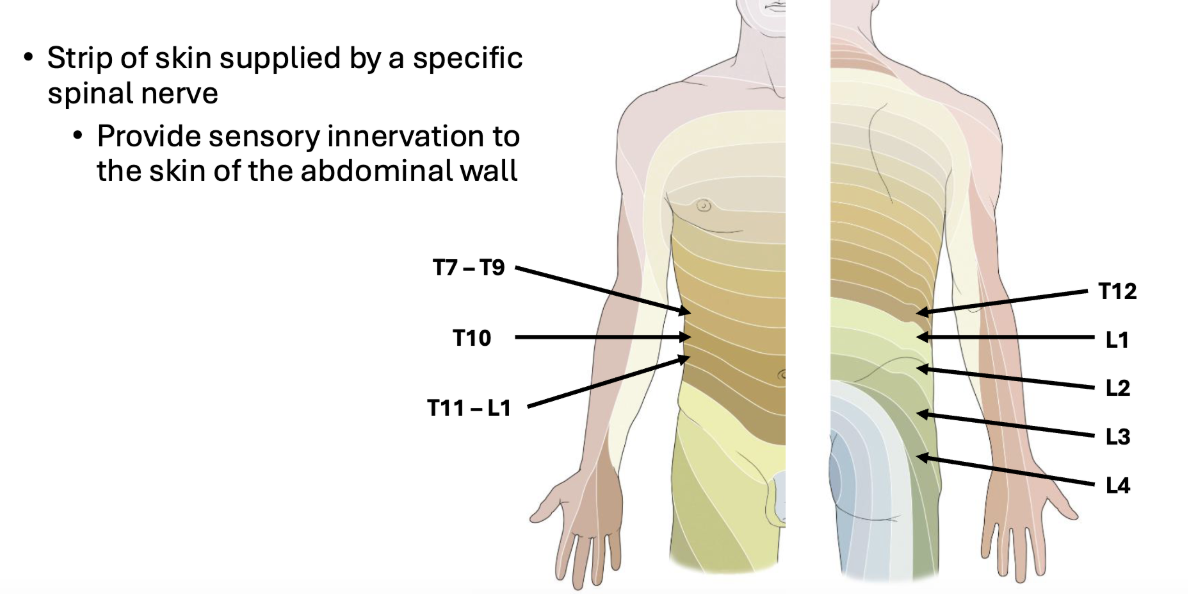
Abdominal Wall Innervation comes from...
1. Intercostal nerves
2. The lumbar plexus
3. Dermatomes
Intercostal nerves
The ventral rami of thoracic nerve roots T2 –T12 form the intercostal nerves
Travel between the ribs
Inferior intercostal nerves (T7 – T12) supply motor innervation to the abdominal wall
Lumbar plexus
Dermatomes
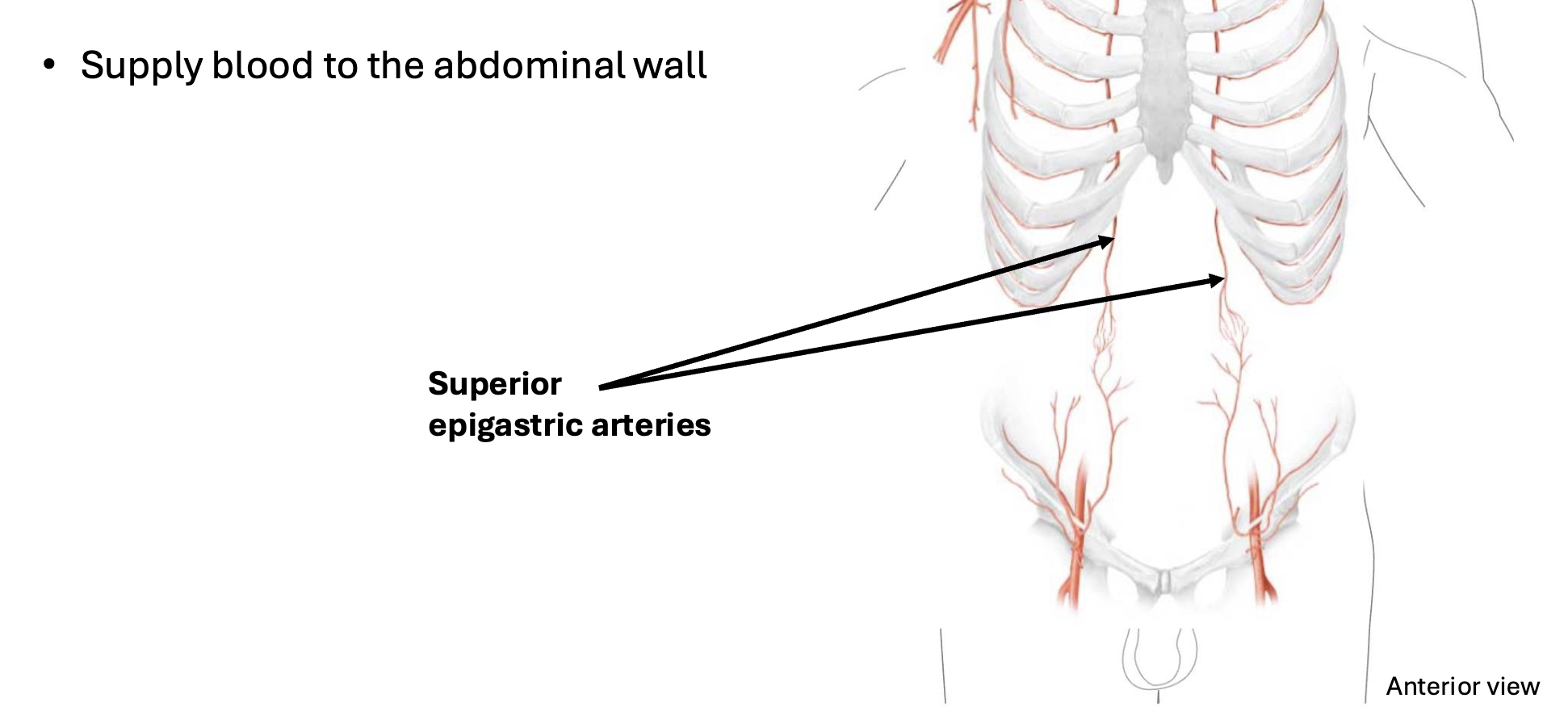
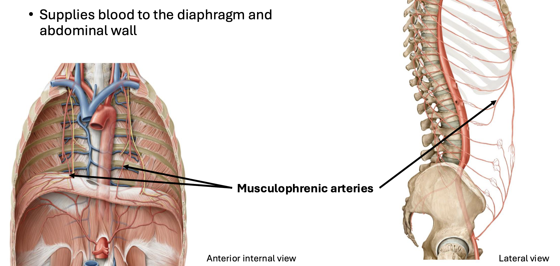
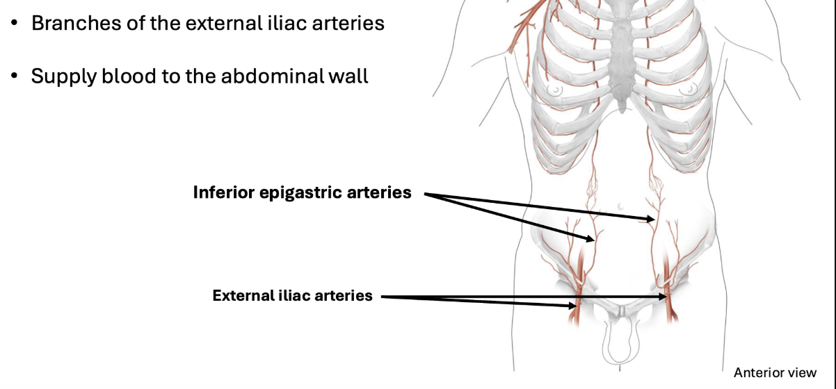
Abdominal Wall Blood is supplied by...
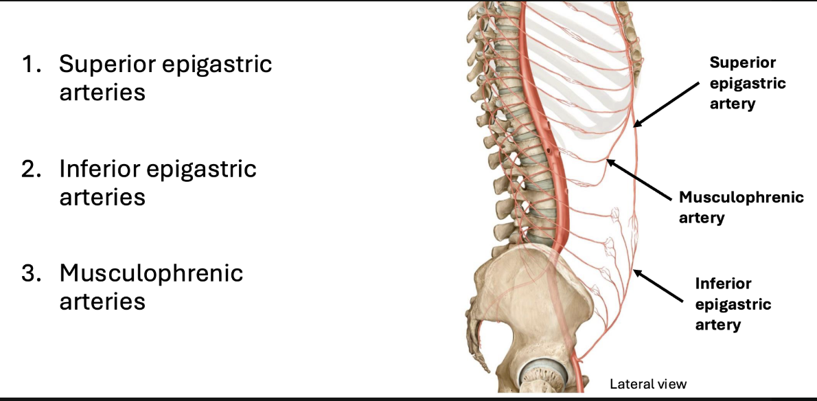
Internal thoracic artery
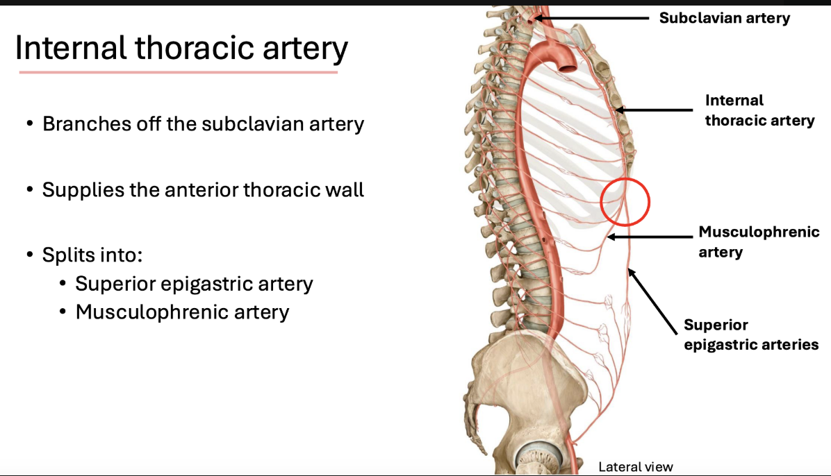
Superior epigastric arteries
Musculophrenic arteries
Inferior epigastric arteries
Abdominal Wall Lymphatic drainage
Lymph from the abdominal wall is filtered in the lumbar and inguinal nodes
Is then drained into the cisterna chyli then into the thoracic duct
Oral Cavity
Digestion:
Ingestion: receives food
Mechanical digestion: breakdown of food (mastication)
Chemical digestion: via salivary secretions
Swallowing: move food to esophagus
Divisions of Oral Cavity
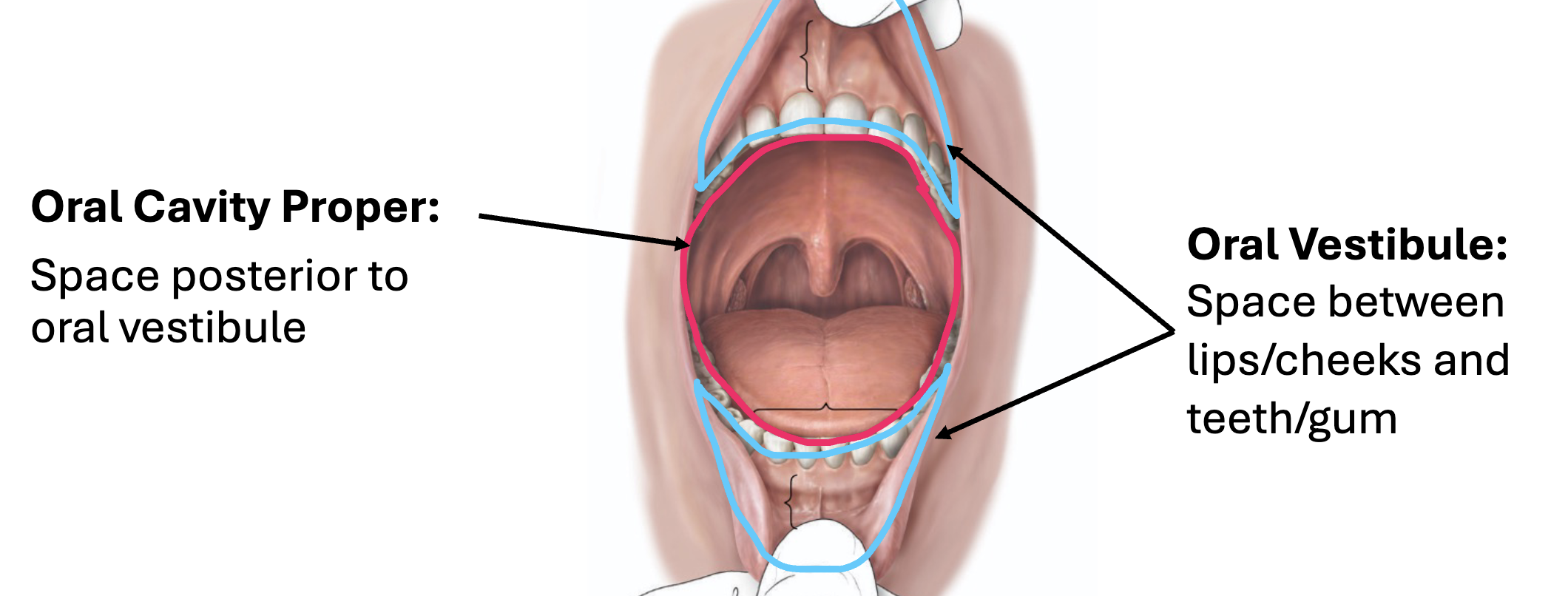
Oral Vestibule
External border: lips and cheeks
Internal border: teeth and gingiva
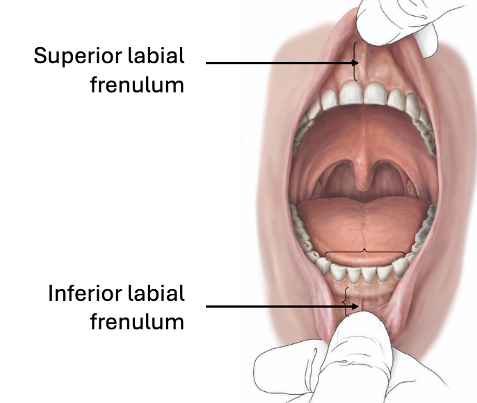
Labial frenulum:
Tissue that connects the lips and gingiva (gums)
Oral Cavity Proper - borders
Roof: hard & soft palate
Floor: tongue & floor of mouth
Posterior border: oropharyngeal isthmus
Anterior border: teeth
Oral Cavity Proper - Arches
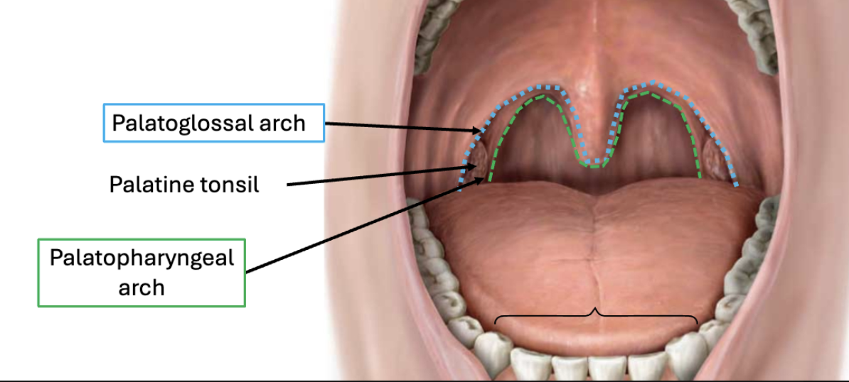
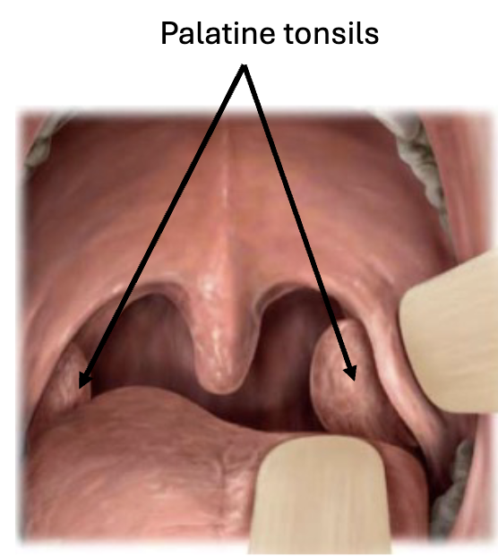
Palatine Tonsils
Lymphatic tissue located between palatoglossal & palatopharyngeal arches
Immune function
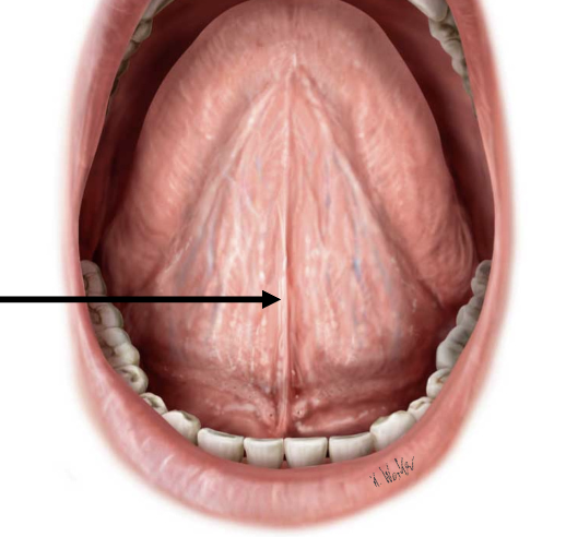
Oral Cavity Proper - Lingual frenulum
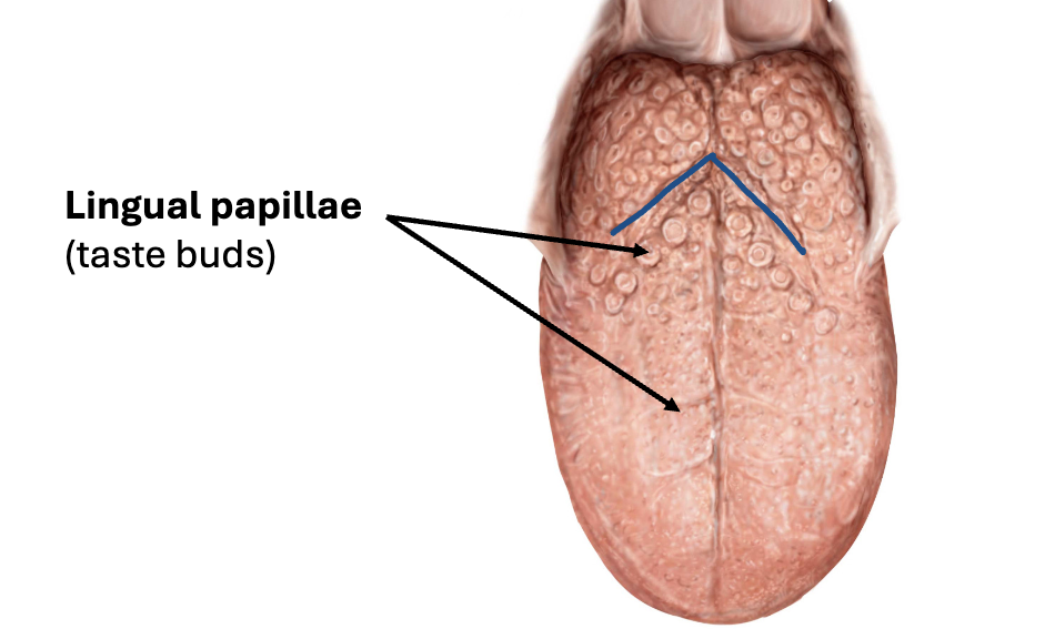
The Tongue
Bundle of skeletal muscle
Functions:
Positions food between teeth
Mix food with saliva
Forms bolus
Initiates swallowing
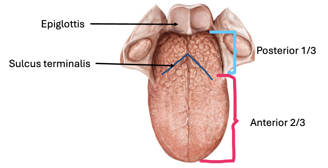
Lingual papillae (taste buds)
Taste receptor cells
Sensation Innervation:
Posterior 1/3: CN IX
Anterior 2/3: CN VII