Osteology, Arthrology and Myology of the Lower Limb
1/139
There's no tags or description
Looks like no tags are added yet.
Name | Mastery | Learn | Test | Matching | Spaced |
|---|
No study sessions yet.
140 Terms
Name and Classification.
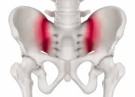
Sacroiliac joint. Synovial plane (diarthrosis).
Knee Joint Classification.
Synovial diarthrosis, modified hinge joint. Mostly uniaxial.
Joint name and classification
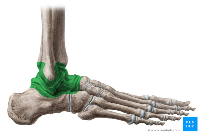
Ankle (Talocrural) joint. Synovial, diarthrosis, hinge (uniaxial)
Intertarsal joint classification.
Synovial (plane), diarthrosis (multiaxial).
Tarsometatarsal joint classification.
Synovial (plane), diarthrosis (multiaxial)
Metatarsophalangeal joint classification
Synovial (condyloid), diarthrosis (biaxial)
Interphalangeal joint classification
Synovial (hinge), diarthrosis (uniaxial)
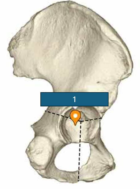
1
Acetabular fossa

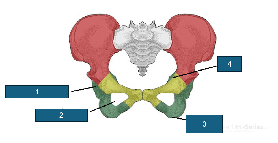
Red
Ilium

Yellow
Pubis

Green
Ischium

1
Acetabulum

2
Obturator foramen
3
Ischial tuberosity

4
Ischial spine

1
Intertrochanteric line

2
Greater trochanter

3
Head

4
Fovea

5
Neck

6
Intertrochanteric crest

8
Trochanteric fossa

9
Lesser trochanter

10
Gluteal tuberosity

11
Pectineal line

12
Shaft

13
Linea aspera

14
Medial epicondyle

15
Lateral condyle

16
Medial condyle

17
Lateral epicondyle
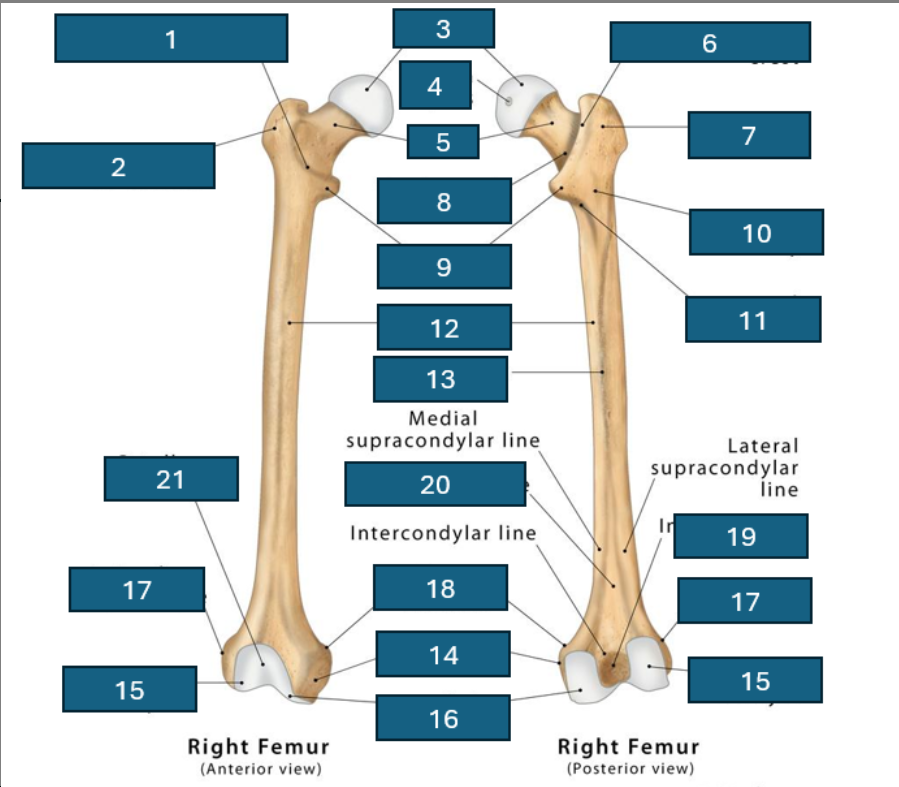
18
Adductor tubercle

19
Intercondylar fossa

20
Popliteal surface

21
Patellar surface

Blue
Intercondylar eminence

Green
Tibial tuberosity

Purple
medial malleolus

Yellow
Superior articular surface (including medial and lateral condyles)
Red
Soleal line
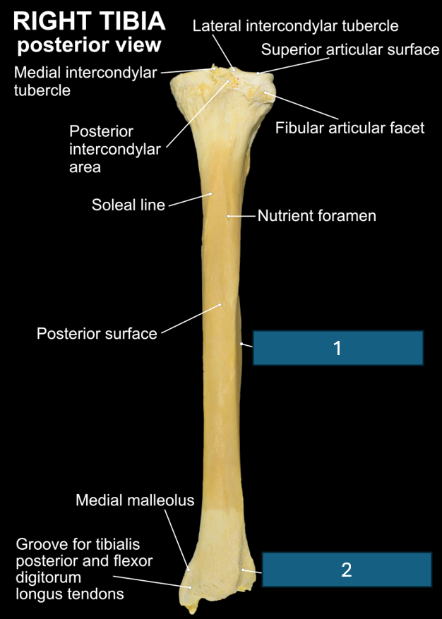
1
Interosseous border

2
Fibular notch

Purple
Calcaneus

Yellow
Talus

Blue
Navicular

Green
Cuboid
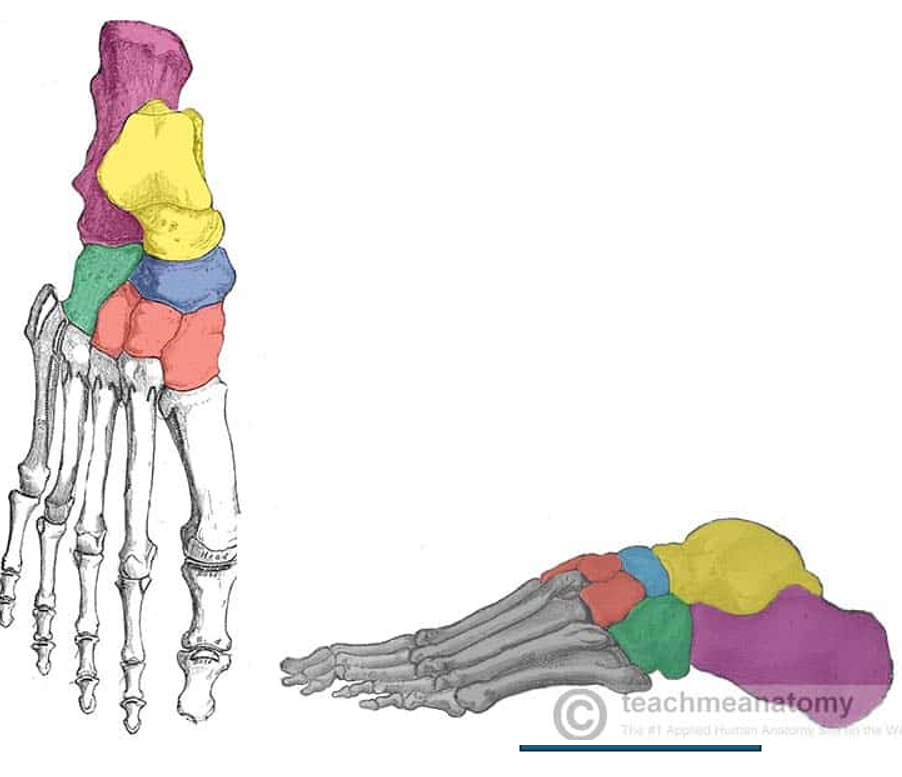
Red
Cuneiforms
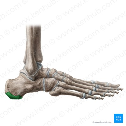
Calcaneal tuberosity
Sustentaculum tali
Trochlea of Talus
Smooth upper joint surface of the talus (links with lower part of tibia)
Head of talus
Rounded anterior part of the talus bone that articulates with the navicular bone.
Navicular tuberosity
Tuberosity on the medial surface of the navicular bone. ONLY THING ON NAVICULAR BONE.
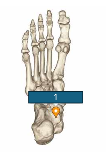
Patella tendon

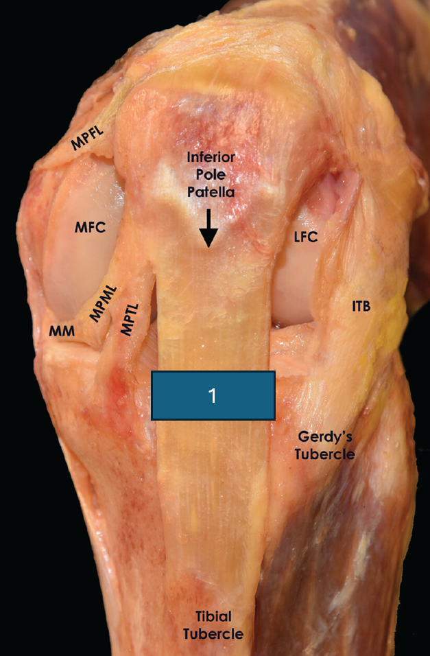
1
Anterior cruciate ligament

2
Medial meniscus

3
Lateral meniscus

4
Lateral (fibular) collateral ligament
5
Posterior cruciate ligament

6
Medial (tibial) collateral ligament
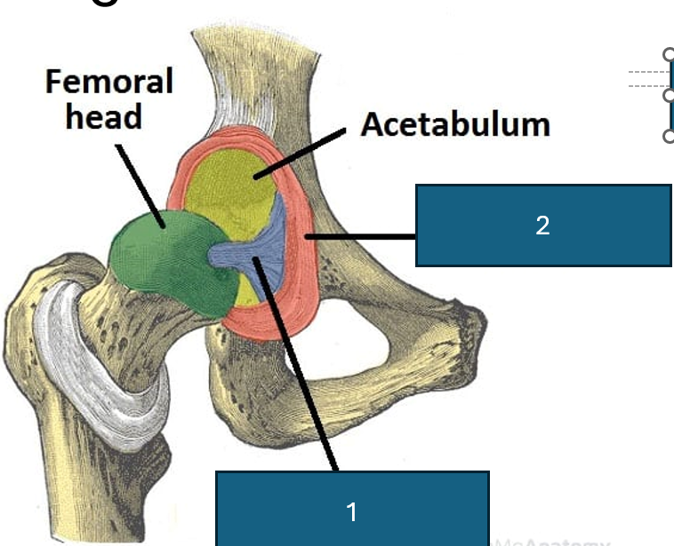
1
Ligament of the femur head (ligamentum teres)

2
Acetabular labrum
Ligaments of the hip
Ischiofemoral, Iliofemoral, Pubofemoral. Look for what part of the hip bone the ligament is coming from to identify.

1
Calcaneofibular ligament

2
Anterior talofibular ligament
3
Posterior talofibular ligament
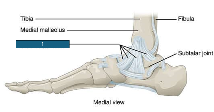
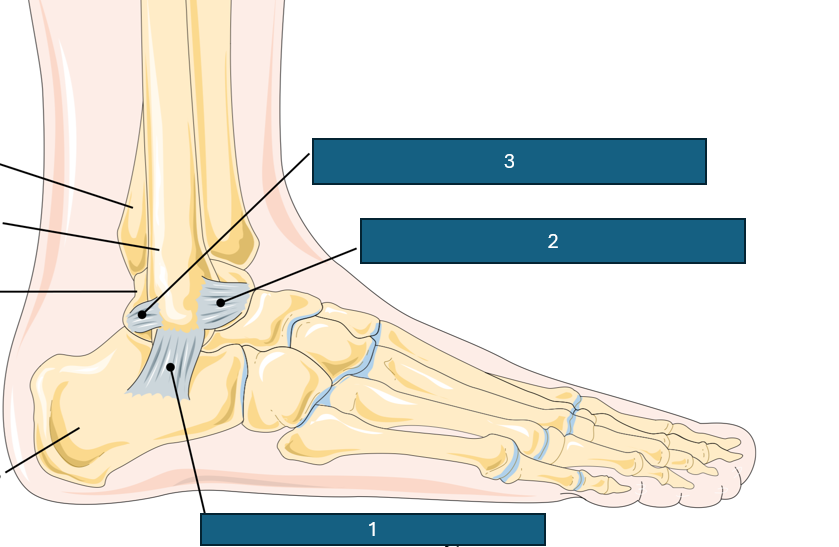
1
Deltoid ligament
1
Short plantar ligament

2
Plantar calcaneonavicular ligament

3
Long plantar ligament
Superior tibiofibular joint classification
Synovial (plane), diarthrosis (nonaxial)
Inferior tibiofibular joint classification
Fibrous (syndesmosis), amphiarthrosis
Middle tibiofibular joint
Fibrous (syndesmoses), amphiarthrosis
Tibiofemoral joint
Synovial (modified hinge), diarthrosis (mostly uniaxial, with slight biaxial movement)
Patellofemoral joint
Synovial (plane), diarthrosis (nonaxial)
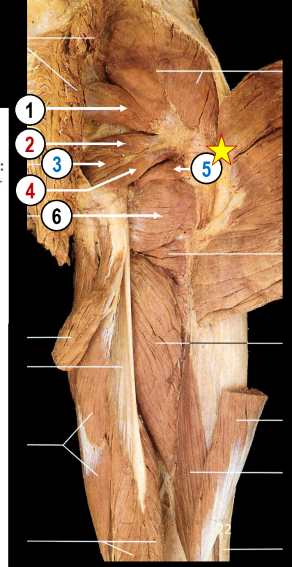
Muscles of the Anterior Hip Group
Iliacus, Psoas major, Psoas minor

1
Iliacus.
Origin: Iliac fossa
Insertion: Distal to lesser trochanter
Function: Hip flexor

2
Psoas Major
Origin: Anterior surfaces/bodes and transverse processes of T12-L5.
Insertion: Lesser trochanter
Function: Hip flexor. Can affect posture.
3
Psoas minor
Origin: Anterior surfaces and sides of vertebral bodies T12-L1
Insertion: Pectineal line - pelvic bone.
Function: DOESNT AFFECT HIP. More to do with posture
What are the superficial gluteal muscles?
Gluteus maximus, medius and minimus and tensor fascia latae.
What are the deep gluteal muscles?
Piriformis, Gemelli superior/inferior, Obturator internus/externus, quadratus femoris
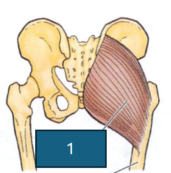
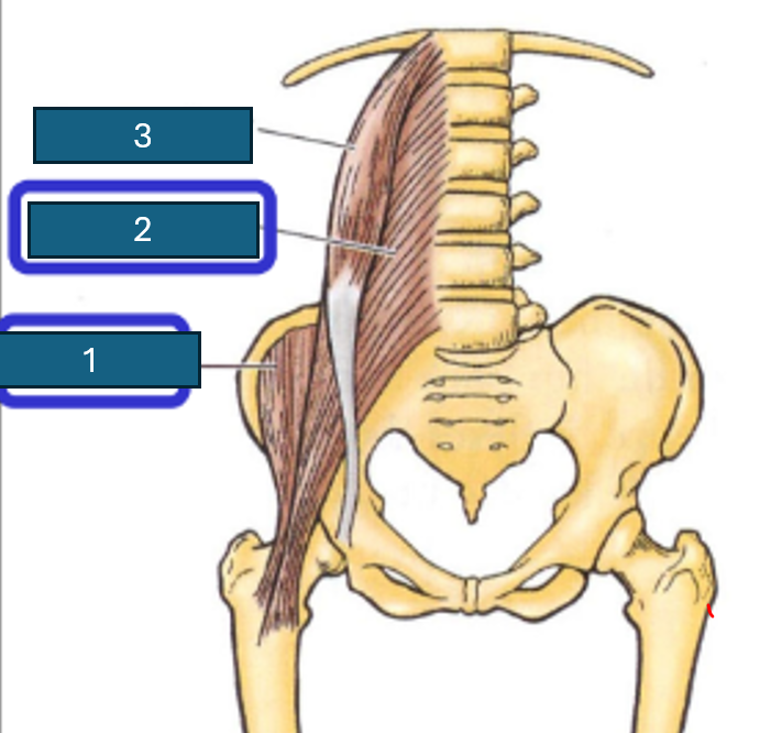
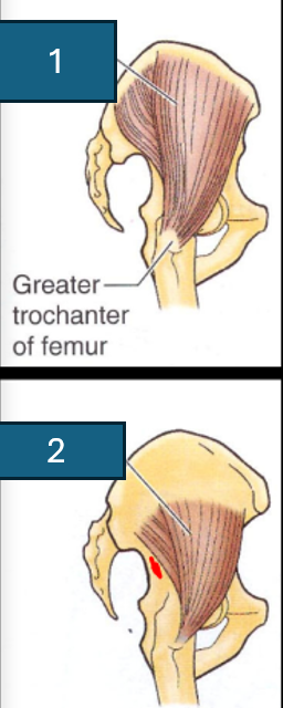
1
Gluteus Maximus
Origin: Dorsal ilium, sacrum and coccyx.
Insertion: Gluteal tuberosity of femur and iliotibial tract
Action: Extension of the hip and slight lateral rotation.

2
Gluteus Minimus
Origin: Between anterior and inferior gluteal lines on the lateral ilium.
Insertion: Anterior border of greater trochanter of femur.
Action: Abduction of femur and slight medial rotation of femur.
1
Gluteus Medius
Origin: Anterior and posterior gluteal lines on lateral ilium
Insertion: Lateral aspect of greater trochanter of femur
What are the general actions of the anterior hip group?
Flex the hip joint (and lumbar vertebral column)
What are the general actions of the gluteal region muscles?
Mostly extend, abduct and laterally rotate the femur.
What is the general action of the anterior thigh group?
Mostly flex the hip joint and/or extend the knee joint.
General action of posterior thigh?
Mostly extend hip and/or flex knee
General action medial thigh group?
Mostly adduction of femur

1
Tensor Fasciae Latae
Origin: Anterior ilium - anterior iliac crest and lateral surface of ASIS
Insertion: Iliotibial tract
Action: Tenses fasciae latae.
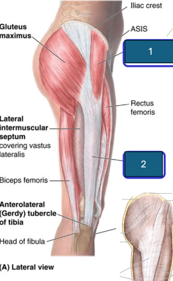
2
Iliotibial tract
1
Piriformis
Origin: Anterolateral sacrum
Insertion: Greater trochanter
Action: Lateral rotation of hip joint

2
Gemelli Superior
Origin: Superior ischial spine (SIS)
Insertion: Greater trochanter
Action: lateral rotation of the hip

3
Obturator internus
Origin: Obturator membrane (inner side)
Insertion: Greater trochanter
Action: Lateral rotation of the hip

4
Gemelli Inferior
Origin: Ischial tuberosity
Insertion: Greater trochanter
Action: Lateral rotation of the hip joint

5
Obturator Externus
Origin: Obturator membrane (outer side)
Insertion: Trochanteric fossa
Action: Lateral rotation of the hip joint
NOT ALWAYS VISIBLE

6
Quadratus femoris
Origin: Ischial tuberosity
Insertion: Intertrochanteric crest
Action: Lateral rotation of the hip
What are the anterior thigh muscles?
Quadriceps femoris (rectus femoris, vastus medialis, vastus lateralis, vastus intermedius) and sartorius
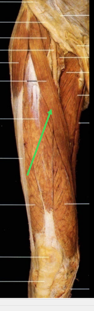
Sartorius
Origin: ASIS
Insertion: Medial surface of proximal tibia (after winning behind the medial aspect of the lower thigh).
Action: Flexion of hip and knee, lateral rotation

1
Rectus Femoris (quasi-fusiform, bipennate)
Origin: Anterior ilium - AIIS and superior margin of the acetabulum. (ONLY MUSCLE TO CROSS HIP)
Insertion: Patella and tibial tuberosity
Action: Extension of knee and flexion of hip.
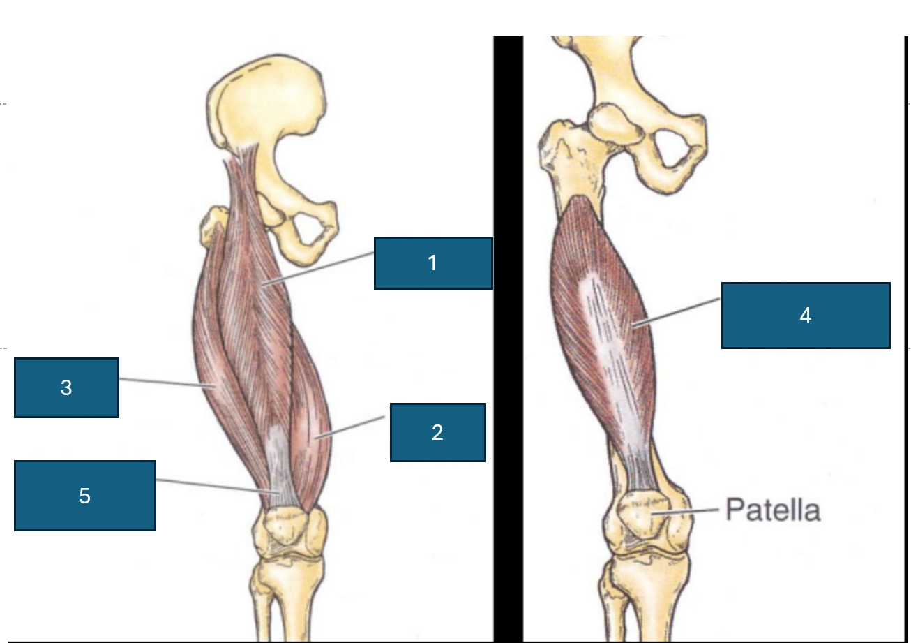
2
Vastus medialis
Origin: Linea aspera, medial supracondylar ridge, intertrochanteric line.
Insertion: Patella and tibial tuberosity.
Action: Extend the knee joint (and stabilise patella)