(21.5.6) Humoral immune response & Cellular immune response

0.0(0)
studied byStudied by 0 people
0.0(0)
full-widthCall Kai
learnLearn
examPractice Test
spaced repetitionSpaced Repetition
heart puzzleMatch
flashcardsFlashcards
GameKnowt Play
Card Sorting

1/69

encourage image

There's no tags or description

Looks like no tags are added yet.

Study Analytics
Name
Mastery
Learn
Test
Matching
Spaced

No study sessions yet.

70 Terms

1
New cards

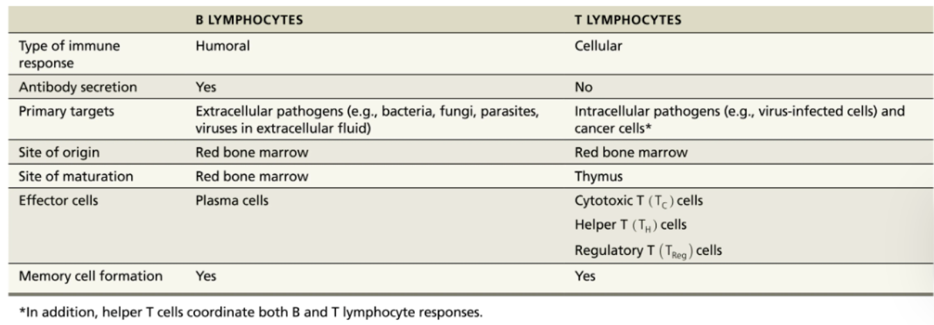
Define Humoral Immune Response

When B cell encounters target antigen → antibodies specific for that particular antigen are then produced 

<p>When <strong><mark data-color="blue" style="background-color: blue; color: inherit;">B cell</mark></strong> encounters target antigen → <span style="color: blue;"><strong>antibodies</strong></span> specific for that particular antigen are then produced&nbsp;</p>
2
New cards

What is meant by the clonal expansion of a B cell?

An activated B cell divides into cells that give rise to memory B cells and plasma cells.

3
New cards

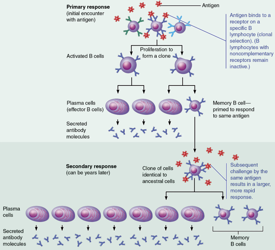
Describe the process of clonal selection of a B cell

  1. Immunocompetent but NAIVE B lymphocyte is ACTIVATED when antigens bind to its surface receptors

  2. The activated B lymphocyte begins clonal selection → the process of the B cell growing and multiplying to form an army of cells that are capable of recognizing the same antigen

  3. Most cells of the clone develop into plasma cells → the antibody-secreting cells of the humoral response 

  4. The cells of the clone that do not become plasma cells develop into memory cells

4
New cards

Compare and Contrast Primary Immune Response & Secondary Immune Response

Humoral Responses

PRIMARY

  • Cell proliferation and differentiation upon exposure to antigen for the first time 

    • LAG period: 3-6 days 

    • Peak levels of plasma antibody are reached in 10 days 

    • Antibody levels then decline 

SECONDARY

  • Re-exposure to same antigen gives faster, more prolonged, more effective response

    • Sensitized memory cells provide immunological memory

    • Respond within HOURS, not days

    • Antibody levels peak in 2 to 3 days at much higher levels

    • Antibodies bind with GREATER affinity

    • Antibodies level can remain HIGH for weeks to months

5
New cards

Discuss the roles of plasma cells and memory cells in humoral immunity

  • Plasma cells → Members of a B cell clone

    • Effector B cells specialized to produce and release antibodies

  • Memory cellsMembers of T cell and B cell clones

    • Provide for immunological memory.

<ul><li><p><strong>Plasma cells </strong>→&nbsp;<span>Members of a </span><span style="color: blue;"><strong><span>B cell </span></strong></span><span style="color: rgb(0, 0, 0);"><span>clone</span></span></p><ul><li><p><span>Effector B cells specialized to produce and release antibodies</span></p></li></ul></li><li><p><strong>Memory cells</strong> → <span>Members of </span><span style="color: red;"><strong><span>T cell </span></strong></span><span>and </span><span style="color: blue;"><strong><span>B cell</span></strong></span><span> clones</span></p><ul><li><p><span>Provide for immunological memory.</span></p></li></ul></li></ul><p></p>
6
New cards

What roles do memory B cells play when a patient is re-exposed to an antigen?

Memory B cells trigger a secondary immune response, which is faster, more prolonged, and more effective than the first immune response

  1. Memory cells to that specific virus are stored in the lymph nodes for many years

  2. When the patient comes into contact with this specific virus, these memory cells quickly divide and differentiate into antibody-producing plasma cells

  3. The antibodies will prevent the virus from reaching an infectious titer

7
New cards

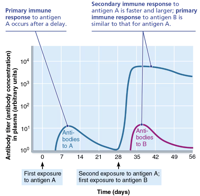
KNOWN Primary and Secondary Humoral Responses Graph 

  • The primary response to antigen A generates memory cells that give rise to the enhanced secondary response to antigen A

  • The response to antigen B is independent of the response to antigen A.

<ul><li><p>The <strong>primary response</strong> to antigen A <span style="color: red;"><strong>generates memory cells</strong></span> that give rise to the enhanced <strong>secondary response</strong> to antigen A</p></li><li><p>The response to antigen B is independent of the response to antigen A.</p></li></ul><p></p>
8
New cards

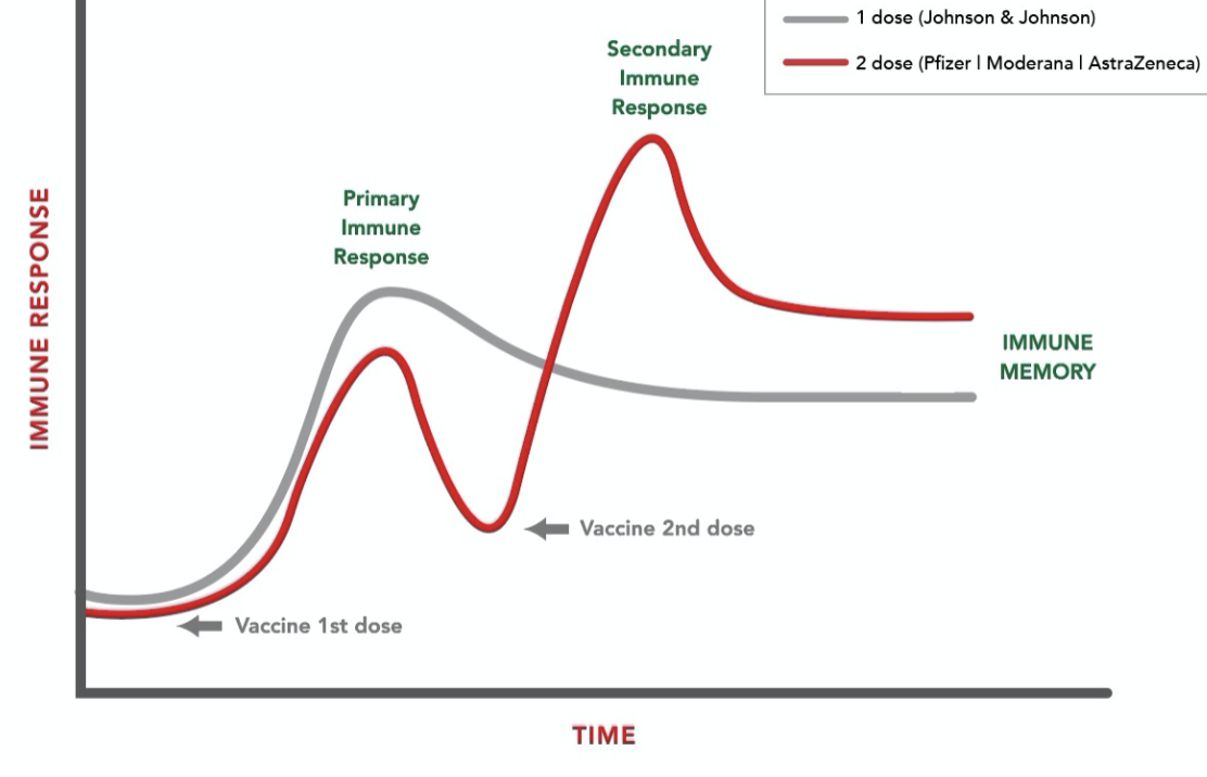
KNOWN Primary and Secondary Humoral Responses with Vaccies Graph 

knowt flashcard image
9
New cards

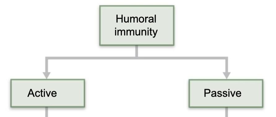
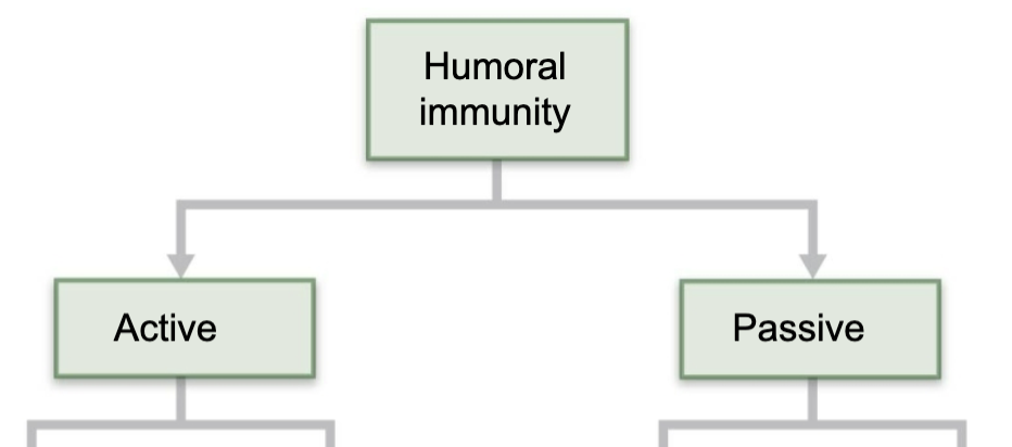
Define Active Humoral Immunity 

  • Occurs when B cells encounter antigens and produce specific antibodies against them 

  • Two types:

    1. Naturally acquired

    2. Artificially acquired

<ul><li><p><span style="color: red;"><strong>Occurs when B cells encounter antigens and produce specific antibodies against them&nbsp;</strong></span></p></li><li><p><strong>Two types:</strong></p><ol><li><p><strong>Naturally acquired</strong></p></li><li><p><strong>Artificially acquired</strong></p></li></ol></li></ul><p></p>
10
New cards

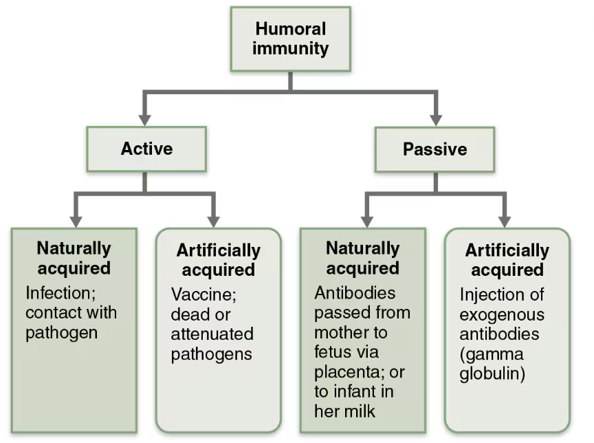
Compare and Contrast Naturally acquired & Artificially acquired Active Humoral Immunity

  1. Naturally acquired 

    • Formed in response to actual bacterial or viral infection

  2. Artificially acquired

    • Formed in response to vaccine of dead or attenuated pathogens

11
New cards

Benefits from Vaccines

  1. Provide antigenic determinants that are immunogenic and reactive

  2. Spare us symptoms of primary response

12
New cards

Define Passive Humoral Immunity 

  • Occurs when ready-made antibodies are introduced into body

    • B cells are not challenged by antigens

    • Immunological memory does not occur 

    • Protection end when antibodies degrade 

  • Two Types: 

    1. Naturally acquired

    2. Artificially acquired

<ul><li><p><span style="color: blue;"><strong><span>Occurs when ready-made antibodies are introduced into body</span></strong></span></p><ul><li><p>B cells are not challenged by antigens</p></li><li><p>Immunological memory does not occur&nbsp;</p></li><li><p>Protection end when antibodies degrade&nbsp;</p></li></ul></li></ul><ul><li><p><strong>Two Types:&nbsp;</strong></p><ol><li><p><strong>Naturally acquired</strong></p></li><li><p><strong>Artificially acquired</strong></p></li></ol></li></ul><p></p>
13
New cards

Compare and Contrast Naturally acquired & Artificially acquired Passive Humoral Immunity

  • Naturally acquired

    • Occurs when a mother’s antiboides enter fetal circulation 

  • Artificially acquired

    • Occurs when a person is given preformed antibodies that have been harvested from another person 

14
New cards

Compare and Contrast Active and Passive humoral immunity

ACTIVE

  • Occurs when B cells encounter antigens and produce specific antibodies against them 

PASSIVE

  • Occurs when ready-made antibodies are introduced into body

<p><span style="color: red;"><strong>ACTIVE</strong></span> </p><ul><li><p><span><strong><span>Occurs when</span><u><span> </span></u><span>B cells encounter antigens and </span><u><span>produce specific antibodies</span></u><span> against them&nbsp;</span></strong></span></p></li></ul><p><span style="color: blue;">PASSIVE</span> </p><ul><li><p><span><strong><span>Occurs when </span><u><span>ready-made antibodies</span></u><span> are introduced into body</span></strong></span></p></li></ul><p></p>
15
New cards

Define Antibodies 

  • Immunoglobulins (Igs) 

  • Proteins secreted by plasma cells 

    • Make up gamma globulin protein of blood 

  • Capable of binding specifically with antigens detected by B cells 

  • Group into one of 5 Ig classes

16
New cards

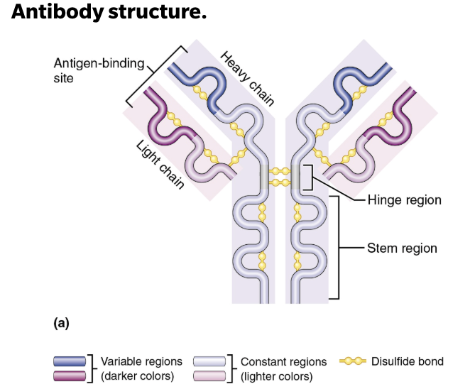
Describe the Structure of Antibodies

  • T- or Y-shaped antibody monomer consists of 4 looping polypeptides chains linked by disulfide bonds

    • 2 identical heavy (H) chains

    • 2 identical shorter, light (L) chains

  • Each chain has a variable region at one end, which varies depending on the antigen it binds, and a constant region at the other end, which is nearly identical among all members of a given class of antibodies

<ul><li><p>T- or Y-shaped <span style="color: blue;"><strong>antibody monomer</strong></span> consists of 4 looping polypeptides chains linked by disulfide bonds </p><ul><li><p>2 identical heavy (H) chains </p></li><li><p>2 identical shorter, light (L) chains </p></li></ul></li><li><p>Each chain has a variable region at one end, which varies depending on the antigen it binds, and a constant region at the other end, which is nearly identical among all members of a given class of antibodies </p></li></ul><p></p>
17
New cards

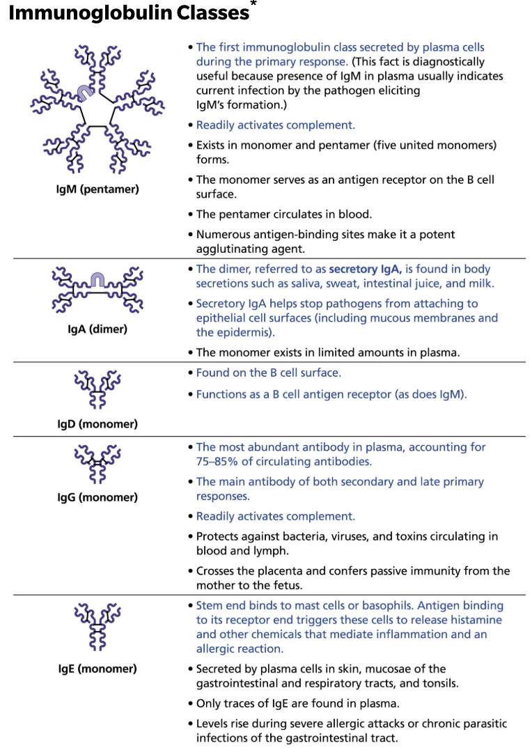
Describe the structure and functions of antibodies and name the five antibody classes.

Antibodies are divided into 5 classes based on their structure 

  • IgM

    • 1st immunoglobulin class secreted by plasma cells during primary response

    • Readily fixes and activates complement

  • IgA

    • The dimer, referred to as secretory IgA, is found in body secretions such as saliva, sweat, intestinal juice, and milk

    • Secretory IgA helps stops pathogens from attaching to epithelial cell surfaces (inculding mucous membranes and the epidermis)

  • IgD

    • Found on the B cell surface

    • Functions as B cell antigen receptor (as does IgM)

  • IgG

    • The most abundant antibody in plasma, accounting 75-85% of circulating antibodies

    • The main antibody of both secondary and late primary responses

    • Readily fixes and activates complement

  • IgE

    • Stem end binds to mast cells or basophils

    • Antigen binding to its receptor end triggers these cells to release histamine and other chemicals that mediate inflammation and an allergic reaction

<p><strong><em>Antibodies are divided into 5 classes based on their structure&nbsp;</em></strong></p><ul><li><p><strong>IgM</strong></p><ul><li><p><span style="color: purple;"><strong><mark data-color="purple" style="background-color: purple; color: inherit;">1st immunoglobulin class secreted by plasma cells during primary response </mark></strong></span></p></li><li><p>Readily fixes and activates complement </p></li></ul></li><li><p><strong>IgA</strong></p><ul><li><p>The dimer, referred to as secretory IgA, is <span style="color: purple;"><strong><mark data-color="purple" style="background-color: purple; color: inherit;">found in body secretions such as saliva, sweat, intestinal juice, and milk </mark></strong></span></p></li><li><p>Secretory IgA helps stops pathogens from attaching to epithelial cell surfaces (inculding mucous membranes and the epidermis) </p></li></ul></li><li><p><strong>IgD</strong></p><ul><li><p><span style="color: purple;"><strong><mark data-color="purple" style="background-color: purple; color: inherit;">Found on the B cell surface </mark></strong></span></p></li><li><p>Functions as B cell antigen receptor (as does IgM) </p></li></ul></li><li><p><strong>IgG</strong></p><ul><li><p><span style="color: purple;"><strong><mark data-color="purple" style="background-color: purple; color: inherit;">The most abundant antibody in plasma</mark></strong></span>, accounting 75-85% of circulating antibodies </p></li><li><p>The main antibody of both secondary and late primary responses </p></li><li><p>Readily fixes and activates complement </p></li></ul></li><li><p><strong>IgE </strong></p><ul><li><p><span style="color: purple;"><strong><mark data-color="purple" style="background-color: purple; color: inherit;">Stem end binds to mast cells or basophils </mark></strong></span></p></li><li><p>Antigen binding to its receptor end triggers these cells to release histamine and other chemicals that mediate inflammation and an allergic reaction </p></li></ul></li></ul><p></p>
18
New cards

Cause and Treatment of Ascaris and Schistosoma  

  • CAUSE  

    • Parasitic infections by worms, require different immune attack strategies  

  • TREATMENT

    • IgE antibodies still play a critical role in worm’s destruction by binding to surface of worm, marking it for destruction by eosinophils

    • Eosinophils bind to exposed stems of IgE → which triggers eosionphils to release their toxic contents onto prey, lysing it from the outside

19
New cards

Describe Antibody Targets and Function(s) 

Antibody do not destroy antigens → they inactivate and tag them

  • Form antigen-antibody (immune) complexes

<p><strong><em><u>Antibody do not destroy antigens → they </u></em></strong><span style="color: red;"><strong><em><u>inactivate</u></em></strong></span><strong><em><u> and tag them</u></em></strong></p><ul><li><p>Form <span style="color: blue;"><strong><span>antigen-antibody (immune) complexes</span></strong></span></p></li></ul><p></p>
20
New cards

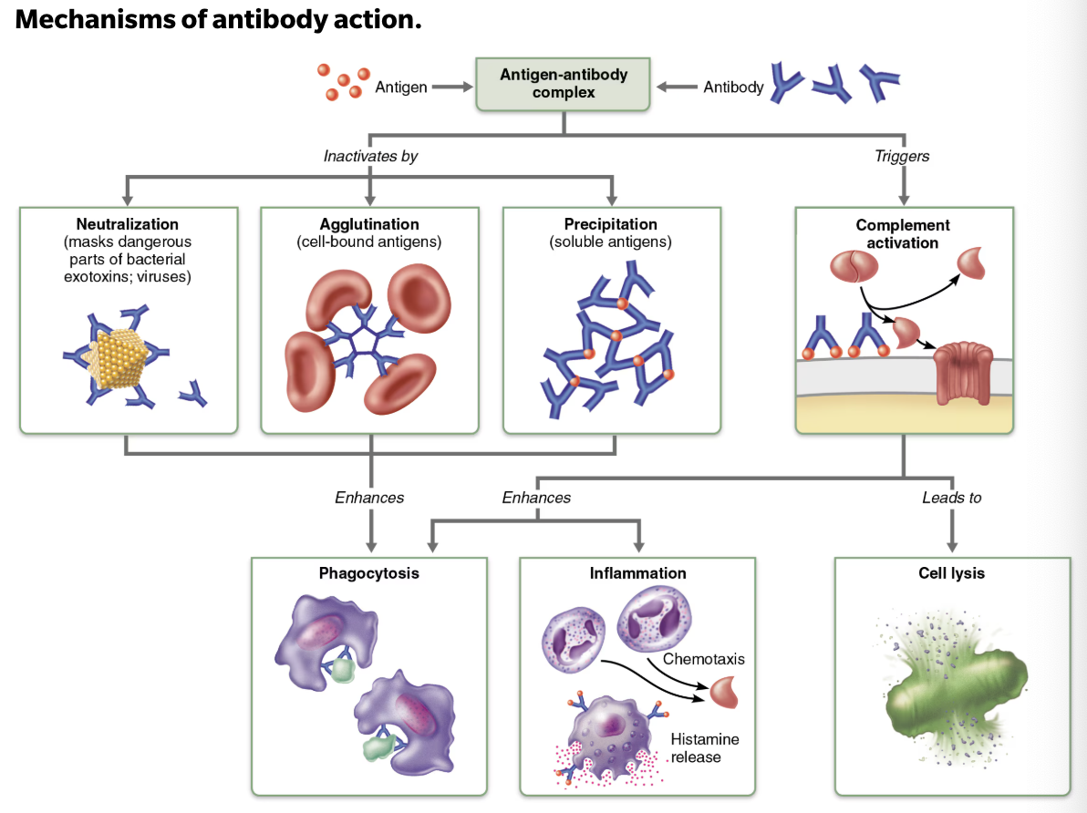
List and Describe Defensive Mechanism Used by Antibodies

  1. Neutralization 

    • Occurs when antibodies block specific sites on viruses or bacterial exotoxins → causing them to lose their toxic effects

  2. Agglutination 

    • Occurs when antibodies cross-link to cells-bound antigens→ causing clumping

  3. Precipitation 

    • Occurs when soluble molecules are cross-linked into large complexes that settle out of solution

  4. Complement activation

    • Occurs when complement binds to antibodies attached to antigens →leads to lysis of the cell

<ol><li><p>N<strong>eutralization&nbsp;</strong></p><ul><li><p>Occurs when antibodies block specific sites on <span style="color: green;"><strong><span>viruses or bacterial exotoxins</span></strong></span> → causing them to lose their toxic effects</p></li></ul></li><li><p><strong>Agglutination&nbsp;</strong></p><ul><li><p>Occurs when antibodies cross-link to <span style="color: green;"><strong><span>cells-bound antigens</span></strong></span>→ causing clumping</p></li></ul></li><li><p><strong>Precipitation&nbsp;</strong></p><ul><li><p>Occurs when <span style="color: green;"><strong><span>soluble molecules</span></strong></span> are cross-linked into large complexes that settle out of solution</p></li></ul></li><li><p><strong>Complement activation</strong></p><ul><li><p>Occurs when complement binds to <span style="color: green;"><strong><span>antibodies attached to antigens</span></strong></span> →leads to lysis of the cell</p></li></ul></li></ol><p></p>
21
New cards

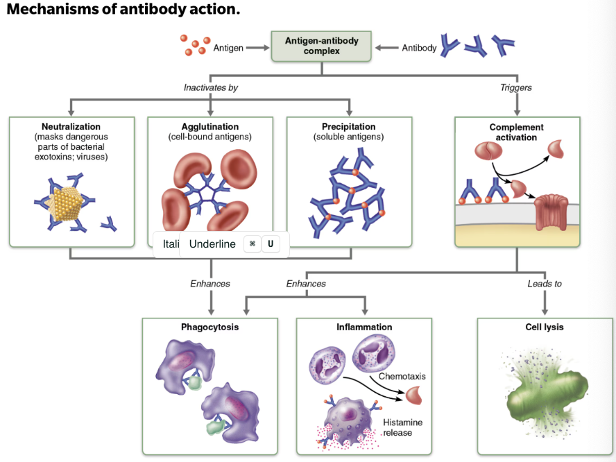
Summary of Antibody Actions

  • Antigen-antibody complexes do not destroy antigens → they prepare them for destruction by innate defenses

  • Antibodies go after extracellular pathogens → they do not invade solid tissue unless lesion is present

    • EXCEPTION → antibodies can act intracellularly if attached to virus before it enters cell

22
New cards

Define Monoclonal Antibodies

  • Clinical and research tools

  • Commercially prepared pure antibodies that are specific for a single antigenic determinant

23
New cards

Define Cellular Immunity

  • T cells provide defense against intracellular antigens

  • EX: cells infected with viruses or bacteria, cancerous or abnormal cells, foreign (transplanted) cells

24
New cards

Function of T cells

  1. Directly kill cells

  2. Others release chemicals that regulate immune response

25
New cards

T/F: T cells are more complex than B cells both in classification and function

→ True

26
New cards

Name the Major Populations of T cells

Based on which cell differentiation glycoprotein receptors are displayed on their surface

  1. CD4+ T cells 

  2. CD8+ T cells 

<p><strong><em>Based on which cell differentiation </em></strong><span style="color: blue;"><strong><em>glycoprotein</em></strong></span><strong><em> </em></strong><span style="color: blue;"><strong><em>receptors</em></strong></span><strong><em> are displayed on their </em></strong><span style="color: purple;"><strong><em><mark data-color="purple" style="background-color: purple; color: inherit;">surface</mark></em></strong></span></p><ol><li><p><strong>CD4<sup>+</sup> T cells&nbsp;</strong></p></li><li><p><strong>CD8<sup>+</sup> T cells&nbsp;</strong></p></li></ol><p></p>
27
New cards

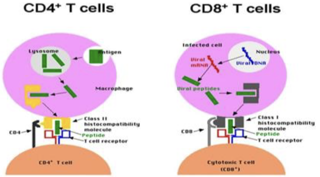
Function of CD4 cells

  • CD4 cells become helper T cells (TH) that can activate B cells, other T cells, and macrophages

    • Direct adaptive immune response

    • Can also become memory T cells

<ul><li><p><strong><mark data-color="blue" style="background-color: blue; color: inherit;">CD4 cells</mark></strong> become<span style="color: rgb(0, 0, 0);"><mark data-color="blue" style="background-color: blue; color: inherit;"> </mark><strong><mark data-color="blue" style="background-color: blue; color: inherit;">helper</mark></strong></span><span style="color: rgb(0, 0, 0);"><strong><mark data-color="blue" style="background-color: blue; color: inherit;"><span> T cells (T</span><sub><span>H</span></sub><span>)</span></mark></strong></span><span style="color: blue;"><strong><span> </span></strong></span>→ <span style="color: red;"><strong><span>that can activate B cells, other T cells, and macrophages</span></strong></span></p><ul><li><p>Direct adaptive immune response</p></li><li><p>Can also become <span style="color: blue;"><strong><span>memory T cells</span></strong></span></p></li></ul></li></ul><p></p>
28
New cards

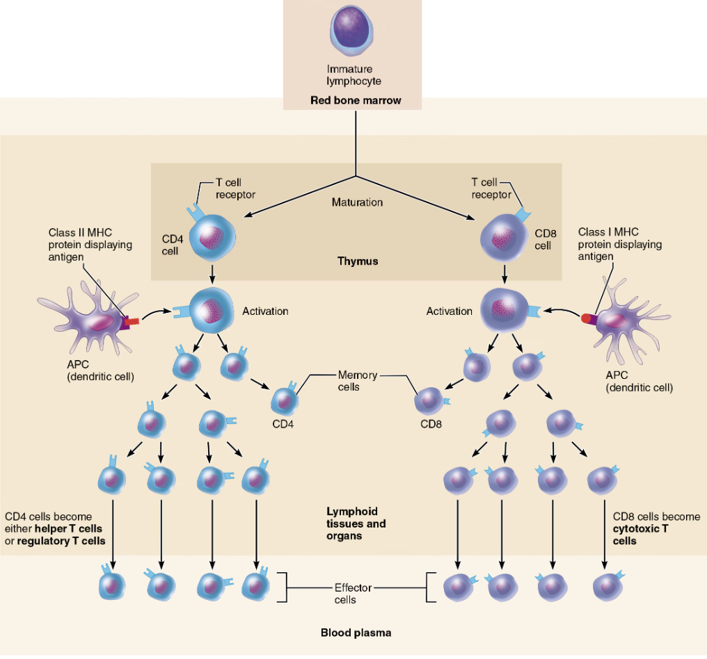
Function of CD8 cells

  • CD8 cells become cytotoxic T cells (TC)that are capable of destroying cells haboring foreign antigens

    • Can also become memory T cells

<ul><li><p><strong><mark data-color="red" style="background-color: red; color: inherit;">CD8 cells</mark></strong><mark data-color="red" style="background-color: red; color: inherit;"> </mark>become <span style="color: rgb(0, 0, 0);"><strong><mark data-color="red" style="background-color: red; color: inherit;"><span>cytotoxic T cells (TC)</span></mark></strong></span> → <span style="color: red;"><strong><span>that are capable of destroying cells haboring foreign antigens</span></strong></span></p><ul><li><p>Can also become <span style="color: blue;"><strong><span>memory T cells</span></strong></span></p></li></ul></li></ul><p></p>
29
New cards

Role of Helper, Cytotoxic, & Regulatory T cells

Activated T cells

<p>Activated T cells </p>
30
New cards

Define Naive T cells 

Simply termed CD4 or CD8 cells 

<p>Simply termed CD4 or CD8 cells&nbsp;</p>
31
New cards

Role of MHC proteins

Antigen presentation through the use of MHC (Major Histocompatibility Complex) proteins is necessary for BOTH activation and functioning of T cells

32
New cards

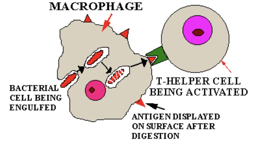
Where does most exogenous antigen presentation take place?

  • Most exogenous antigen presentation to T cells occurs in lymphoid tissues and organs located throughout the body

  • Dendritic cells are primarily responsible for presenting antigens to T cells in these sites.

<p></p><ul><li><p><strong>Most exogenous antigen presentation to T cells occurs in </strong><span style="color: purple;"><strong><mark data-color="purple" style="background-color: purple; color: inherit;">lymphoid tissues and organs</mark></strong></span><strong> located throughout the body</strong></p></li><li><p><strong>Dendritic cells are primarily responsible for presenting antigens to T cells in these sites.</strong></p></li></ul><p></p>
33
New cards

T/F: T cells can ONLY be activated by APCs

→ True

34
New cards

Explain the Activation of T cells 

T cells respond only to processed fragments of antigens displayed on surfaces of cells by Major Histocompatibility Complex (MHC) proteins

<p>T cells respond only to processed fragments of antigens displayed on surfaces of cells by <span style="color: blue;"><strong><span>Major Histocompatibility Complex (MHC) proteins</span></strong></span></p><p></p>
35
New cards

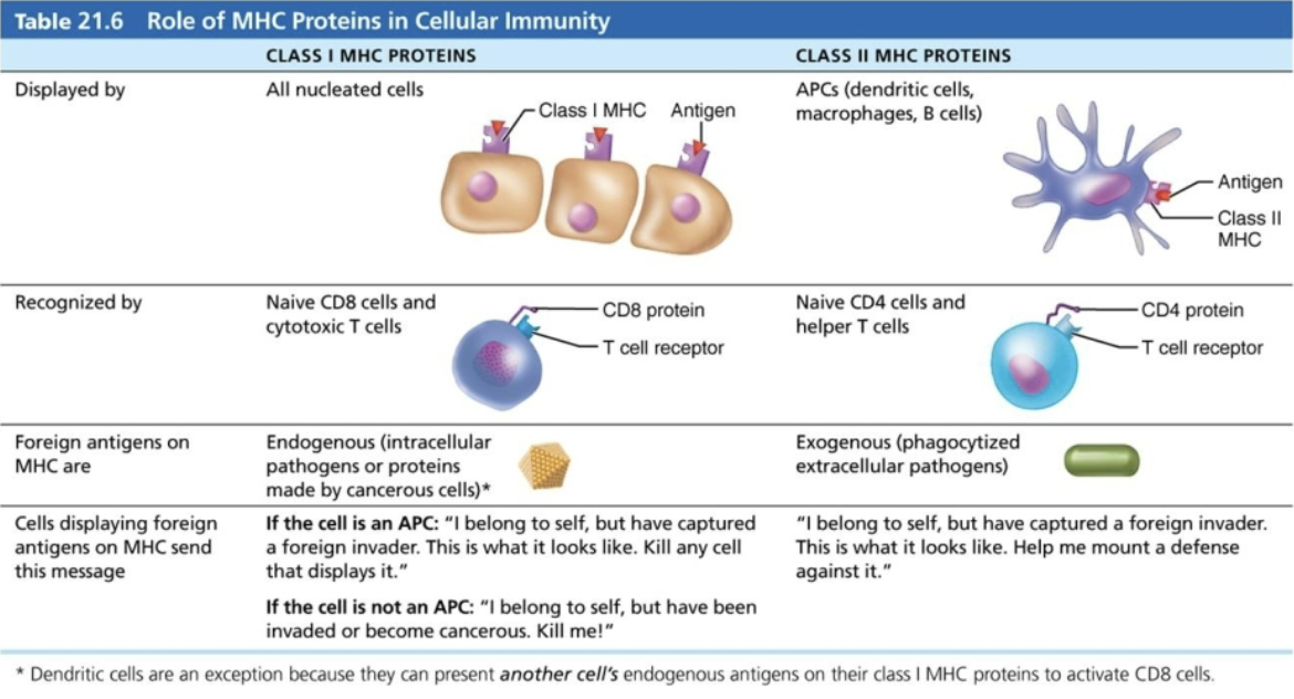
Name and Describe Two Classes of MHC Proteins 

  1. Class I MHC proteins

    • Displayed by ALL cells except RBCs

      • Antigens synthesized from within the cell 

      • If infected → fragments of foreign antigens 

    • FUNCTION → crucial for CD8 cell activation

      • Act as antigen holders → form “self” part that T cells recognize

  1. Class II MHC proteins

    • Displayed by APCs (dendritic cells, macrophages, and B cells)

      • Antigens arising from outside the cell that are engulfed by the displaying cell  

    • FUNCTION → recognized by helper T cells

      • Signal CD4 cells that help is required

BOTH types are synthesized in ER and bind to peptide fragments

36
New cards

Which of the following types of cells display protein fragments produced by the cancer within  them?

ALL nucleated body cells bring pieces of endogenous proteins to the surface to display on the MHC protein

37
New cards

Class II MHC proteins are found on which of the following cell types?

Only on antigen-presenting cells

38
New cards

Which class of MHC proteins presents exogenous antigens?

II MHC proteins present antigens that originated from outside the cell (phagocytized extracellular pathogens)

39
New cards

Class I MHC proteins are recognized by which of the following cell types (that are destined to become Tcells)?

I MHC proteins are recognized by CD8 cells

40
New cards

T/F: BCR and TCR interact

→ FALSE

  1. CD4 and MHC-II

  2. BCR and epitope

  3. CD8 and MHC-I

41
New cards

SUMMARY of Role of MCH Proteins in Cellular Immunity

knowt flashcard image
42
New cards

Explain MCH Restriction 

  • CD4 and CD8 cells have different requirements for MHC protein that presents antigens to them 

    • CD4 cells that become TH are restricted to binding to ONLY II MHC (typically on APC surfaces) → Once activated, cytotoxic T cells seek out same antigen on class I MHC proteins on ANY cell 

43
New cards

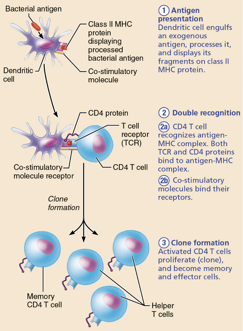
Describe the process of Activation and Clonal Selection of T cells.

Simultaneous recognition of self and non-self

  1. Antigen presentation

    • Dendritic cell engulfs an exogenous antigen, processes it, and displays it fragments on class II MHC protein 

  2. Double recognition 

    • CD4 T cell recognizes antigen-MHC complex → Both TCR and CD4 proteins bind to antigen-MHC complex 

    • T cell must bind one or more co-stimulatory signals present on the antigen-presenting cell

  3. Clone formation 

    • Activated CD4 T cells proliferate (clone), and become memory and effector cells 

<p><strong><em>Simultaneous recognition of self and non-self</em></strong></p><ol><li><p><strong>Antigen presentation</strong></p><ul><li><p><span style="color: blue;"><strong><mark data-color="blue" style="background-color: blue; color: inherit;"><span>Dendritic cell</span></mark></strong></span> engulfs an exogenous antigen, processes it, and displays it fragments on class<span style="color: blue;"><strong><span> II MHC protein&nbsp;</span></strong></span></p></li></ul></li><li><p><strong>Double recognition&nbsp;</strong> </p><ul><li><p><strong><mark data-color="blue" style="background-color: blue; color: inherit;">CD4 T cell</mark></strong> recognizes <span style="color: blue;"><strong>antigen-MHC complex</strong></span> → Both TCR and CD4 proteins bind to antigen-MHC complex&nbsp;</p></li><li><p>T cell must bind one or more <span style="color: blue;"><strong>co-stimulatory signals</strong></span> present on the antigen-presenting cell </p></li></ul></li><li><p><strong>Clone formation&nbsp;</strong></p><ul><li><p><strong><mark data-color="blue" style="background-color: blue; color: inherit;">Activated CD4 T cells</mark></strong> proliferate (clone), and become <span style="color: blue;"><strong><mark data-color="blue" style="background-color: blue; color: inherit;"><span>memory</span></mark></strong></span> and <span style="color: blue;"><strong><mark data-color="blue" style="background-color: blue; color: inherit;"><span>effector cells&nbsp;</span></mark></strong></span></p></li></ul></li></ol><p></p>
44
New cards

Name the Major Groups of Effector T cells

  1. Helper T cells

  2. Cytotoxic T cells

  3. Regulatory cells 

45
New cards

Function of Helper T (TH) cells

  • Activate both Humoral and Cellular arms 

  • Once primed by APC presentation of antigen, helper T cells

    1. Help activate B cells and other T cells 

    2. Induce T and B cell proliferation 

    3. Secrete cytokines that recruit other immune cells 

46
New cards

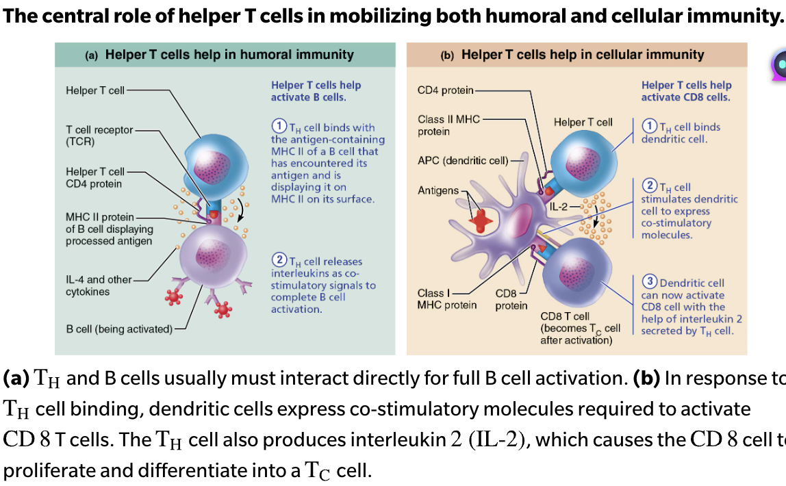
Which lymphocytes act as the bridge between the cellular and humoral responses?

Helper T cells are lymphocytes that organize the cellular and humoral immune response branches of the immune system.

47
New cards

Central Role of Helper T cells in Mobilizing Both Humoral and Cellular Immunity

Humoral Immunity

  1. TH cell binds with the self-nonself complexes of a B cell that has encountered its antigen and is displaying it on MHC II on its surface

  2. TH cell releases interleukins as co-stimulatory signals to complete B activation 

Cellular Immunity

  1. TH cell binds dendritic cell

  2. TH cells stimulates dendritic cell to express co-stimulatory molecules 

  3. Dendritic cell can now activate CD8 cell with the help of interleukin 2 secreted by TH cell

<p><strong>Humoral Immunity</strong></p><ol><li><p><span style="color: rgb(0, 0, 0);"><strong><span>T</span><sub><span>H</span></sub><span> cell</span></strong></span><span style="color: rgb(0, 0, 0);"> </span>binds with the <span style="color: purple;"><strong><span>self-nonself complexes of a B cell</span></strong></span> that has encountered its antigen and is displaying it on <span style="color: blue;"><strong><span>MHC II</span></strong></span> on its surface</p></li><li><p><strong>T<sub>H</sub> cell r</strong>eleases <span style="color: green;"><strong><span>interleukins</span></strong></span> as co-stimulatory signals to <span style="color: blue;"><strong><u><span>complete B activation</span></u></strong></span>&nbsp;</p></li></ol><p><strong>Cellular Immunity</strong></p><ol><li><p><span style="color: rgb(0, 0, 0);"><strong><span>T</span><sub><span>H</span></sub><span> cell</span></strong></span> binds <span style="color: purple;"><strong><span>dendritic cell</span></strong></span></p></li><li><p><strong>T<sub>H</sub> cells </strong>stimulates <span style="color: blue;"><strong><mark data-color="blue" style="background-color: blue; color: inherit;"><span>dendritic cell</span></mark></strong></span> to express co-stimulatory molecules&nbsp;</p></li><li><p><span style="color: blue;"><strong><mark data-color="blue" style="background-color: blue; color: inherit;"><span>Dendritic cell</span></mark></strong></span> can now <span style="color: red;"><strong><u><span>activate CD8 cell</span></u></strong></span> with the help of <span style="color: green;"><strong><span>interleukin 2</span></strong></span> secreted by<strong>&nbsp;T<sub>H</sub> cell</strong></p></li></ol><p></p>
48
New cards

Function of Cytotoxic T (TC) cells

  1. Activated TC cells circulate in blood and lymph and lymphoid organs in search of body cells displaying antigen they recognize

  2. Directly attack and kill other cells 

49
New cards

List Activated TC cells Targets 

  1. Virus-infected cells 

  2. Cells with intraceullar bacteria or parasites 

  3. Cancer cells 

  4. Foreign cells (transfusion or transplants)

<ol><li><p>Virus-infected cells&nbsp;</p></li><li><p>Cells with <em><u>intraceullar</u></em> bacteria or parasites&nbsp;</p></li><li><p>Cancer cells&nbsp;</p></li><li><p>Foreign cells (transfusion or transplants) </p></li></ol><p></p>
50
New cards

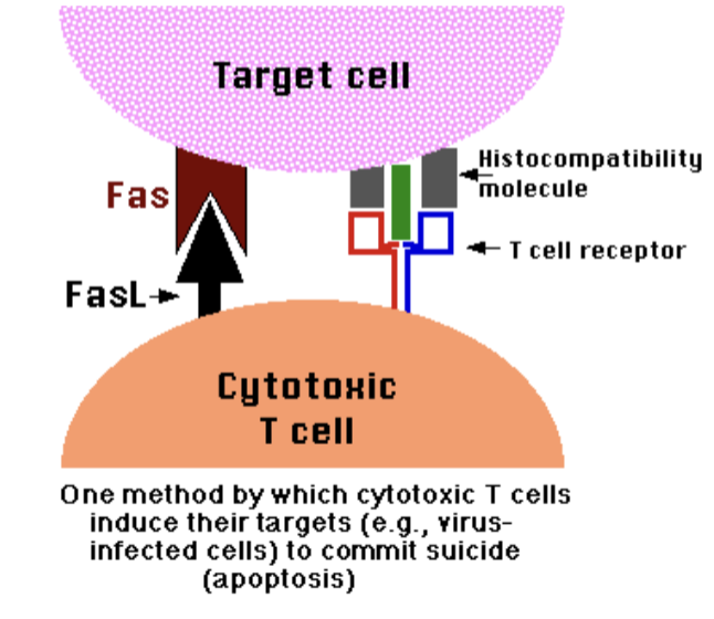
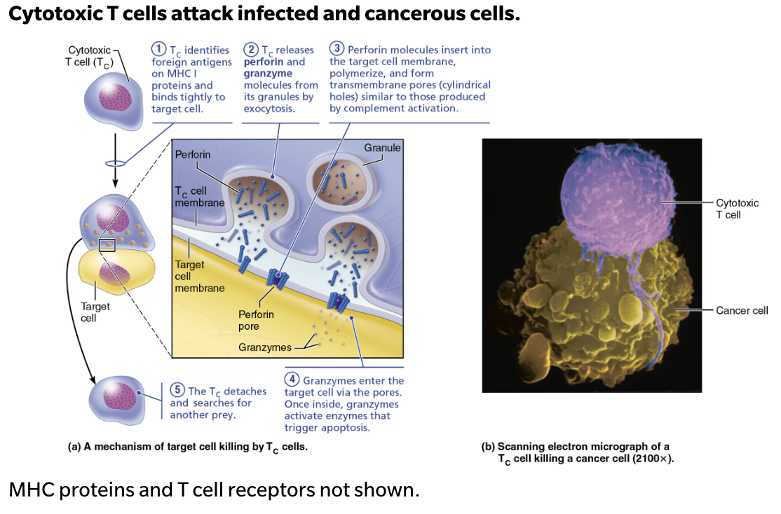
Describe Steps Cytotoxic T cells Attack Mechanism 

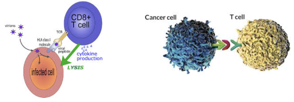
  1. TC identifies foreign antigens on MHC I proteins and binds tightly to target cell 

  2. TC releases perforin and granzyme molecules from its granules by exocytosis 

  3. Perforin molecules insert into the target cell membrane, polymerize, and form transmembrane pores (cylindrical holes) similar to those produced by complement activation 

  4. Granzymes enter the target cell via the pores → Once inside, granzymes activate enzymes that trigger apoptosis 

  5. The TC detaches and searches for another prey 

<ol><li><p><strong>T<sub>C</sub></strong> identifies foreign antigens on <span style="color: red;"><strong>MHC I proteins</strong></span> and binds tightly to target cell&nbsp;</p></li><li><p><strong>T<sub>C</sub></strong> releases <span style="color: green;"><strong>perforin</strong></span> and <span style="color: green;"><strong>granzyme molecules</strong></span> from its granules by <span style="color: blue;"><strong><em>exocytosis</em></strong></span>&nbsp;</p></li><li><p><span style="color: green;"><strong>Perforin</strong></span> molecules insert into the target cell membrane, polymerize, and form transmembrane pores (cylindrical holes) <span style="color: blue;"><strong>similar to those produced by complement activation</strong></span>&nbsp;</p></li><li><p><span style="color: green;"><strong>Granzymes</strong></span> enter the target cell via the pores → Once inside, granzymes activate enzymes that trigger <span style="color: blue;"><strong>apoptosis</strong></span>&nbsp;</p></li><li><p>The <strong>T<sub>C</sub></strong> detaches and searches for another prey&nbsp;</p></li></ol><p></p>
51
New cards

T/F: A step used by cytotoxic T cells to kill infected host is recognition of infected host cell using its CD4 glycoprotein

FALSE

  1. The cytotoxic T cell uses its CD8 glycoprotein to bind to the MHC-I of an infected host cell.

  2. Recognition of infected host cell using its TCR

  3. Secretion of granzyme

  4. Secretion of perforin

52
New cards

T/F: T cells can become effector cells and memory cells 

FALSE 

  • T cells can become effector cells OR memory cells 

  • Similar like B cells 

<p>→ <strong>FALSE</strong>&nbsp;</p><ul><li><p>T cells can become <strong>effector cells</strong> OR <strong>memory cells</strong>&nbsp;</p></li><li><p><strong>Similar like B cells&nbsp;</strong></p></li></ul><p></p>
53
New cards

Describe the roles of different types of T cells

  1. Helper T cells

    • Effector CD4 T cell

    • Central to both humoral and cellular immunity Stimulate proliferation of other T cells and B cells that have already become bound to antigen

  2. Cytotoxic T cells

    • Effector CD8 T cell

    • ONLY T cells that can directly attack and kill other cells displaying antigen to which they have been sensitized, through the use of perforin and granzymes, or by triggering apoptosis of the target cell

  3. Regulatory cells 

    • Effector CD4 T cell

    • Either by direct inhibition or by causing the release of cytokines, suppress the activity of both B cells and other types of T cells

54
New cards

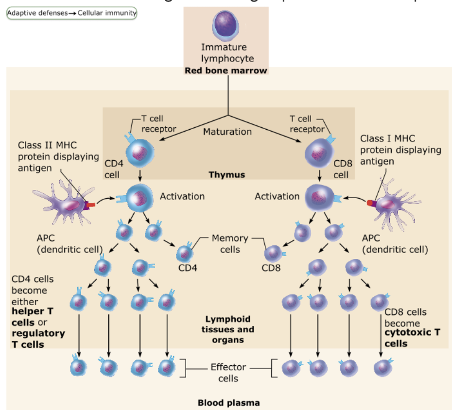
Sequence of Cellular Immunity  

  1. Dendritic cells activate CD4 cells

  2. Activated CD4 cells form a clone of Helper T (TH) cells and memory cells

  3. Helper T cells and dendritic cells activate CD8 cells

  4. Activated CD8 cells form a clone of Cytotoxic T (TC) cells and memory cells

55
New cards

A person who has AIDS contracts rare and often life-threatening infections because their helper T cell count is so low. Which of the following components of the immune response still respond to antigen despite the low helper T cell count?

Clonal selection of B cells → However, without a helper T cell, clonal expansion and antibody production will not occur

56
New cards

Define Allograft 

Most common type of organ transplant (from same species)  

57
New cards

What determines Transplant Success Depends?

  • Success depends on similarity of tissues

    • ABO, Other blood antigens, MHC antigens are matched as closely as possible 

  • WHY → Cytotoxic T cells, NK cells, and antibodies work to destroy foreign tissues

58
New cards

Explain Immunosuppressive Therapy

  • After surgery 

    • Patient treated with immunosuppressive therapy  → to suppress rejection  

    • Many of these therapies have severe side effects → weakened immune system 

59
New cards

What happens when Patient’s immune system is suppressed

  • Cannot protect body from foreign agents such as bacterial and viral infections → Leading cause of death among transplant recipients 

  • Best circumstances → rejection after 10 years in 

60
New cards

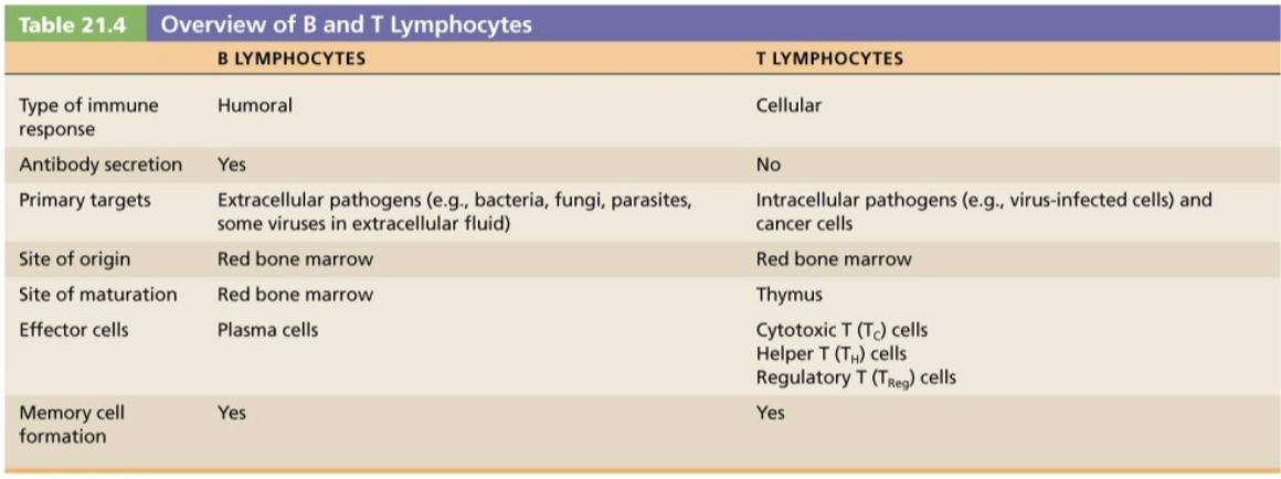
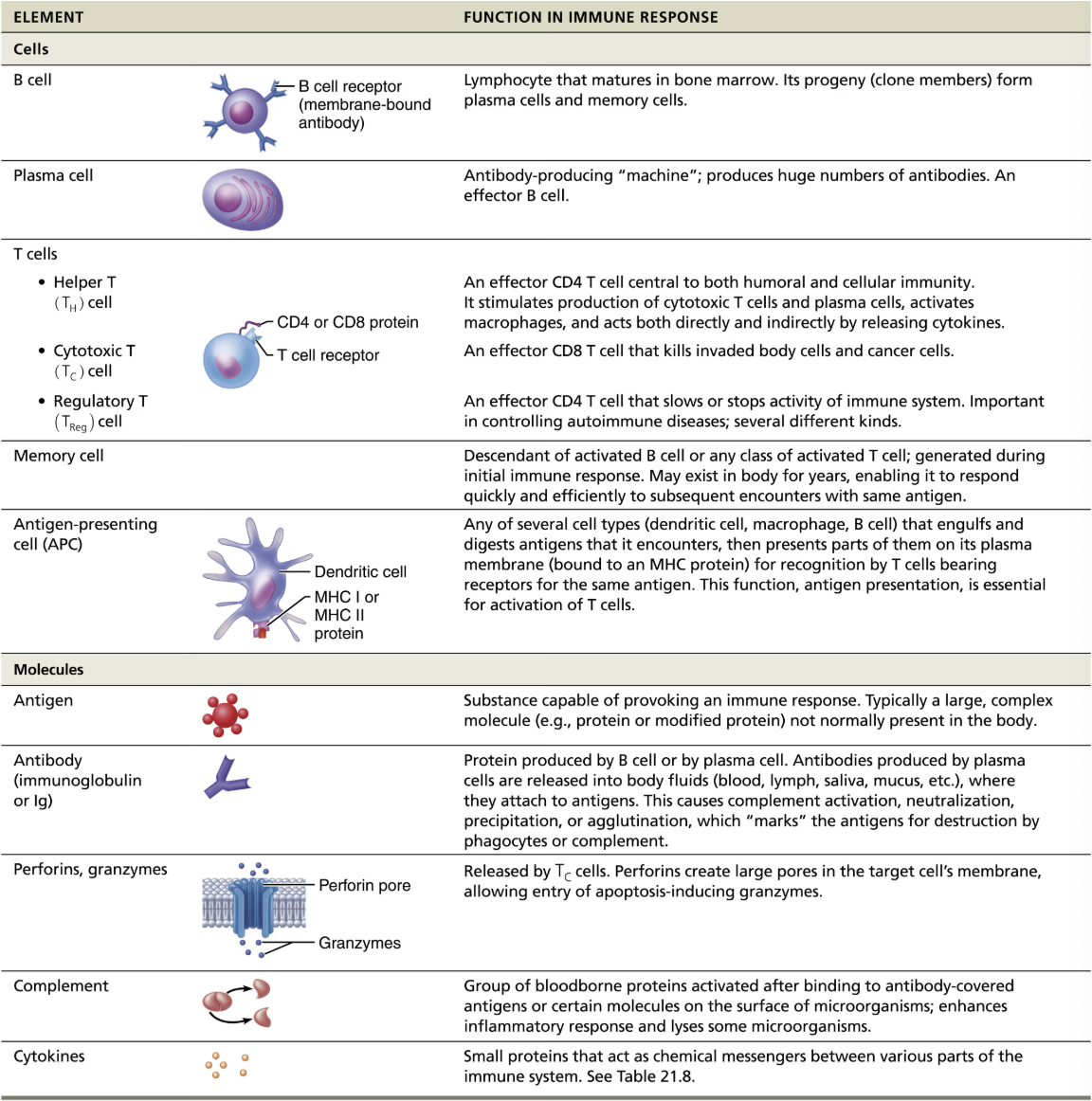
SUMMARY of Cells and Molecules of the Adaptive Immune Response

knowt flashcard image
61
New cards

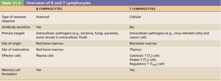
SUMMARY B & T Lymphocytes

knowt flashcard image
62
New cards

Which specific type of cell produces antibodies?

Activated plasma cells

  • When activated by the presence of antigens, B lymphocytes proliferate into plasma cells

  • Plasma cells generate antigen-specific antibodies.

63
New cards

How do cytotoxic cells directly attack target cells?

Cytotoxic cells bind to the target cell and secrete chemicals that induce apoptosis

  • Cytotoxic cells are able to dock with antigens on the target cell membrane

  • Once docked, the cytotoxic cell releases perforins and granzymes, which weaken the cell membrane and induce apoptosis

64
New cards

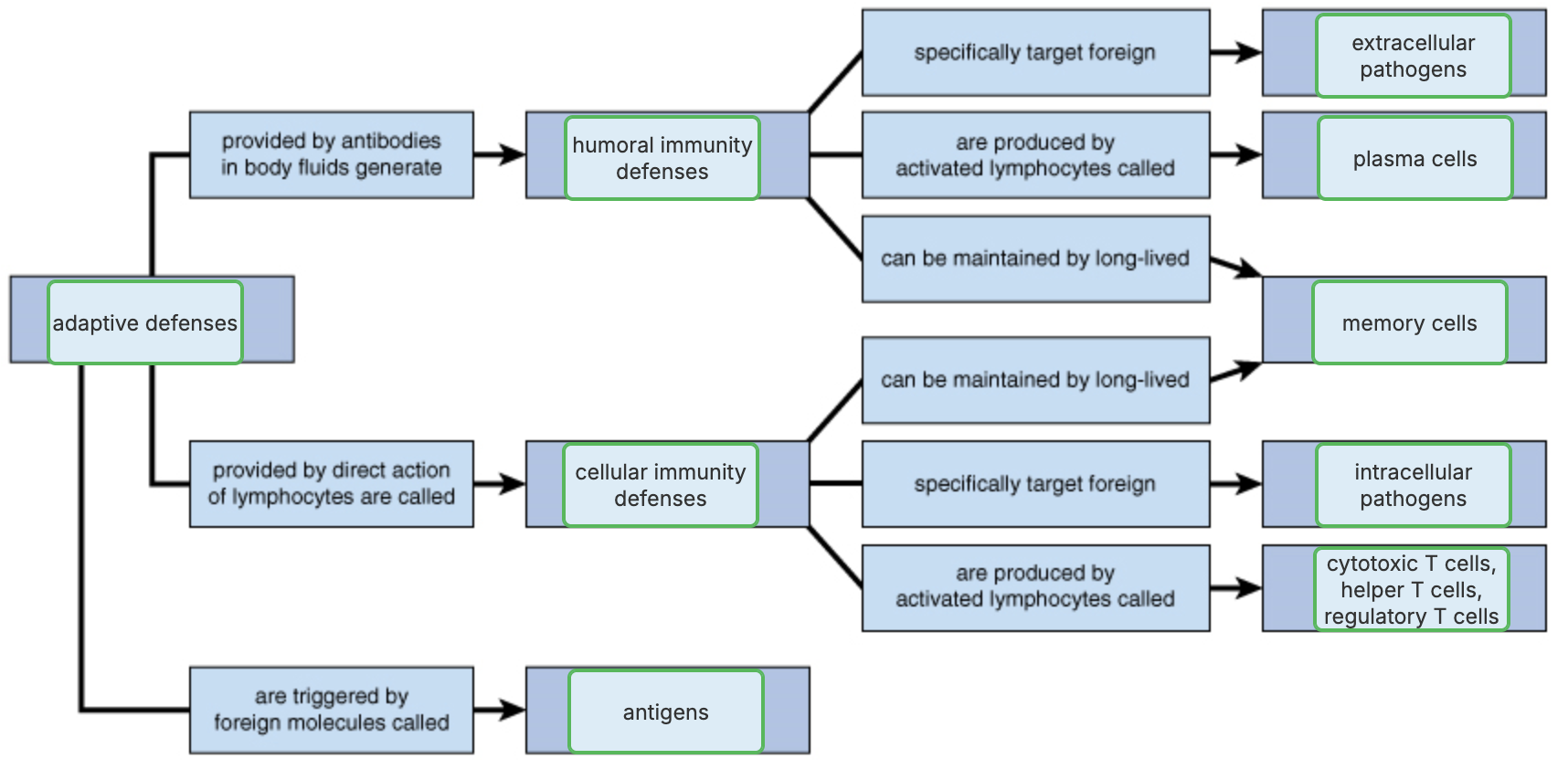
SUMMARY of ADAPTIVE DEFENSES

knowt flashcard image
65
New cards

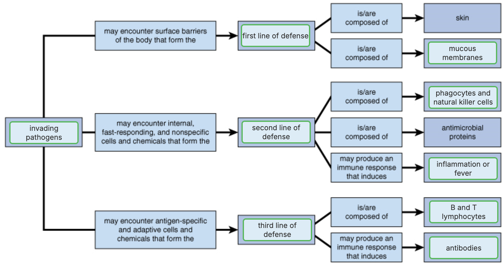
SUMMARY of 1st, 2nd, 3rd Line of Defense

knowt flashcard image
66
New cards

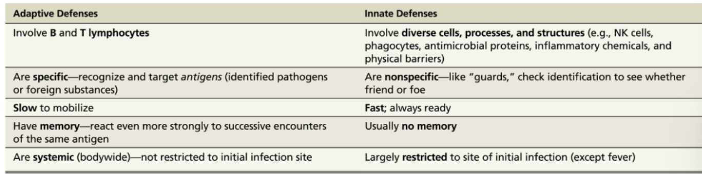
SUMMARY of Key Differences between Adaptive & Innate Defenses

knowt flashcard image
67
New cards

Tears and mucous membranes would be a part of which defense system?

Innate external defenses →  (surface barriers) are the first line of defense and include tears, mucous membranes, and the skin

68
New cards

Phagocytotic cells such as macrophages identify a variety of enemies by recognizing markers unique to pathogens.  They would be classified as which type of defense system?

Innate internal defenses

69
New cards

What type of immunity can be transferred by bodily fluids from one person to another, thus conferring immunity to the recipient?

Humoral immunity → involves antibodies that can be transferred from one person to another.

70
New cards

If a virus attacks a cell, which type of immunity would be activated?

Cell-mediated immunity → involves an intracellular pathogen such as a virus

Explore top flashcards

Rats Exam
Updated 532d ago
flashcards Flashcards (43)
Los deportes
Updated 990d ago
flashcards Flashcards (40)
Apush Amsco 24
Updated 1055d ago
flashcards Flashcards (32)
S2L6-Vocabulario
Updated 537d ago
flashcards Flashcards (93)
APUSH Unit 5
Updated 1034d ago
flashcards Flashcards (111)
Rats Exam
Updated 532d ago
flashcards Flashcards (43)
Los deportes
Updated 990d ago
flashcards Flashcards (40)
Apush Amsco 24
Updated 1055d ago
flashcards Flashcards (32)
S2L6-Vocabulario
Updated 537d ago
flashcards Flashcards (93)
APUSH Unit 5
Updated 1034d ago
flashcards Flashcards (111)