Oral Anatomy Lecture: Occlusion
1/129
There's no tags or description
Looks like no tags are added yet.
Name | Mastery | Learn | Test | Matching | Spaced |
|---|
No study sessions yet.
130 Terms
MD Central Incisors and MX 3rd Molars
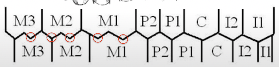
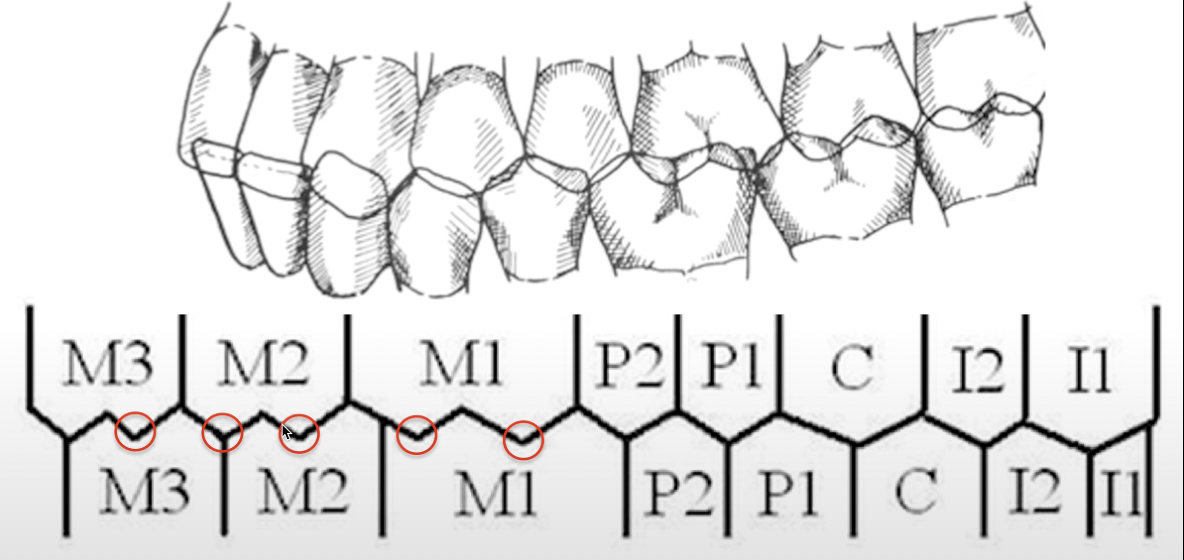
Which teeth occlude with only 1 tooth in the opposing arch?
MX lateral opposes incisal edge of MD lateral & canine but opposes no teeth at its incisal edge.
Explanation:
In dental occlusion, the term "opposes" refers to how the maxillary (upper) and mandibular (lower) teeth interact or come into contact during biting and chewing.
The maxillary lateral incisor (MX lateral) primarily opposes the incisal edges of the mandibular lateral incisor (MD lateral) and the mandibular canine.
However, the incisal edge of the MX lateral incisor does not directly oppose any other tooth incisal edge-to-incisal edge. Instead, it typically comes into contact at a point just below its edge with the mandibular teeth during occlusion.
This setup helps prevent direct edge-to-edge contact, which would otherwise lead to faster wear on the incisal edges. Instead, the teeth interlock in a way that distributes forces more efficiently along their surfaces.
MX lateral opposes incisal edge of which teeth?
Facial embrasure of MD teeth
Cusp tip of MX canine is in direct alignment of which anatomical item?
MD 1st and 2nd PMs
MX 1st PM opposes which teeth?
MD 2nd PM and 1st M
MX 2nd PM opposes which tooth?
Marginal ridge, Central and distal fossa
Lingual cusps of MX posterior teeth oppose which anatomical items of which teeth?
Buccal grooves and embrasures
Buccal cusps of MX posterior teeth oppose which anatomical items of which teeth?
Buccal groove of MD 1st M
Mesiobuccal cusp of MX 1st M opposes what anatomical item of which tooth?
Developmental groove of MD 1st M
The distobuccal cusp of which tooth opposes which anatomical feature?

MX Central Incisors
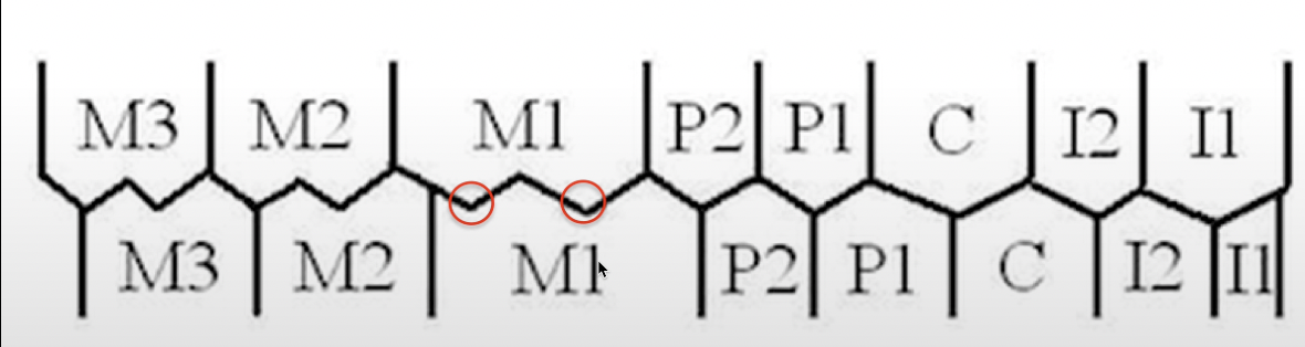
MD central incisors contact which teeth during protrusion and lateral protrusion?
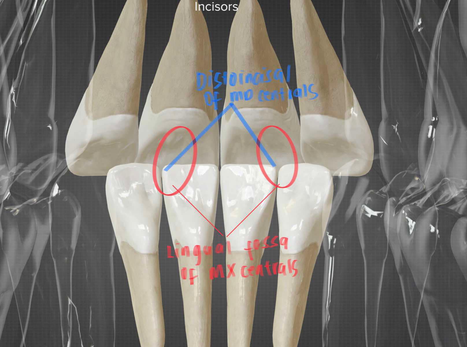
MX central incisors only — Distoincisal aspect of MD central opposes the lingual fossa of MX central
Explanation:
During protrusion, the mandibular central incisors contact only the maxillary central incisors, with the distoincisal aspect of the mandibular centrals opposing the lingual fossa of the maxillary centrals.
In lateral protrusion, this contact may also extend to the maxillary lateral incisors depending on the direction of movement.
MD Central opposes which aspect of which tooth?
Protrusion
When the lower jaw moves forward
Lateral protrusion
When the jaw moves forward and to the side
MMR area of MX 1st PM
Which anatomical feature does the buccal cusp of the mandibular first premolar contact on the opposing tooth?
The mesiobuccal cusp of the mandibular first molar contacts the mesiomarginal ridge (MMR) of the maxillary first molar and the distomarginal ridge (DMR) of the maxillary second molar.
Which anatomical features of the opposing teeth does the mesiobuccal cusp of the mandibular molar contact?
6
Lingual surfaces of maxillary incisors and canines
6
Labial surface of mandibular incisors and canines
16
Triangular ridges of maxillary buccal cusps of premolars and molars
16
Triangular ridges of lingual cusps of mandibular premolars and molars
8
Buccal embrasure of mandibular premolars and molars
10
Lingual embrasures of maxillary premolars and molars (including the canine and first premolar embrasure accommodating the mandibular premolar)
16
Lingual cusp points of maxillary premolars and molars
8
Distal fossae of premolars
12
Central fossae of the molars
6
Mesial fossae of the mandibular molars
6
Distal fossae of the maxillary molars
6
Lingual grooves of the maxillary molars
6*
Buccal grooves of the mandibular molars
16
Buccal cusp points of mandibular premolars and molars
Ideal Occlusion
↳ A point
↳ Preconceived concept – models at the stores.
↳ Perfect or optimal occlusion
↳ Theoretical concept of occlusal, structural, and functional relationship that includes idealized principle.
↳ Never found in natural dentition.
Normal Occlusion
↳ A range (EX. 4 - 6)
↳ Perfect and still has the right occlusion but not IDEAL.
↳ May spacing, may ikot, at may nakalabas.
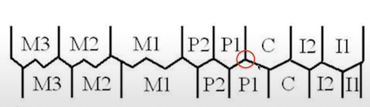
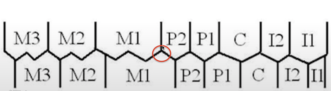
Interdigitation of the Teeth
↳ How are the teeth related to one another.
Plane of Occlusion
The _______ can be established by aligning it from the incisal surfaces of the maxillary and mandibular incisors up to the occlusal surfaces of the molars.
Occlusal Plane
We can define the _______ as an imaginary line that extends from the incisal edges of the front teeth and passes through the tips of the cusps of the posterior teeth.
Superior Labial Frenulum
Midline and parallel to RAL
MD central and MX 2nd M
Teeth occlude only in one teeth in primary
MD central and MX 3rd M
Teeth occlude only in one teeth in permanent
ellipse
Maxillary teeth describe the segment of an _____ and is larger than the segment following the same surfaces on mandibular teeth.
3 y.o.
All the roots and teeth should be fully formed if the child is ___ y.o.
year
A _____ or so after the teeth have fully erupted and have assumed their respective positions in the arches, the rapid development of the jaws is sufficient to create an interdental space or physiologic spacing, between some of them. Spaces in primary occlusion.
Primate Spaces
Anthropoid
Simian Spaces
Interdental Space
Physiologic Spacing
Spaces in primary occlusion
MX lateral to distal of MX canine
Primary spaces in maxilla
MD canine (distal) and MD 1st molar (mesial)
Primary spaces in mandible
crowding & misalignmentS
These spaces are essential in accommodating the larger permanent teeth, which helps prevent _____ and ______ as the child grows.
4-5 y.o
Primary spaces or separation of teeth usually start at the age of?
First permanent molars
After normal jaw growth has resulted in considerable separation, the occlusion is supported and made more efficient by the eruption and occlusion of the __________ immediately distal to the primary molars. (6 years of age)
Deciduous teeth
________ are important space retainers to accommodate the perm. teeth.
Occlusal Contact
Opposing Occlusal Surfaces: In posterior teeth (premolars and molars), the cusps and grooves on the occlusal (chewing) surfaces meet and fit into the opposing teeth in the opposite arch.
Proximal Contact
Mesial and Distal Contacts: Teeth have contact points on their mesial (front) and distal (back) surfaces where they meet adjacent teeth.
Incisal Contact
Opposing Incisal Edges: In anterior teeth (incisors and canines), the incisal edges or tips contact the incisal or occlusal surfaces of the opposing teeth.
2 y.o
The primary teeth should be in normal alignment and occlusion shortly after the age of?
A. Degree of bruxism
B. Factors related to the development of contact relations at the time of eruption of teeth:
Position of the tooth germ
Presence of permanent teeth
Development of condyles
Cuspal inclines
Neuromuscular influences
C. Tooth-tooth observations: ML cusp of MX molar occludes w/ the central fossa of deciduous MD molar.
Contact relations of teeth tend to vary with:
Normal TMJ relationship
Normal cusp relationship
TMD
Disease in TMJ
Centric relationship
Jaw to jaw relationship
Centric occlusion
Teeth to teeth
9 months
Arch form and width has been largely established by the age of?
Alveolar bone and basal bone
Determinants of the shape of dental arches
None
Primary Teeth: > 6mm
1 in 5
Primary Teeth: 3-5mm
1 in 2
Primary Teeth: > 3mm
2 in 3
No spacing
1 in 1
Crowded
Distal surface of the primary 2nd molar
Terminal plane naka-base sa?
Flush terminal plane
The distal surface of maxillary second molar is flush or coincides with the distal surface of mandibular 2nd molar.
Likely Class II
Distal step in deci. teeth
56% Class I, 44% Class II
Flush terminal plane in deci. teeth
More likely Class I, less likely Class III
Mesial step in deci. teeth
Physiologic Occlusion
Occlusion that deviates in one or more ways from ideal yet it is well adapted to that particular environment is esthetic & shows no pathologic manifestations.
Physiological Occlusion (Normal Occlusion)
This refers to a normal, ideal alignment where the teeth interdigitate (fit together) properly, with no malocclusion or misalignment issues.
Functional/Working Occlusion
An arrangement of teeth which will provide highest efficiency during excursive movements (moving the jaws from right to left and vice versa) of the mandible which is necessary during function.
Balanced/Non-working Occlusion
An occlusion in which balance & equal contacts are maintained throughout the entire arch during all excursions of the mandible.
Therapeutic Occlusion
It is a treated occlusion employed to counteract structural interrelationship. It is an approach to adjusting or modifying a patient's occlusion (bite) with the goal of improving function, comfort, or stability of the teeth and surrounding structures. This form of occlusion is often used as part of treatment for conditions affecting the masticatory system, such as temporomandibular joint disorders (TMD), bruxism, or periodontal disease.
Initial Contact
This is the point where, upon biting, only a specific area of the teeth makes contact. If this point of contact is unbalanced, it can lead to issues like traumatic occlusion.
Slide in Centric
This refers to the slight movement or sliding of the teeth as the patient adjusts their bite to achieve a more stable or complete contact (centric occlusion). It occurs when the initial contact point is off, and the bite slides to distribute forces more evenly across the teeth.
Freedom in Centric
Once the occlusion has been adjusted and treated (therapeutic occlusion), the teeth should reach a state of _________.
This means that when the patient bites down, there is no sliding, and centric occlusion.
The bite is stable and free from unbalanced forces, promoting long-term health of the periodontal structures and minimizing trauma.
Traumatic Occlusion
An occlusion judged to be causative factors in the formation of traumatic lesions of disturbance in the orofacial complex.
Trauma from Occlusion
It is defined as periodontal tissue injury caused by occlusal forces through abnormal occlusal contacts
Non Supporting, Guiding, Shear Cusp
Found on the buccal cusp of maxillary teeth and lingual cusp of mandibular posterior teeth.
Supporting, Centric Holding, Stamp Cusp
Found on the lingual cusp of maxillary teeth and buccal cusp of mandibular posterior teeth.
Condylar Guidance
is the posterior determinant of occlusion in the TMJ (temporomandibular joint).
Incisal/ANTERIOR Guidance
Refers to the anterior determinant of occlusion where the incisal edges of the lower incisors contact the lingual slopes of the maxillary (upper) incisors.
Canine Guided/Canine Protected Occlusion
In lateral movement, canine guidance occurs when only the cusp tips of the maxillary and mandibular canines make contact. For example, during a rightward jaw shift, the only teeth that touch are the cusp tips of the maxillary and mandibular canines, with no other cusps involved. This selective contact protects the posterior teeth by ensuring that only the canines bear the lateral forces, reducing wear on the other teeth.
Grouped Lateral/ Group Function Occlusion
Posterior teeth on the working be also contacted during lateral movement the mandible.
Protrusive Functional Occlusion
When the jaw moves forward the 6 mandibular- anterior teeth contact along the lingual inclines of the maxillary anterior teeth while the posterior disocclude.
Mastication, Speech, Appearance, and Stability
Importance of normal occlusion
KEY 1: Molar Relationship
Distal surface of the disto-buccal cusp of the upper first permanent molar occludes with the mesial surface of the mesio-buccal cusp of the lower second molar.
Or sometimes between the embrasures of max first and man 2nd molar.
KEY 2: Crown Angulation (MD Angulation)
(mesio-distal tip): the gingival part of the long axis of the crown is distal to the incisal part of the axis. The extent of angulation varies according to tooth type.
Perpendicular to 90 degree pero medyo offset of about 5 degree.
KEY 3: Crown Inclination
(labio- lingual, bucco-lingual): the incisors are at a sufficient angulation to prevent overeruption.
Upper posterior teeth: the lingual tip is constant and similar from canine to second premolar and increased in the molars.
Lower posterior teeth: the lingual tip increases progressively from the canines to the
The crown should be inclined labially.
KEY 4: No Rotation
Rotations are not present.
KEY 5: Presence of Tight Contact
There are no interdental spaces or no diastema present.
IN permanent teeth - pathologic spacing.
Pag malaki spaces - physiologic yun and that’s diastema.
KEY 6: Flat Curve of Spee
There is a flat plane of occlusion – normal occlusion.
KEY 7: Bolton Ratio
This key helps in analyzing the proportionality of the maxillary and mandibular teeth.
Each tooth is measured mesiodistally to ensure that each has the correct size for proper occlusion between the upper and lower teeth. This proportionality is based on the alignment of interproximal contacts between mesial and distal surfaces of the teeth.
MBDG OF MANDIBULAR
MBC OF MAXI 1ST MOLAR OPPOSES WHICH ANATOMICAL ITEM? - Class I
MBC EMBRASURES OF MANDI 2ND PM AND 1ST MOLAR
MBC OF MAXI 1ST MOLAR OPPOSES WHICH ANATOMICAL ITEM? - Class II
STATIC CONCEPT/STATIONARY OCCLUSION/FIXED
Contact between upper and lower closed, enclosed proximity
Stability and placement of the teeth
Dynamic Occlusion
Functions of stomatognathic system (teeth, muscles, nerves, TMJ and bones) as a whole
Mouth, Jaw
STOMA - ____
MATHIC - _____
2-3 mm
Normal measurement of OJ
1-2 mm
Normal measurement of OJ (Primary)